. 2004 Jan;15(1):294-309.
doi: 10.1091/mbc.e03-05-0352. Epub 2003 Oct 31.

Mechanism of human dermal fibroblast migration driven by type I collagen and platelet-derived growth factor-BB

Affiliations

Mechanism of human dermal fibroblast migration driven by type I collagen and platelet-derived growth factor-BB

Wei Li et al. Mol Biol Cell. 2004 Jan.

Abstract

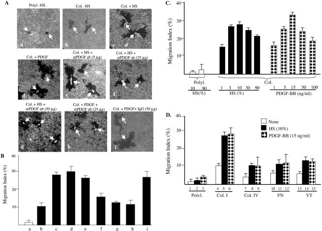
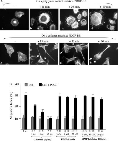
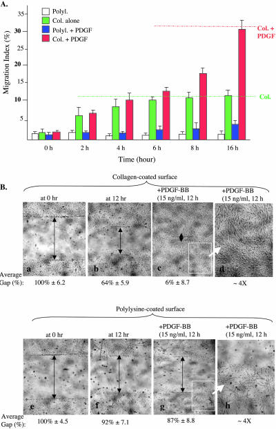
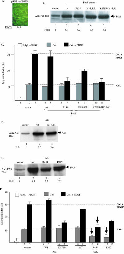
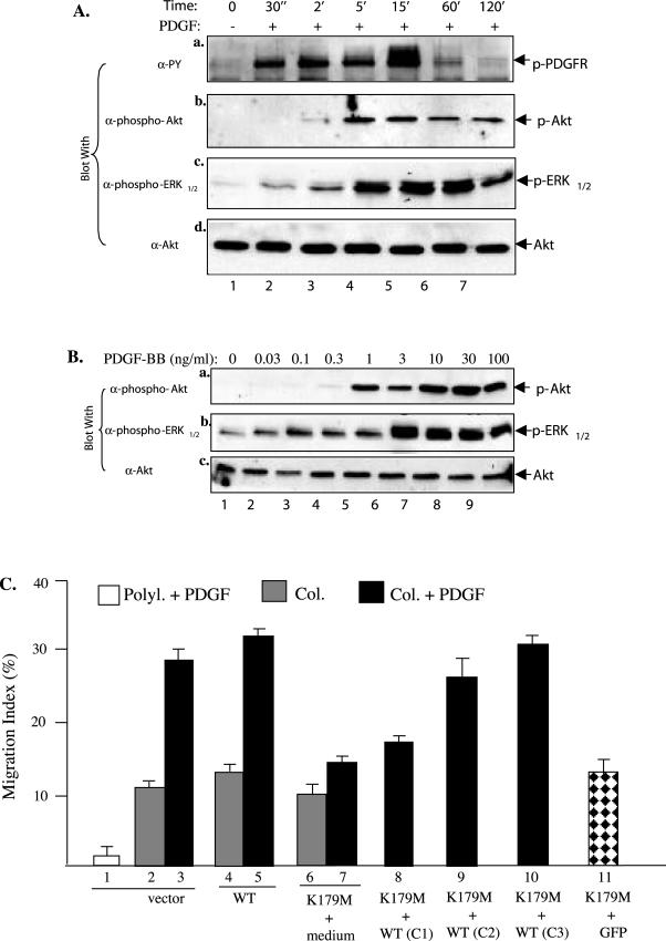
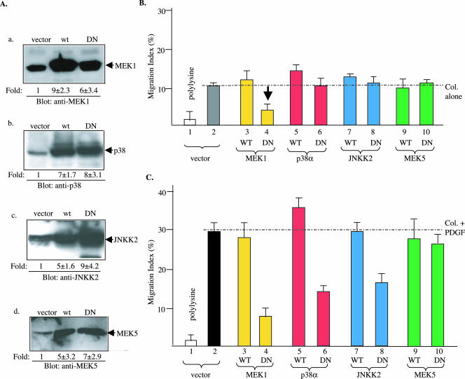
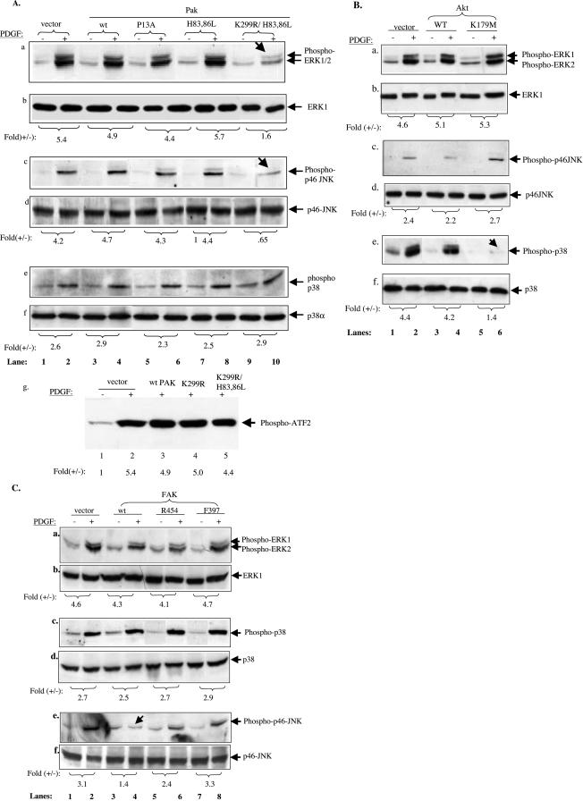
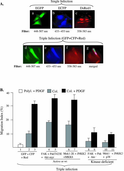
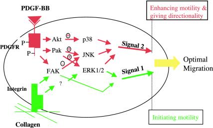
Migration of human dermal fibroblasts (HDFs) is critical for skin wound healing. The mechanism remains unclear. We report here that platelet-derived growth factor-BB (PDGF-BB) is the major promotility factor in human serum for HDF motility on type I collagen. PDGF-BB recapitulates the full promotility activity of human serum and anti-PDGF neutralizing antibodies completely block it. Although collagen matrix initiates HDF migration without growth factors, PDGF-BB-stimulated migration depends upon attachment of the cells to a collagen matrix. The PDGF-BB's role is to provide directionality and further enhancement for the collagen-initiated HDF motility. To study the collagen and PDGF-BB "dual signaling" in primary HDF, we establish "gene cassettes" plus lentiviral gene delivery approach, in which groups of genes are studied individually or in combination for their roles in HDF migration. Focal adhesion kinase, p21(Rac,CDC42)-activated kinase and Akt are grouped into an upstream kinase gene cassette, and the four major mitogen-activated protein kinases (extracellular signal-regulated kinase 1/2, p38, c-Jun NH2-terminal kinase, and extracellular signal-regulated kinase 5) are grouped into a downstream kinase gene cassette. The experiments demonstrate 1) the genes' individual roles and specificities, 2) their combined effects and sufficiency, and 3) the mechanisms of their intermolecular connections in HDF migration driven by collagen and PDGF-BB.


Figures

Figure 1.
Figure 1.
PDGF-BB is the major promotility factor in human serum for HDFs. Primary neonatal HDFs, serum starved for 18 h, were seeded onto colloidal gold coverslips, which were coated with either poly-l-lysine (Polyl., 10 μg/ml) as a nonphysiological control matrix or type I collagen (Col, 45 μg/ml) for 2 h. Cells were incubated in media with or without indicated concentrations of either human serum (HS, 10% or as indicated) or human recombinant PDGF-BB (15 ng/ml or as indicated). To block PDGF-BB' action in humans serum, human serum-containing media were added with either anti-PDGF neutralizing antibodies (5, 25, or 50 μg/ml) or control IgG for 45 min before being used in migration assays. HDF migration caused colloidal gold-free areas (“tracks”), which were quantified by a computer-assisted analysis as MI (see MATERIALS AND METHODS). (A) The representative images of single-cell migration tracks under indicated conditions. (B) Computer-assisted quantification of 15 randomly selected microscopic fields per condition. (C) HDF migration in response to various doses of HS or PDGF-BB. (D) Comparison of HDF motility on four indicated ECMs without or with HS or PDGF-BB. Col. IV, collagen IV, FN, fibronectin, VT, vintronectin. The results represent those of three independent experiments.
Figure 3.
Figure 3.
PDGF-BB stimulates polarization of HDFs on collagen and MMP participation. (A) HDFs were seeded on polylysine or on collagen-coated coverslips. After attachment, cells were serum starved overnight and then untreated or treated with PDGF-BB (15 ng/ml) for the indicated time. Cells were fixed and stained with rhodamine-conjugated phalloidin (see MATERIALS AND METHODS). Morphological differences of the cells on either polylysine (a–d) or on collagen (f–i) in the absence (a and f) or presence (b, c, d and g, h, i) of PDGF-BB were analyzed under a fluorescent microscope and the representative images photographed. Approximately 80 randomly selected cells per condition were examined and quantified by taking the percentage of polarized HDFs. Less than 14% and >78% of HDFs were polarized in the absence or presence of PDGF-BB, respectively. (B) Serum-starved HDFs were subjected to colloidal gold migration assays in the absence or presence of indicated concentrations of MMP inhibitors, GM6001, TIMP-1 and MMP Inhibitor III. After 14 h of incubation, migration was analyzed as described previously. Results from one of two similar experiments are presented.
Figure 2.
Figure 2.
Collagen initiates and PDGF further enhances HDF migration. Serum-starved HDFs were subjected to colloidal gold migration assay or in vitro wound healing assay. (A) Time course of HDF migration under four well-defined conditions: i) on polylysine (white bars), on collagen (green bars), on polylysine plus PDGF-BB (blue bars), and on collagen plus PDGF-BB (red bars). Migration was stopped at the indicated time points by cell fixation and subjected to computer-assisted analyses (for MIs). Representative results of three independent experiments are shown. (B) Serum-starved HDFs were plated in six-well tissue culture dishes precoated with either polylysine or collagen. After cell attachment (∼2 h), “wounds” (scratches) were made at the middle of the wells, and free cells were removed. Media were then changed to fresh ones with or without PDGF-BB and incubated for additional 14 h. Mitomycin C (to block proliferation) was present in all wells. Closure of wound was photographed and quantified as described in MATERIALS AND METHODS. d and h, amplified partial images of c and g, respectively, to closely visualize changes of the cells on polylysine or collagen after PDGF-BB treatment. This experiment was repeated four times and similar results were obtained.
Figure 4.
Figure 4.
Specificities of cassette I genes in collagen- and PDGF-BB–driven HDF motility HDFs were infected with lentiviruses carrying wild-type (wt) or mutants of Pak, Akt, and FAK genes or a control EGFP gene. Infection was stopped after 6 h and cells were incubated in growth media for additional 48 h. After serum starvation, cells were subjected to Western blot analyses for protein expression and colloidal gold migration assays. (A) Fluorescence-activated cell sorting analysis of EGFP-positive cells over total cells (%). (B, D, and E) Immunoblots of equal amounts of cell lysates (50 μg of total proteins) with indicated antibodies. Intensity of the bands was quantified by scanning densitometry and estimated as fold increases in reference with their endogenous expressions (lanes 1). (C and F) MIs of the cells on polylysine or collagen with or without PDGF-BB (15 ng/ml) are presented. (C) Effects of wt or mutants of Pak1. (F) Effects of wt or mutant Akt and FAK on collagen- or collagen plus PDGF-stimulated HDF migration. A representative experiment of four independent experiments is shown.
Figure 5.
Figure 5.
Akt is a primary target of PDGF-BB signaling. (A) Kinetics of PDGF-BB–stimulated activation of PDGFR, Akt and ERK1/2 in HDFs. HDFs were serum starved and treated without or with PDGF-BB (15 ng/ml) for the indicated times. Equalized cell lysates (50 μg of total proteins) were immunoblotted with the indicated antibodies. The anti-Akt blot was included as a loading control. (B) Comparison of the sensitivities of Akt and ERK1/2 activations in response to increasing concentrations of PDGF-BB. HDFs were treated with the indicated concentrations of PDGF-BB for 5 min and analyzed for activation of Akt and ERK1/2 by blotting with the indicated antibodies, as described above. Akt protein levels were used as a control for equal loadings. (C) Reversal of Akt-K179M' inhibition by overexpressing wt Akt. HDFs were infected with vector (1–3), wt Akt (4 and 5), Akt-K179M or Akt-K179M mixed with increasing amounts of wt Akt. The total volume of infection was 2 ml, in which 1 ml of virus was mixed with 1 ml of medium for all single infections. For coinfections, 1 ml of Akt-K179M virus was mixed with 1 ml of 25% (C1), 50% (C2), and 75% (C3) of wt Akt viruses. The cells were then subjected to colloidal gold migration assays. This experiments was repeated twice and similar results were obtained.
Figure 6.
Figure 6.
Strong specificities of cassette II MAPKs between collagen- and PDGF-BB–driven HDF migration HDFs were infected with lentivirus carrying either a control EGFP gene (vector) or the wild-type or mutant genes of four MAPK pathways as indicated. (A) Equalized lysates (50 μg of total proteins) of the infected cells were analyzed for expression of the corresponding MAPK pathway genes 48 h after infection by Western blots using indicated antibodies. The fold increases in intensity over the endogenous expression are estimated by densitometry scanning. (B) MIs of HDF migration on collagen in the absence of PDGF-BB showing the effects of MAPK genes. (C) MIs of HDF migration on collagen plus PDGF-BB showing the effects of MAPK genes. Open bar, polylysine control; all other bars, Col alone or Col plus PDGF-BB. A representative experiment of three independent experiments is presented.
Figure 7.
Figure 7.
Cassette I kinases connect to distinct cassette II downstream MAPK cascades. Serum-starved HDFs expressing wt or mutants of Pak1, Akt, or FAK were either untreated (-) or treated (+) for 10 min with PDGF-BB (15 ng/ml). Equalized cell extracts (50 μg of total proteins) were subjected to analyses for activation of ERK1/2, p38, and JNK by using corresponding anti-phospho-MAPK antibodies. Duplicate blots were probed with anti-ERK1/2, anti-p38, and anti-JNK antibodies to show the loaded protein levels. (A) Effects of wt and mutants of Pak1 on PDGF-stimulated activation of ERK1/2 (a and b), JNK (c and d), and p38 (e and f). Each of the top panels (a, c, and e) was blotted with anti-phospho-MAPK antibodies, and the bottom panels (b, d, and f) with corresponding anti-MAPK protein antibodies. (g) The same set of cell extracts was immunoprecipitated with an anti-p38 antibody, and the immunocomplexes were tested for p38 kinase activity toward purified ATF-2 by using an in vitro p38 kinase assay kit (#9820; Cell Signaling Technology). (B and C) HDFs, expressing the wt or mutants of Akt or FAK were subjected to similar experiments as in A with indicted antibodies. Fold (+/-) referred to PDGF-stimulated over unstimulated activation of the indicated MAPKs. The data were quantified by densitometry scanning of the phosphorylated bands against their corresponding protein control bands. Calculations were based on references to each of their corresponding MAPK protein bands. Four independent experiments were carried out for Pak-, Akt-, and FAK-infected HDFs. In statistical evaluations comparing before and after PDGF stimulation, all Pak, Akt, or FAK mutants gave p < 0.05–0.01 by paired t tests.
Figure 8.
Figure 8.
Combined effects of cassette I and cassette II genes after multiple lentiviral coinfections. (A) HDFs were either infected with a single or coinfected with three lentiviruses carrying an EGFP, an ECFP, or a DsRed gene. Individual expression of the three genes shows the expected colors of fluorescence (a–c). Simultaneous expression of three genes in the same cells shows the feasibility of introducing multiple genes into cells by the lentiviral system. (B) HDFs were triple infected with either the three fluorescent genes (1–3), wt FAK plus PAK-T423E plus Akt-myr (4 and 5), or Mek1–2E plus wt JNKK2 plus MKK6 (6 and 7), or kinase-dead FAK, Pak, and Akt (8 and 9), or kinase-dead Mek1, JNKK2, and p38 (10 and 11). Cells were then subjected to colloidal gold migration assays in the absence or presence of PDGF-BB (15 ng/ml), as described.
Figure 9.
Figure 9.
A schematic dual signaling model of collagen and PDGF-BB in the control of HDF migration. Collagen matrix initiates HDF migration (signal 1, green). PDGF-BB enhances and provides directionality for the collagen-driven migration (signal 2, red). We propose that collagen and PDGF-BB together determine the optimal HDF motility. Collagen matrix and PDGF-BB use specific and common signaling pathways. Arrows do not implicate direct activations.

Similar articles

Cited by

References

    1. Albrecht-Buehler, G. (1977). The phagokinetic tracks of 3T3 cells. Cell 11, 395-404. - PubMed
    1. Adelmann-Grill, B.C., Wach, F., Cully, Z., Hein, R., and Krieg, T. (1990). Chemotactic migration of normal dermal fibroblasts towards epidermal growth factor and its modulation by platelet-derived growth factor and transforming growth factor-beta. Eur. J. Cell Biol. 51, 322-336. - PubMed
    1. Bagrodia, S., Derijard, B., Davis, R.J., and Cerione, R.A. (1995). Cdc42 and PAK-mediated signaling leads to Jun kinase and p38 mitogen-activated protein kinase activation. J. Biol. Chem. 270, 27995-27998. - PubMed
    1. Bauer, E.A., Cooper, T.W., Huang, J.S., Altman, J., and Deuel, T.F. (1985). Stimulation of in vitro human skin collagenase expression by platelet-derived growth factor. Proc. Natl. Acad. Sci. USA 82, 4132-4136. - PMC - PubMed
    1. Blatti, S.P., Foster, D.N., Ranganathan, G., Moses, H.L., and Getz, M.J. (1988). Induction of fibronectin gene transcription and mRNA is a primary response to growth-factor stimulation of AKR-2B cells. Proc. Natl. Acad. Sci. USA 85, 1119-1123. - PMC - PubMed

Publication types

MeSH terms

LinkOut - more resources