. 2004 Jan;15(1):397-406.
doi: 10.1091/mbc.e03-05-0281. Epub 2003 Oct 31.

Novel membrane protein shrew-1 targets to cadherin-mediated junctions in polarized epithelial cells

Affiliations

Novel membrane protein shrew-1 targets to cadherin-mediated junctions in polarized epithelial cells

Sanita Bharti et al. Mol Biol Cell. 2004 Jan.

Abstract

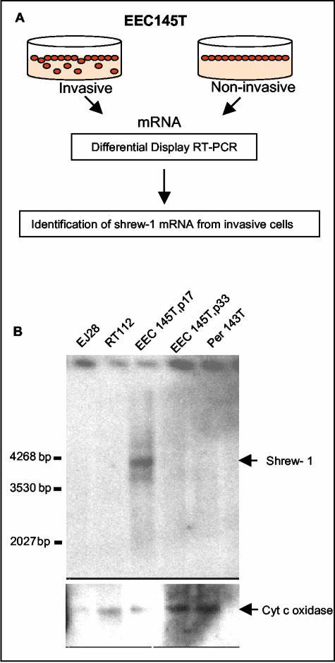
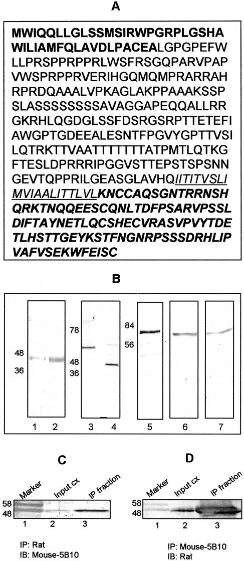
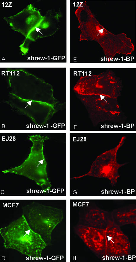
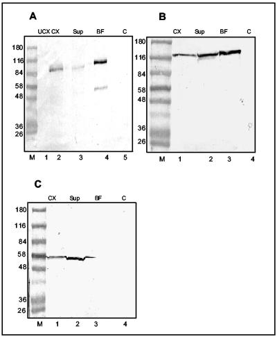
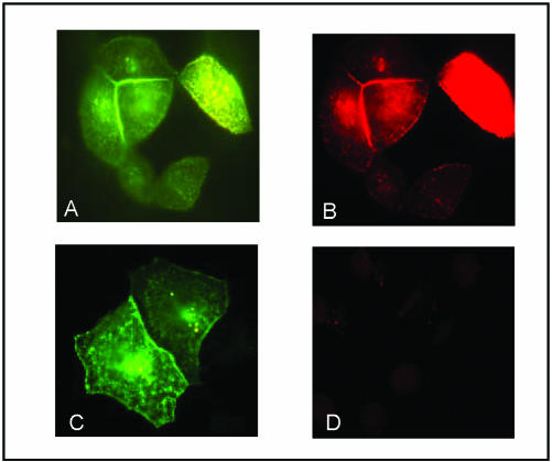
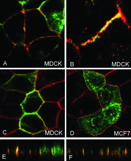
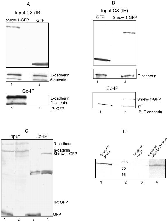
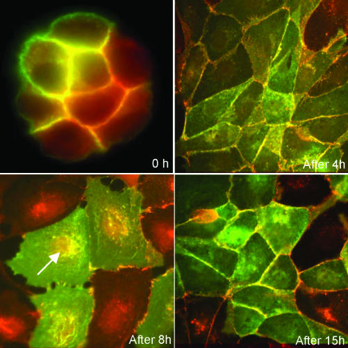
While searching for potential candidate molecules relevant for the pathogenesis of endometriosis, we discovered a 2910-base pair cDNA encoding a novel putative 411-amino acid integral membrane protein that we called shrew-1. The putative open-reading frame was confirmed with antibodies against shrew-1 peptides that labeled a protein of approximately 48 kDa in extracts of shrew-1 mRNA-positive tissue and also detected ectopically expressed shrew-1. Expression of epitope-tagged shrew-1 in epithelial cells and analysis by surface biotinylation and immunoblots demonstrated that shrew-1 is indeed a transmembrane protein. Shrew-1 is able to target to E-cadherin-mediated adherens junctions and interact with the E-cadherin-catenin complex in polarized MCF7 and Madin-Darby canine kidney cells, but not with the N-cadherin-catenin complex in nonpolarized epithelial cells. Direct interaction of shrew-1 with beta-catenin in in vitro pull-down assay suggests that beta-catenin might be one of the proteins that targets and/or retains shrew-1 in the adherens junctions. Interestingly, shrew-1 was partially translocated in response to scatter factor (ligand of receptor tyrosine kinase c-met) from the plasma membrane to the cytoplasm where it still colocalized with endogenous E-cadherin. In summary, we introduce shrew-1 as a novel component of adherens junctions, interacting with E-cadherin-beta-catenin complexes in polarized epithelial cells.


Figures

Figure 1.
Figure 1.
(A) Diagram depicting DDRT-PCR performed with invasive and noninvasive passages of the endometriotic cell line EEC145T, leading to the identification of shrew-1 mRNA. (B) The 391-base pair cDNA was used as a probe to test for the presence of shrew-1 mRNA in endometriotic and carcinoma cell lines. Poly A+ RNA was prepared from the cell lines EJ28 (invasive bladder carcinoma), RT112 (noninvasive bladder carcinoma), EEC145T (p17, invasive passage 17; p33, noninvasive passage 33 of the endometriotic cell line), and Per 143T (peritoneal cells immortalized with simian virus 40 T antigen). A Northern blot probed with 32P-labeled shrew-1 probe detected an mRNA of ∼4 kb in the invasive endometriotic cell line. Bottom, membrane was reprobed with cytochrome c oxidase to check the integrity and loading of the RNA samples. p17 and p33 are on the same blot at the same exposure-same as cytochrome c oxidase control.
Figure 2.
Figure 2.
(A) The complete 411-amino acid sequence of the shrew-1 protein. The putative signal peptide is depicted in bold letters and the transmembrane domain is underlined. (B) Lanes 1 and 2 show the endogenous expression of shrew-1 protein in pancreas and uterus sections, respectively, as detected by immunoblotting by using the mAb against shrew-1; lanes 3 and 4 show the autoradiography of in vitro-translated luciferase control cDNA and shrew-1-BP, respectively, after separation by SDS-PAGE; lane 5 depicts shrew-1-GFP expressed in MCF7 cells, as detected by monoclonal GFP antibody, lane 6 shows shrew-1-GFP detected by the rat polyclonal antibody against shrew-1 cytoplasmic peptide, and lane 7 shows shrew-1-GFP detected by the mAb generated against shrew-1 peptide of the putative ectodomain. (C) Immunoprecipitation of endogenous shrew-1 from uterus cell extracts by using shrew-1 polyclonal antibody and detection with the mAb. Lane 2 depicts the input cell extract and lane 3 shows the immunoprecipitated protein detected with the mAb. (D) Immunoprecipitation of endogenous shrew-1 from uterus cell extracts using shrew-1 mAb and detection with the polyclonal antibody. Lane 2 depicts the input cell extract and lane 3 depicts the immunoprecipitated protein detected with the polyclonal antibody. Input is 10% of the total cell extract. Lane 1 in each C and D depicts the marker proteins.
Figure 3.
Figure 3.
Membrane localization of shrew-1. Shrew-1 tagged with GFP (shrew-1-GFP) or BP (shrew-1-BP) was expressed in the eukaryotic epithelial cells 12Z (human invasive endometriotic cell line), RT112 (human bladder carcinoma cell line, noninvasive), EJ28 (human bladder carcinoma cell line, invasive), and MCF7 (human breast carcinoma cell line, noninvasive). A–D show shrew-1-GFP fluorescence and E–H show immunofluorescence signals by using a mouse mAb against the BP tag visualized by a mouse-specific fluorochrome-conjugated secondary antibody. The arrows indicate the expression of shrew-1 at the membrane. The arrows depict the areas of cell-cell contact where shrew-1 is concentrated. The cells shown are transfected cells which border a few nontransfected cells.
Figure 4.
Figure 4.
Cell surface biotinylation of MCF7 cells transfected with shrew-1-GFP. The biotinylated cell surface proteins were pulled down with neutravidin-coupled beads. The proteins present in various cell extract fractions were analyzed by Western blots. (A) Shrew-1-GFP was detected by anti-GFP antibody (lanes 1–5). (B) E-cadherin, a positive control membrane protein, was detected by a mAb against E-cadherin (lanes 1–4). (C) Pyruvate kinase, a negative control cytosolic protein, was detected with a specific antibody (lanes 1–4). UCX, untransfected cell extract; CX, transfected cell extract; sup, supernatant after pull down of the biotinylated fraction; BF, pulled-down biotinylated fraction; C, control of neutravidin beads bound to nonbiotinylated cell extract.
Figure 5.
Figure 5.
Carboxy terminus of shrew-1 is cytoplasmic. Shrew-1-GFP–transfected MCF7 cells were permeabilized (A and B) or not permeabilized (C and D), as described under MATERIALS AND METHODS and then subjected to immunofluorescence staining with anti-GFP antibody and Alexa 594-labeled secondary goat anti-mouse antibody (B and D, red fluorescence). Intrinsic GFP fluorescence is green (A and C). Shrew-1-GFP could be detected in permeabilized cells by immunostaining with anti-GFP antibody (B) but not if the cells were not permeabilized (D).
Figure 6.
Figure 6.
Colocalization of shrew-1-GFP with endogenous E-cadherin (red) at the membrane in MDCK cells (A–C); and in MCF7 cells (D) along the xy-axis as seen in the confocal microscope. Colocalization at the junctions is seen along the xz-axis with the confocal microscope (E and F).
Figure 7.
Figure 7.
Interaction between shrew-1 and E-cadherin shown by coimmunoprecipitation. (A) MCF7 cells transfected with shrew-1-GFP (lanes 1 and 3) or with GFP (lanes 2 and 4) were subjected to immunoprecipitation with anti-GFP. To test antigen content, 10% of the total cell extract (input) was immunoblotted (IB) with anti-GFP (top) and anti-E-cadherin plus anti-β-catenin (middle) antibodies. Coimmunoprecipitations (CoIP) were performed with anti-GFP antibody and the immunoprecipitates subjected to immunoblotting with anti-E-cadherin and then anti-β-catenin antibodies (lanes 3 and 4). (B) In the reverse experiment, the cell extracts from MCF7 cells transfected with GFP (lanes 1 and 3) or shrew-1-GFP (lanes 2 and 4) were subjected to IP with anti-E-cadherin antibody. Input panels depict 10% of the cell extracts immunoblotted with anti-GFP antibody (top) or endogenous E-cadherin protein immunoblotted with anti-E-cadherin antibody (bottom). CoIP were performed with E-cadherin antibody, and shrew-1 was detected by immunoblotting with anti-GFP antibody as seen in lane 4. CX denotes the total cell extract. (C) Coimmunoprecipitation of N-cadherin and shrew-1-GFP. A, EJ28 cells were transfected with GFP (lanes 1 and 3) or shrew-1-GFP (lanes 2 and 4). Input shows 10% of the total cell extracts (lanes 1 and 2). Immunoprecipitation (CoIP) was performed with GFP-antibody (lanes 3 and 4). Immunoblotting was performed with antibodies against GFP, N-cadherin, and β-catenin. No interaction of shrew-1 with N-cadherin and β-catenin was observed. (D) Direct interaction of β-catenin with the cytoplasmic domain of shrew-1 (GST-CPD-shrew) in an in vitro pull-down assay. Full-length β-catenin was translated in vitro using [35S]methionine. GST and GST-CPD-shrew were purified on glutathione-Sepharose beads, and then incubated at RT for 1 h with radioactively labeled β-catenin. After washing the beads, samples prepared as described under MATERIALS AND METHODS, were subjected to SDS-PAGE and autoradiography. Lane 1, radioactive β-catenin as input; lane 2, the marker; lane 3; GST alone with β-catenin; and lane 4, GST-CPD-shrew with β-catenin.
Figure 8.
Figure 8.
Effect of SF on MDCK cells. MDCK cells transfected with shrew-1-GFP were seeded at very low density on coverslips and were grown until formation of small colonies. SF/HGF was added at a concentration of 20 ng/ml to these cells, and the effect was monitored on coverslips from the same culture dish at 0, 4, 8, and 15 h. After 8 h, cell-cell contacts were disrupted but colocalization of shrew-1-GFP (green) and endogenous E-cadherin (red) could also be seen in cytoplasmic vesicles. After 15 h, shrew-1-GFP and endogenous E-cadherin colocalized again predominantly at the plasma membrane.

References

    1. Aberle, H., Butz, S., Stappert, J., Weissig, H., Kemler, R., and Hoschuetzky, H. (1994). Assembly of the cadherin-catenin complex in vitro with recombinant proteins. J. Cell Sci. 107, 3655-3663. - PubMed
    1. Aberle, H., Schwartz, H., Hoschuetzky, H., and Kemler, R. (1996). Single amino acid substitutions in proteins of the armadillo gene family abolish their binding to alpha-catenin. J. Biol. Chem. 271, 1520-1526. - PubMed
    1. Akhtar, N., and Hotchin, N.A. (2001). RAC1 regulates adherens junctions through endocytosis of E-cadherin. Mol. Biol. Cell 12, 847-862. - PMC - PubMed
    1. Anastasiadis, P.Z., Moon, S.Y., Thoreson, M.A., Mariner, D.J., Crawford, H.C., Zheng, Y., and Reynolds, A.B. (2000). Inhibition of RhoA by p120 catenin. Nat. Cell Biol. 2, 637-644. - PubMed
    1. Birchmeier, W., Weidner, K.M., and Behrens, J. (1993). Molecular mechanisms leading to loss of differentiation and gain of invasiveness in epithelial cells. J. Cell Sci. Suppl. 17, 159-164. - PubMed

Publication types

MeSH terms