. 2003 Nov 11;42(44):12805-12.
doi: 10.1021/bi035370p.

Lecithin retinol acyltransferase is a founder member of a novel family of enzymes

Affiliations

Lecithin retinol acyltransferase is a founder member of a novel family of enzymes

Wan Jin Jahng et al. Biochemistry. .

Abstract

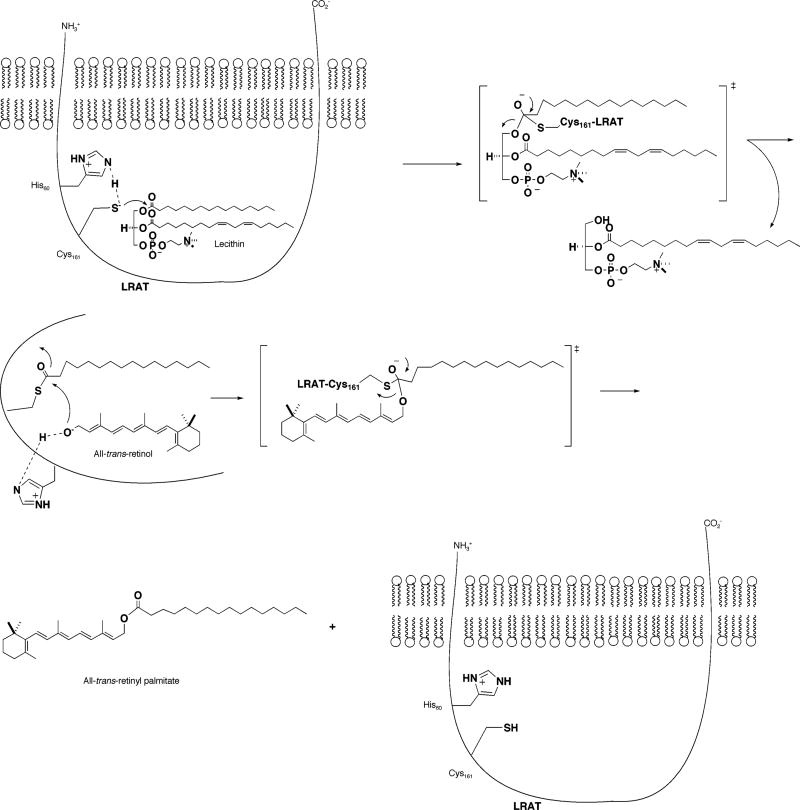
Lecithin retinol acyltransferase (LRAT) catalyzes the reversible esterification of vitamin A using lecithin as the acyl donor. LRAT is the founder member of a new class of enzymes, which include class II tumor suppressors, proteins essential for development, and putative proteases. All of these proteins possess Cys and His residues homologous to C161 and H60 of LRAT. These two residues are shown here to be essential for LRAT activity and are part of a catalytic dyad reminiscent of that found in thiol proteases. However, the local primary sequence contexts of C161 and H60 of LRAT and family are not at all homologous to those found in the approximately 20 thiol protease families. Moreover, LRAT shows pKs of 8.3 and 10.8, compared to approximately 4.0 and 8.5 observed in the thiol proteases. LRAT also contains Gln177 and Asp67 residues, which are largely conserved in the homologues. However, neither of these residues is essential for catalysis. Thiol proteases often contain catalytically essential Asp or Gln residues. It is concluded that LRAT is the founder member of a new class of Cys-His enzymes with diverse functions.


Figures

Figure 1
Figure 1
tLRAT labeling by affinity labeling reagents. (A) HPLC chromatogram of [11,12-3H2]RCA (2) labeled tLRAT peptide. RPHPLC was performed on a microcapillary C18 column (Vydac 218TP52). The peptides were eluted with a linear gradient from 0 to 70% acetonitrile with 0.1% TFA. Fractions were collected every minute (50 µL), and radioactivity was monitored by scintillation counting (Beckman LS6500) using Ultima Gold scintillation cocktail. (B) tLRAT labeling by alkali-cleavable affinity reagent BRCA (3). tLRAT was visualized by Coomassie blue staining, Western blot by anti-LRAT antibody/anti-rabbit Ig-HRP/ECL, and biotin blot by avidin-HRP/ ECL. The 40 kDa band represents the tLRAT homodimer. (C) MALDI TOF analysis of the BACMK (4) labeled tLRAT peptide. One peptide (MW = 1315) represents NNCEHF (763) + BACMK - Cl - Boc (552).
Figure 2
Figure 2
Steady-state kinetic analysis of LRAT. Retinyl ester formation by WT LRAT (○) and H57Q (□) is shown. The same numbering system based on full-length LRAT is used for tLRAT as well to avoid confusion. (A) Full-length LRAT and the mutants. H57Q and H60Q mutants were generated by the overlapping extension method. Expression and membrane protein preparation were performed as described in Methods. The calculated Vmax values of WT and H57Q are 122.58 ± 4.92 and 67.50 ± 3.65 nmol min−1 mg−1, respectively. The KMs are 0.65 ± 0.03 and 0.29 ± 0.08 µM, respectively. (B) tLRAT and the mutants. Mutagenesis was conducted using the QuikChange site-directed mutagenesis kit (Stratagene Inc.). The Vmax values of tLRAT and H57Q are 16.41 ± 0.38 and 12.70 ± 0.67 mmol min−1 mol−1, respectively. The KMs are 1.67 ± 0.24 and 2.73 ± 0.37 µM, respectively. No activity was observed in H60Q and the empty vectors in both (A) and (B).
Figure 3
Figure 3
Normalized all-trans-retinyl ester formation by tLRAT mutants using all-trans-retinol as substrate. Esterification activity of tLRAT was monitored by following the formation of all-trans-retinyl esters using 3H-labeled all-trans-retinol (0.2 µM) along with DPPC (220 µM), 0.6% BSA (0.6%), EDTA (1 mM), DTT (2 mM), and CHAPSO (0.1%) in Tris buffer (100 mM, pH 8.3). The values are the average of triplicate measurements.
Scheme 1
Scheme 1
LRAT Mechanism
Scheme 2
Scheme 2
Affinity Labeling Reagents of LRAT
Scheme 3
Scheme 3
Multiple Sequence Alignment of the LRAT Familya a Multiple sequence alignment of the LRAT family was performed by using CLUSTAL W 1.82 (http://www.ebi.ac.uk/clustalw/). Fully conserved His, Asn, and Cys are in red, the predicted transmembrane domain is in blue, and highly conserved Asn and Gln are in pink. Accession numbers are as follows: human LRAT (GP:AF 071510), bovine LRAT (GP:AF 275344), mouse LRAT (GP:AF 255061), rat LRAT (GP:AF 255060), mouse Hrev107 (AAH24581), rat Hrev107 (X76453), human Hrev107-3 (P53816), tazarotene-induced gene protein (TIG3) (AF060228), echovirus 23, human parechovirus 1 (L02971), human parechovirus 2 (AJ005695), avian encephalomyelitis virus (AJ225173), EGL26 (NP493652), and aichivirus (AB010145).
Scheme 4
Scheme 4
Active Site Labeling of tLRAT by BRCA 3

Similar articles

Cited by

References

    1. Fulton B, Rando RR. Biochemistry. 1987;26:7938–7945. - PubMed
    1. MacDonald PN, Ong DE. J. Biol. Chem. 1988;263:12478–12482. - PubMed
    1. Saari JC, Bredberg DL. J. Biol. Chem. 1989;264:8636–8640. - PubMed
    1. Rando RR. Chem. Rev. 2001;101:1881–1896. - PubMed
    1. Zolfaghari R, Ross AC. J. Lipid Res. 2000;41:2024–2034. - PubMed

Publication types

MeSH terms