. 2003 Nov 11;100(23):13615-20.
doi: 10.1073/pnas.2336064100. Epub 2003 Nov 3.

Delayed K+ clearance associated with aquaporin-4 mislocalization: phenotypic defects in brains of alpha-syntrophin-null mice

Affiliations

Delayed K+ clearance associated with aquaporin-4 mislocalization: phenotypic defects in brains of alpha-syntrophin-null mice

Mahmood Amiry-Moghaddam et al. Proc Natl Acad Sci U S A. .

Abstract

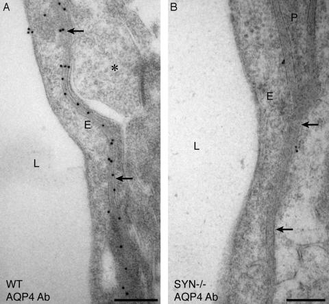
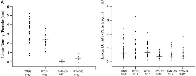
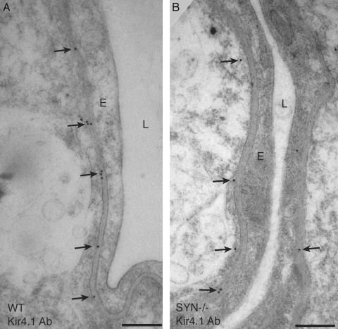
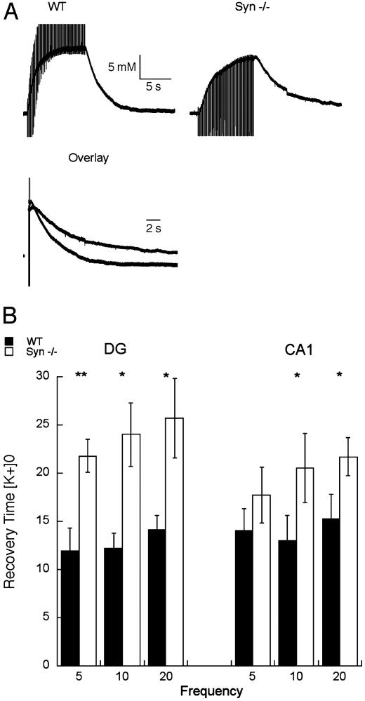
Recovery from neuronal activation requires rapid clearance of potassium ions (K+) and restoration of osmotic equilibrium. The predominant water channel protein in brain, aquaporin-4 (AQP4), is concentrated in the astrocyte end-feet membranes adjacent to blood vessels in neocortex and cerebellum by association with alpha-syntrophin protein. Although AQP4 has been implicated in the pathogenesis of brain edema, its functions in normal brain physiology are uncertain. In this study, we used immunogold electron microscopy to compare hippocampus of WT and alpha-syntrophin-null mice (alpha-Syn-/-). We found that <10% of AQP4 immunogold labeling is retained in the perivascular astrocyte end-feet membranes of the alpha-Syn-/- mice, whereas labeling of the inwardly rectifying K+ channel, Kir4.1, is largely unchanged. Activity-dependent changes in K+ clearance were studied in hippocampal slices to test whether AQP4 and K+ channels work in concert to achieve isosmotic clearance of K+ after neuronal activation. Microelectrode recordings of extracellular K+ ([K+]o) from the target zones of Schaffer collaterals and perforant path were obtained after 5-, 10-, and 20-Hz orthodromic stimulations. K+ clearance was prolonged up to 2-fold in alpha-Syn-/- mice compared with WT mice. Furthermore, the intensity of hyperthermia-induced epileptic seizures was increased in approximately half of the alpha-Syn-/-mice. These studies lead us to propose that water flux through perivascular AQP4 is needed to sustain efficient removal of K+ after neuronal activation.


Figures

Fig. 1.
Fig. 1.
Electron micrographs showing immunogold labeling of AQP4 in perivascular astrocyte end-feet of hippocampus. Corresponding ultrathin sections of brain from WT mouse (A) and α-Syn-/- (B) mouse after immunogold labeling show markedly reduced labeling of AQP4 in the latter. L, lumen; E, endothelial cell; P, pericyte; arrows, perivascular membrane; *, unlabeled nerve terminal. (Bars = 0.5 μm.)
Fig. 2.
Fig. 2.
Scatter plots showing the linear densities of immunogold labeling of AQP4 and Kir4.1 in perivascular astrocyte end-feet of hippocampus. (A) AQP4 labeling densities of preparations from two WT mice and two Syn-/- mice are visibly different (P < 0.001 for each animal by post hoc Scheffé test). (B) Kir4.1 labeling densities of preparations from three WT and three Syn-/- mice overlap with no statistically significant differences (P = 0.4-0.8). Each dot (y axis) represents the linear density of gold particles around a single capillary (n noted below x axis) in preparations from WT or Syn-/- mice; horizontal bars represent median values.
Fig. 3.
Fig. 3.
Electron micrographs showing immunogold labeling of Kir4.1 in perivascular astrocyte end-feet of hippocampus. Corresponding ultrathin sections of brain from WT mouse (A) and α-Syn-/- mouse (B) after immunogold labeling show a modest reduction of Kir4.1 labeling in the latter. L, lumen; E, endothelial cell; arrows, perivascular membrane. (Bars = 0.5 μm.)
Fig. 4.
Fig. 4.
Prolongation of the recovery of [K+]o after repetitive orthodromic stimulation of isolated hippocampal slices. (A) Representative tracings of [K+]o signal during and after a 10-Hz train delivered to the Schaffer collaterals from WT and α-Syn-/- mice shown separately or superimposed in overlay. (B) Pooled data from measurements taken from five WT mice and six α-Syn-/- mice. Plots represent time, in seconds, elapsed during recovery from 90% of peak value to the 10% level in CA1 and the dentate gyrus (DG). Data are shown as mean ± SEM. *, P < 0.05; **, P < 0.001.

References

    1. Hodgkin, A. L. & Huxley, A. F. (1952) J. Physiol. (London) 117 500-544; reprinted ( - PMC - PubMed
    2. 1990) Bull. Math. Biol. 52 25-71. - PubMed
    1. Karwoski, C. J., Lu, H. K. & Newman, E. A. (1989) Science 244 578-580. - PMC - PubMed
    1. Paulson, O. B. & Newman, E. A. (1987) Science 237 896-898. - PMC - PubMed
    1. Newman, E. A., Frambach, D. A. & Odette, L. L. (1984) Science 225 1174-1175. - PMC - PubMed
    1. Holthoff, K. & Witte, O. W. (1996) J. Neurosci. 16 2740-2749. - PMC - PubMed

Publication types

MeSH terms