. 2003 Nov 11;100(23):13567-72.
doi: 10.1073/pnas.1834876100. Epub 2003 Nov 3.

Refractory nature of normal human diploid fibroblasts with respect to oncogene-mediated transformation

Affiliations

Refractory nature of normal human diploid fibroblasts with respect to oncogene-mediated transformation

Tsuyoshi Akagi et al. Proc Natl Acad Sci U S A. .

Abstract

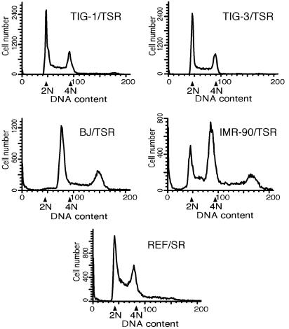
Human cells are known to be more refractory than rodent cells against oncogenic transformation in vitro. To date, the molecular mechanisms underlying such resistance remain largely unknown. The combination of simian virus 40 early region and H-Ras V12 has been effective for transformation of rat embryo fibroblasts, but not for human cells. However, the additional ectopic expression of the telomerase catalytic subunit (hTERT) was reported to be capable of causing transformation of normal human cells. In this study, however, we demonstrate that the combined expression of the above-mentioned three genetic elements is not always sufficient to transform normal human diploid fibroblasts (HDF). Although the expression and function of these introduced genetic elements were essentially the same, among four HDF, TIG-1 and TIG-3 were resistant to transformation. The other two (BJ and IMR-90) showed transformed phenotypes, but they were much restricted compared with rat embryo fibroblasts in expressing simian virus 40 early region and H-Ras V12. In correlation with these phenotypes, TIG-1 and TIG-3 remained diploid after the introduction of these genetic elements, whereas BJ and IMR-90 became highly aneuploid. These results strongly suggest that the lack of telomerase is not the sole reason for the refractory nature of HDF against transformation and that normal human cells have still undefined intrinsic mechanisms rendering them resistant to oncogenic transformation.


Figures

Fig. 1.
Fig. 1.
Expression of hTERT, SV40 T antigens, and Ras. Total cell lysates were prepared from four HDF (TIG-1, TIG-3, BJ, and IMR-90) infected with retroviral vectors expressing hTERT (T), SV40 ER (S), and H-RasV12 (R), from REF infected with retroviral vectors expressing S and R, and from uninfected TIG-3. Ten micrograms of each total cell lysate was subjected to immunoblot analysis with the antibodies indicated on the right.
Fig. 2.
Fig. 2.
Elongation of telomeres in human fibroblasts expressing hTERT. Genomic DNA was prepared from four HDF (TIG-1, TIG-3, BJ, and IMR-90) infected with retroviral vectors expressing hTERT (T), SV40 ER (S), and HRasV12 (R), from REF infected with retroviral vectors expressing S and R, and from uninfected TIG-3. Two micrograms of each genomic DNA was digested with HinfI and RsaI and hybridized with a telomere-specific oligonucleotide probe.
Fig. 3.
Fig. 3.
Association of SV40 large T antigen with p53 and Rb. Total cell lysates were prepared from four HDF (TIG-1, TIG-3, BJ, and IMR-90) infected with retroviral vectors expressing hTERT (T), SV40 ER (S), and H-RasV12 (R), from REF infected with retroviral vectors expressing S and R, and from uninfected TIG-3. Of each total cell lysate, 250 μg was subjected to immunoprecipitation with anti-SV40 large T antibody, and the precipitates were then subjected to immunoblot analysis with the antibodies indicated on the right.
Fig. 4.
Fig. 4.
Activation of MAPK. Total cell lysates (10 μg) were subjected to immunoblot analysis with anti-phospho-MAPK antibody (Upper) or anti-MAPK antibody (Lower). Lane 1, TIG-1 infected with a retroviral vector expressing hTERT (T); lane 2, TIG-1 infected with retroviral vector expressing T, SV40 ER (S), and H-RasV12 (R); lane 3, TIG-3 infected with a retroviral vector expressing T; lane 4, TIG-3 infected with retroviral vectors expressing T, S, and R; lane 5, BJ infected with a retroviral vector expressing T; lane 6, BJ infected with retroviral vectors expressing T, S, and R; lane 7, IMR-90 infected with a retroviral vector expressing T; lane 8, IMR-90 infected with retroviral vectors expressing T, S, and R; lane 9, REF; lane 10, REF infected with retroviral vectors expressing S and R.
Fig. 5.
Fig. 5.
Morphological changes in fibroblasts. Cell morphologies are shown at ×40 magnification. Shown are TIG-3 (A) and TIG-3 (B) infected with retroviral vectors expressing hTERT (T), SV40 ER (S), and H-RasV12 (R). (C) REF. (D) REF infected with retroviral vectors expressing S and R.
Fig. 6.
Fig. 6.
Photographs of soft-agar colony formation assay. One representative result of the soft-agar colony formation assay summarized in Table 1. Cells were plated and stained as described in the legend of Table 1, and photographs of stained colonies were taken.
Fig. 7.
Fig. 7.
Nude mouse tumorigenicity assays. The four HDF (TIG-1, TIG-3, BJ, and IMR-90) infected with retroviral vectors expressing hTERT (T), SV40 ER (S), and H-RasV12 (R), and REF infected with retroviral vectors expressing S and R, were subjected to nude mouse tumorigenicity assays. HeLa, a human cervical carcinoma cell line, was used as a control. The cells (1 × 10⁶) were injected, and the tumor volumes were calculated as described in Materials and Methods. Each point is the average of the tumor volumes of nine inoculated sites, except for REF/SR (five inoculated sites) and HeLa (four inoculated sites).
Fig. 8.
Fig. 8.
Flow cytometric analysis of DNA content. Four HDF (TIG-1, TIG-3, BJ, and IMR-90) infected with retroviral vectors expressing hTERT (T), SV40 ER (S), and H-RasV12 (R), and REF infected with retroviral vectors expressing S and R, were analyzed for DNA content by using flow cytometry as described in Materials and Methods. Arrows indicate the location of 2N and 4N peaks as determined by using normal human and rat fibroblasts, where N represents the haploid genome.

References

    1. DiPaolo, J. A. (1983) J. Natl. Cancer Inst. 70, 3-8. - PubMed
    1. Holliday, R. (1996) Cancer Surv. 28, 103-115. - PubMed
    1. Kuroki, T. & Huh, N. H. (1993) Jpn. J. Cancer Res. 84, 1091-1100. - PMC - PubMed
    1. Land, H., Parada, L. F. & Weinberg, R. A. (1983) Nature 304, 596-602. - PubMed
    1. Ruley, H. E. (1983) Nature 304, 602-606. - PubMed

Publication types

MeSH terms