. 2003 Nov 11;100(23):13453-8.
doi: 10.1073/pnas.2230460100. Epub 2003 Nov 3.

Critical roles for IFN-beta in lymphoid development, myelopoiesis, and tumor development: links to tumor necrosis factor alpha

Affiliations

Critical roles for IFN-beta in lymphoid development, myelopoiesis, and tumor development: links to tumor necrosis factor alpha

Raj Deonarain et al. Proc Natl Acad Sci U S A. .

Abstract

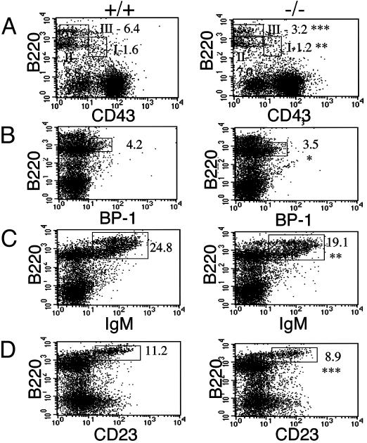
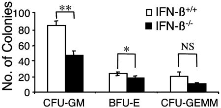
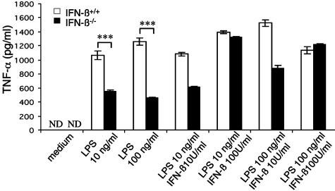
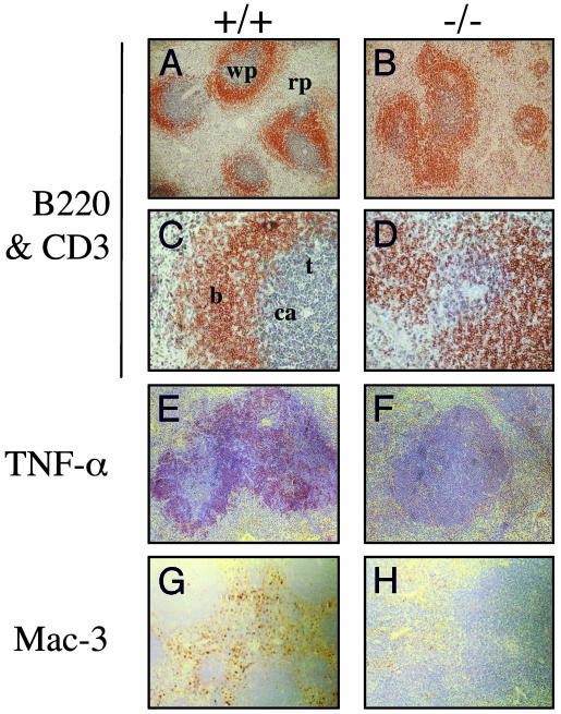
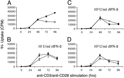
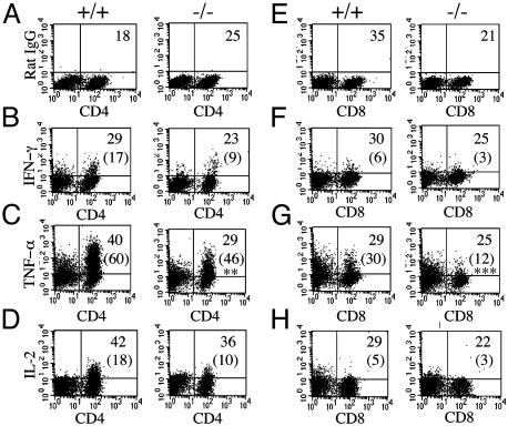
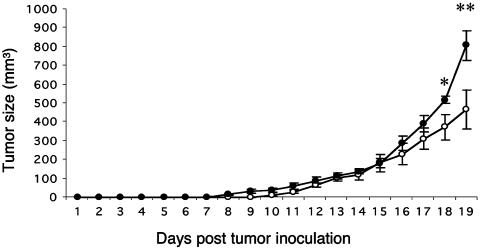
We have generated mice null for IFN-beta and report the diverse consequences of IFN-beta for both the innate and adaptive arms of immunity. Despite no abnormalities in the proportional balance of CD4 and CD8 T cell populations in the peripheral blood, thymus, and spleen of IFN-beta-/- mice, activated lymph node and splenic T lymphocytes exhibit enhanced T cell proliferation and decreased tumor necrosis factor alpha production, relative to IFN-beta+/+ mice. Notably, constitutive and induced expression of tumor necrosis factor alpha is reduced in the spleen and bone marrow (BM) macrophages, respectively, of IFN-beta-/- mice. We also observe an altered splenic architecture in IFN-beta-/- mice and a reduction in resident macrophages. We identify a potential defect in B cell maturation in IFN-beta-/- mice, associated with a decrease in B220+ve/high/CD43-ve BM-derived cells and a reduction in BP-1, IgM, and CD23 expression. Circulating IgM-, Mac-1-, and Gr-1-positive cells are also substantially decreased in IFN-beta-/- mice. The decrease in the numbers of circulating macrophages and granulocytes likely reflects defective maturation of primitive BM hematopoiesis in mice, shown by the reduction of colony-forming units, granulocyte-macrophage. We proceeded to evaluate the in vivo growth of malignant cells in the IFN-beta-/- background and give evidence that Lewis lung carcinoma-specific tumor growth is more aggressive in IFN-beta-/- mice. Taken altogether, our data suggest that, in addition to the direct growth-inhibitory effects on tumor cells, IFN-beta is required during different stages of maturation in the development of the immune system.


Figures

Fig. 1.
Fig. 1.
Defect in BM B cell maturation in IFN-β-/- mice. BM cells, harvested from IFN-β+/+ and IFN-β-/- mice, were depleted of RBC and analyzed by flow cytometry after labeling with the indicated Abs. (A) B220- and CD43-lineage cells. B220-positive cells are shown subdivided into fractions I (B220+ve/CD43+ve), II (B220+ve/low/CD43-ve), and III (B220+ve/high/CD43-ve). (B-D) B220 and BP-1, IgM, and CD23 lineages. Where indicated, numbers represent the gated cell population as a percentage of total cell population. Data represent the mean from groups of five mice. *, P < 0.05; **, P < 0.01; ***, P < 0.001. +/+, IFN-β+/+; -/-, IFN-β-/-.
Fig. 2.
Fig. 2.
Defective maturation of CFU-GM hematopoietic progenitors in the BM of IFN-β-/- mice. BM mononuclear cells from IFN-β+/+ and IFN-β-/- mice were plated in a methylcellulose assay system. The data are expressed as the mean total number of cfu ± SEM from three independent experiments. CFU-GM, burst-forming units (BFU-E), and mixed-lineage colonies (CFUGEMM). *, P = 0.049; **, P = 0.01.
Fig. 3.
Fig. 3.
Impaired LPS-induced TNF-α production from IFN-β-/- BMMs. BMMs from IFN-β+/+ and IFN-β-/- mice were cultured for 24 h after LPS stimulation (10 and 100 ng/ml), in the presence or absence of rIFN-β. Supernatants were assayed for TNF-α in triplicate by ELISA. Data are representative of the mean ± SEM of two independent assays. (IFN-β+/+ n = 5; IFN-β-/- n = 5). ND, not detected; ***, P ≤ 0.0001.
Fig. 4.
Fig. 4.
Comparative analysis of splenic tissues from naïve IFN-β+/+ and IFN-β-/- mice. Paraffin-embedded spleen sections were stained with anti-B220 (brown) and anti-CD3 (blue) (A-D), anti-TNF-α (red) (E and F), or anti-Mac-3 (brown) (G and H) Abs. wp, white pulp; rp, red pulp; b, B cell follicles; t, PALS; ca, central arterioles. +/+, IFN-β+/+; -/-, IFN-β-/-. (Original magnification: A, B, and E-H, ×10; C and D, ×40.)
Fig. 5.
Fig. 5.
Enhanced T cell proliferation in the absence of IFN-β. Lymph node cells from IFN-β+/+ (○) and IFN-β-/- (•) mice were stimulated with murine recombinant anti-CD3 (3 μg/ml)/anti-CD28 (1 μg/ml), in the presence (B-D)or absence (A) of murine rIFN-β (10, 100, or 1,000 units/ml) then pulsed for 18 h with [methyl-3H]thymidine. [3H]Thymidine uptake was measured over 96 h. Data are representative of the mean ± SEM from three independent experiments (P = 0.002).
Fig. 6.
Fig. 6.
Reduced levels of TNF-α in activated IFN-β-/- T cells. Splenocytes were stimulated with mouse recombinant anti-CD3 for 48 h, followed by PMA and ionomycin for 4 h. Cells were then simultaneously labeled with cell-surface Abs for CD4 (A-D) or CD8 (E-H) and stained for either a nonspecific antibody with the same isotype (A and E) or intracellular cytokine antibodies as indicated. Values shown are the lymphocyte population as a percentage of total splenic population. Numbers in parentheses represent the percentage of either CD4- or CD8-positive cytokine-secreting cells. Statistical analysis was by Student's t test; **, P = 0.008; ***, P = 0.001. +/+, IFN-β+/+; -/-, IFN-β-/-. Data are representative from IFN-β+/+ (n = 5) and IFN-β-/- (n = 5) mice.
Fig. 7.
Fig. 7.
Impaired anti-LLC-1 tumor response in IFN-β-/- mice. IFN-β+/+ (○) and IFN-β-/- (•) mice were inoculated s.c. with 106 LLC-1 cells and monitored daily for tumor growth, as indicated. Each point is representative of the average tumor size for the group ± SEM. *, P ≤ 0.05; **, P ≤ 0.01.

References

    1. Stark, G. R., Kerr, I. M., Williams, B. R. G., Silverman, R. H. & Schreiber, R. D. (1998) Annu. Rev. Biochem. 67, 227-264. - PubMed
    1. Diaz, M. O., Pomykala, H. M., Bohlander, S. K., Maltepe, E., Malik, K., Brownstein, B. & Olopade, O. I. (1994) Genomics 22, 540-552. - PubMed
    1. LaFleur, D. W., Nardelli, B., Tsareva, T., Mather, D., Feng, P., Semenuk, M., Taylor, K., Buergin, M., Chinchilla, D., Roshke, V., et al. (2001) J. Biol. Chem. 276, 39765-39771. - PubMed
    1. Kelley, K. A. & Pitha, P. M. (1985) Nucleic Acids Res. 13, 805-823. - PMC - PubMed
    1. Higashi, Y., Sokawa, Y., Watanabe, Y., Kawade, Y., Ohno, S., Takaoka, C. & Taniguchi, T. (1983) J. Biol. Chem. 258, 9522-9529. - PubMed

Publication types

MeSH terms