. 2003 Nov 11;100(23):13332-7.
doi: 10.1073/pnas.2232379100. Epub 2003 Nov 3.

Endosomal compartmentalization in three dimensions: implications for membrane fusion

Affiliations

Endosomal compartmentalization in three dimensions: implications for membrane fusion

J L A N Murk et al. Proc Natl Acad Sci U S A. .

Abstract

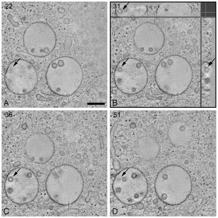
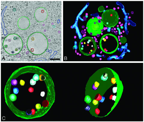
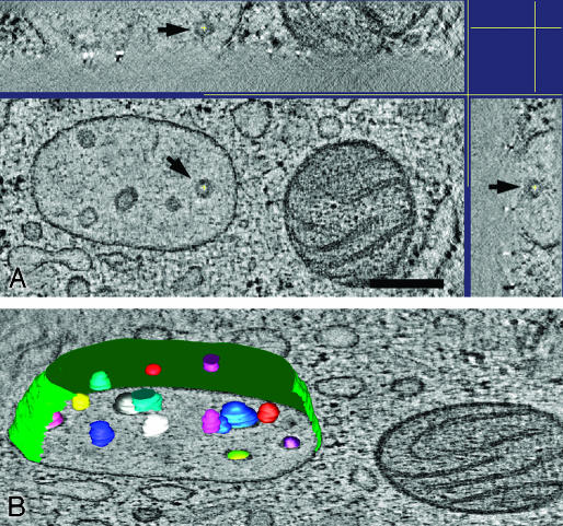
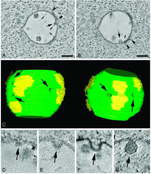
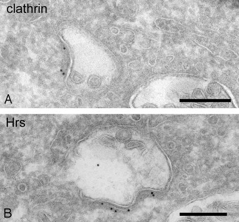
Endosomes are major sorting stations in the endocytic route that send proteins and lipids to multiple destinations in the cell, including the cell surface, Golgi complex, and lysosomes. They have an intricate architecture of internal membrane structures enclosed by an outer membrane. Recycling proteins remain on the outer membrane, whereas proteins that are destined for degradation in the lysosome are sorted to the interior. Recently, a retrograde pathway was discovered whereby molecules, like MHC class II of the immune system, return from the internal structures to the outer membrane, allowing their further transport to the cell surface for T cell activation. Whether this return involves back fusion of free vesicles with the outer membrane, or occurs via the continuity of the two membrane domains, is an unanswered question. By electron tomography of cryo-immobilized cells we now demonstrate that, in multivesicular endosomes of B-lymphocytes and dendritic cells, the inner membranes are free vesicles. Hence, protein transport from inner to outer membranes cannot occur laterally in the plane of the membrane, but requires fusion between the two membrane domains. This implies the existence of an intracellular machinery that mediates fusion between the exoplasmic leaflets of the membranes involved, which is opposite to regular intracellular fusion between cytoplasmic leaflets. In addition, our 3D reconstructions reveal the presence of clathrin-coated areas at the cytoplasmic face of the outer membrane, known to participate in protein sorting to the endosomal interior. Interestingly, profiles reminiscent of inward budding vesicles were often in close proximity to the coats.


Figures

Fig. 1.
Fig. 1.
MVBs contain free internal vesicles. Shown are tomographic slices (X–Y plane) through the 3D volume of an electron tomographic reconstruction of a cluster of MVBs in a high-pressure frozen freeze-substituted RN cell. The arrows in the different slices point at the same internal vesicle: in A and D, the top and bottom of the vesicle are indicated, respectively; B and C show slices through the vesicle. Numbers specify the slice number through the tomographic volume. (B) In addition to the X–Y plane, tomographic slices through the X–Z plane (Upper) and Y–Z plane (Right) are shown at the level of the indicated internal vesicle. The indicated internal vesicle is contained within the whole 3D volume and is a round structure. Each X–Y slice has a thickness in the z axis of ≈4 nm. The total tomographic volume has a thickness of ≈250 nm. (Bar, 250 nm.)
Fig. 2.
Fig. 2.
Three-dimensional model of MVB in RN cell. (A) Membranes in consecutive tomographic slices are manually traced to create a 3D model. Internal vesicles in the models are not perfectly round because of the manual tracing of the membranes. (Bar, 250 nm.) (B) Model view of the 3D reconstruction of the MVBs and surrounding area from Fig. 1. The space surrounding the MVBs is filled with tubules and vesicles. Movie 1 shows a movie of the 3D model. (C) Model view of the MVB tilted along two different angles. In the model on the right, the limiting membrane is partially removed to show the internal vesicles. The vesicles were each given a different color to trace them in each different view of the model.
Fig. 3.
Fig. 3.
Dendritic cell MVB in 3D. (A) Tomographic slice of an electron tomographic reconstruction of a MVB in a high-pressure frozen dendritic cell that shows multiple free internal vesicles. In X–Z(Upper) and Y–Z plane (Right) is shown that the vesicle, indicated by an arrow, is contained within the 3D volume. (Bar, 250 nm.) (B) 3D model of MVB displayed on a tomographic slice showing that the intralumenal membranes are free vesicles.
Fig. 4.
Fig. 4.
Internal vesicle formation in endosomes in RN cells. (A and B) Tomographic slices showing that inward budding profiles of the outer membrane (large arrows) are often situated in close proximity to the clathrin coat (region between arrowheads). Small arrows point to BSA-gold particles that were used in this study as endocytic tracers; their presence demonstrates an early position of the organelle in the endocytic pathway. (Bar, 60 nm.) (C) Model view of MVB from A and B, seen from two different angles, showing the association of the clathrin coat (yellow patches on the surface of the endosome) and the inward budding outer membrane. The arrows point at sites of inward budding profiles. This model is generated from the 3D reconstructions of two serial sections with a total thickness of ≈420 nm. Movie 2, which is published as supporting information on the PNAS web site, shows a movie of the 3D model. (DG) Different stages of internal vesicle formation are shown in tomographic reconstructions of endosomes in RN cells. The arrows point at the site where the outer membrane is forming an inward budding vesicle.
Fig. 5.
Fig. 5.
Clathrin and Hrs in endosomal coat. (A) Early endosome in RN cell immunolabeled for clathrin. Protein A-gold particles are present in the coat at the endosomal outer membrane. (Bar, 200 nm.) (B) Endosomal coat in RN cell immunolabeled for Hrs, indicated by the 10-nm gold particles. (Bar, 200 nm.)

Similar articles

Cited by

References

    1. Mellman, I. (1996) Annu. Rev. Cell Dev. Biol. 12, 575-625. - PubMed
    1. Stoorvogel, W., Strous, G. J., Geuze, H. J., Oorschot, V. & Schwartz, A. L. (1991) Cell 65, 417-427. - PubMed
    1. Mullock, B. M., Bright, N. A., Fearon, C. W., Gray, S. R. & Luzio, J. P. (1998) J. Cell Biol. 140, 591-601. - PMC - PubMed
    1. Raposo, G., Vidal, M. & Geuze, H. J. (1997) in Unusual Secretory Pathways: From Batceria to Man, eds. Kuchler, K., Rubartelli, A. & Holland, B. (R. G. Landes, Washington, DC), pp. 161-184.
    1. Kleijmeer, M., Ramm, G., Schuurhuis, D., Griffith, J., Rescigno, M., Ricciardi-Castagnoli, P., Rudensky, A. Y., Ossendorp, F., Melief, C. J., Stoorvogel, W. & Geuze, H. J. (2001) J. Cell Biol. 155, 53-63. - PMC - PubMed

Publication types