. 2003 Nov 3;198(9):1301-12.
doi: 10.1084/jem.20030182.

A novel endothelial L-selectin ligand activity in lymph node medulla that is regulated by alpha(1,3)-fucosyltransferase-IV

Affiliations

A novel endothelial L-selectin ligand activity in lymph node medulla that is regulated by alpha(1,3)-fucosyltransferase-IV

Christine M'Rini et al. J Exp Med. .

Abstract

Lymphocytes home to peripheral lymph nodes (PLNs) via high endothelial venules (HEVs) in the subcortex and incrementally larger collecting venules in the medulla. HEVs express ligands for L-selectin, which mediates lymphocyte rolling. L-selectin counterreceptors in HEVs are recognized by mAb MECA-79, a surrogate marker for molecularly heterogeneous glycans termed peripheral node addressin. By contrast, we find that medullary venules express L-selectin ligands not recognized by MECA-79. Both L-selectin ligands must be fucosylated by alpha(1,3)-fucosyltransferase (FucT)-IV or FucT-VII as rolling is absent in FucT-IV+VII(-/-) mice. Intravital microscopy experiments revealed that MECA-79-reactive ligands depend primarily on FucT-VII, whereas MECA-79-independent medullary L-selectin ligands are regulated by FucT-IV. Expression levels of both enzymes paralleled these anatomical distinctions. The relative mRNA level of FucT-IV was higher in medullary venules than in HEVs, whereas FucT-VII was most prominent in HEVs and weak in medullary venules. Thus, two distinct L-selectin ligands are segmentally confined to contiguous microvascular domains in PLNs. Although MECA-79-reactive species predominate in HEVs, medullary venules express another ligand that is spatially, antigenically, and biosynthetically unique. Physiologic relevance for this novel activity in medullary microvessels is suggested by the finding that L-selectin-dependent T cell homing to PLNs was partly insensitive to MECA-79 inhibition.


Figures
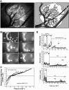
Figure 1.
Figure 1.
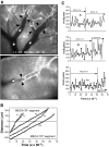
EC adhesiveness and expression of MECA-79 epitope changes abruptly on transition from capillaries to HEVs. (A) CFSE-MECA-79 staining of two venular trees (a and b) in a subiliac PLN (left), with major anatomic features illustrated schematically (right). Blood enters PLN capillaries (not visible) via branches of the superficial epigastric artery (Art). Postcapillary and small collecting HEVs (orders III–V, light gray) are MECA-79+, but low order medullary venules (LOV, orders I–II, dark gray) and extra-lymphoid veins (ELV) are not. The PLN is surrounded by connective tissue (CT). ×40. (B) PLN subcortex showing capillary network (arrows indicate direction of blood flow) draining into an order V HEV (hatched). CFSE-MECA-79 delineates the sharp transition from capillaries to HEV (T = 0 s). A fluorescent lymphocyte (arrowhead) passes through a capillary at high velocity (T = 0.5 – 0.8 s), tethers to MECA-79+ ECs (T = 1.2 s), and rolls slowly along the HEV (T = 7.4 s). The capillary network was subsequently visualized by injecting FITC-dextran (not depicted). ×400. (C) Displacement histograms of three L1-2L-selectin cells (R1, R2, and R3) during transition from capillaries into HEVs. A cell enters the field of view at time 0. The distance of the cell to the transition from MECA-79 ECs to MECA-79+ HEVs was determined for every video frame until the cell disappeared from the field of view. (D) Instantaneous velocity histograms of L1-2L-selectin cells, R1–R3, shown in C during their passage through capillaries and HEVs. See also Video 1, available at http://www.jem.org/cgi/content/full/jem.20030182/DC1.
Figure 2.
Figure 2.
L-selectin transfectants roll throughout the PLN venular tree despite discontinuous expression of MECA-79 epitopes. (A) Micrographs illustrating the abrupt transition from MECA-79+ to MECA-79 ECs in PLN venules in a segment of venular tree a in Fig. 1 A (top panel; ×100), and an order III venule from a different preparation (bottom panel; ×200). Transition from MECA-79 to MECA-79+ ECs (black arrowheads) often coincided with venular bifurcations (top panel), but was also seen within continuous venular segments (bottom panel). White arrows indicate direction of blood flow. (B) Displacement histograms of two noninteracting fast cells (F1 and F2) and three rolling cells (R1–R3) in MECA-79+ and MECA-79 segments of the same venule. Cells were analyzed as in Fig. 1 C. (C) Instantaneous velocity histograms of the three rolling cells, R1–R3, shown in panel B.
Figure 3.
Figure 3.
Fluorescent bead accumulation in PLNs reveals segmental differences in luminal antigen expression between venular branching orders. Binding of nonspecific mAb-coated NR and specific mAb-coated YG fluorescent beads in PLN microvessels 15 min after i.v. injection. NR beads were injected first, followed by an equivalent number of YG beads coated with mAb MECA-79, anti-sLeX/A (HECA-452), anti–ICAM-1, or anti–ICAM-2. The accumulation of specific beads was calculated as described in Materials and Methods. All specific mAb-coated beads bound significantly more than control beads, except for MECA-79–coated beads in order I venules, and both MECA-79 and anti-sLeX/A–coated beads in arterioles. *, P < 0.05; **, P < 0.01; ***, P < 0.001 compared with order I venules. Number of LNs/venules analyzed: 9/54 for ICAM-1; 3/46 for ICAM-2; 5/77 for sLeX/A; and 4/45 for MECA-79.
Figure 4.
Figure 4.
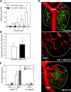
Effect of MECA-79 on L-selectin–dependent lymphocyte homing and rolling in PLNs. (A) The rolling fraction of L1-2L-selectin cells was analyzed before and after MECA-79 treatment (10 mg/kg i.v.) in each branching order of PLN venular trees (n = 6 mice). mAb treatment blocked rolling in order II–V venules (**, P < 0.01; ***, P < 0.001 vs. no mAb), but had minimal effect in order I venules. (B) Tethering fraction of L1-2L-selectin cells in order I and II venules before and after PNAd inhibition. Only free-flowing cells that established the first adhesive contact with LOVs were counted. See also Videos 2–4, available at http://www.jem.org/cgi/content/full/jem.20030182/DC1. (C) L-selectin–mediated homing of naive T cells to PLNs is partially insensitive to MECA-79 inhibition. MECA-79 treatment (10 mg/kg, 15 min before T cell injection) abrogated T cell homing to PLNs harvested 2 h after cell injection (n = 2). 24 h of treatment with MECA-79 (15 min before and 12 h after T cell injection) reduced homing significantly less than anti–L-selectin mAb Mel-14 (100 μg/mouse). Results were normalized to untreated control mice. n = 4 mice/group; *, P < 0.05 versus 24 h MECA-79. (D) 3-D reconstruction of serial confocal micrographs showing homed T cells (green) in inguinal LNs 24 h after treatment with saline (top), anti–L-selectin mAb Mel-14 (middle), or MECA-79 (bottom). The intravascular compartment was delineated by i.v. injection of TRITC-dextran (red). Numerous extravascular T cells are dispersed throughout control and MECA-79–treated PLNs, including in the vicinity of LOVs (order I and II venules). A nearby extralymphoid vein drains blood from the node. LN borders are demarcated by white lines. See also Video 5, available at http://www.jem.org/cgi/content/full/jem.20030182/DC1.
Figure 5.
Figure 5.
Role of FucT-IV and FucT-VII in lymphocyte homing and L-selectin ligand activity in PLN venules. (A) Rolling fractions of rhodamine 6G-labeled leukocytes were assessed in each venular order of PLN from WT and mutant mice. Rolling was absent in FucT-IV+VII−/− PLNs. Compared with WT mice, rolling in FucT-VII−/− PLNs was markedly lower in order III–V HEVs, modestly decreased in order II, and unchanged in order I venules. Rolling in order I–III venules of FucT-IV−/− PLNs was significantly higher than in WT PLNs, whereas rolling in order IV and V venules was similar. *, P < 0.05; **, P < 0.01; ***, P < 0.001 versus WT. (B) Leukocyte rolling in FucT-IV−/− and FucT-VII−/− PLNs before and after injection of anti–L-selectin (100 μg/mouse). ***, P < 0.001 versus control; n = 3 mice/group. (C) Compared with WT mice, naive T cell homing was enhanced in FucT-IV−/− mice and severely compromised in FucT-VII−/− mice. *, P < 0.05 versus WT; ***, P < 0.001 versus WT and FucT-IV−/−; n = 4 mice/group. (D) B cell homing required FucT-VII and was somewhat increased in FucT-IV−/− PLNs (P > 0.05). ***, P < 0.001 versus WT and FucT-IV−/−; n = 4 mice/group.
Figure 6.
Figure 6.
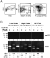
Differential expression of FucT-IV and FucT-VII in PLN ECs. (A) Leukocyte-depleted cell suspensions from PLNs of 60 WT mice were stained for the pan-leukocyte antigen CD45, MECA-79, pan-endothelial CD31, and P-selectin–Ig chimera ligands. After gating on large cells (R1), ECs from high order venules were identified as CD45 MECA-79+ (R3) as well as CD31+ and P-selectin–Ig binding (P-lig+; not depicted). ECs from other vascular segments were isolated from the CD45 MECA-79 fraction (R2) by gating on CD31+ cells. P-lig+ ECs were from LOVs (R4), and nonvenular ECs (MECA-79 P-lig) were from capillaries, arterioles, and possibly lymph vessels (R5). (B) mRNA from equivalent numbers of sorted ECs from LOVs (lanes 1–3), high order venules (lanes 4–6), and nonvenular vessels (lanes 7–9) was subjected to RT-PCR with and without addition of reverse transcriptase (RT) followed by competitive PCR amplification (37 cycles) of pairs of cDNAs for FucT-IV, FucT-VII, and the housekeeping gene β-actin. The intensity ratios of amplified bands in each lane are shown. Intensity ratios could not be determined (ND) in lanes 8 and 9 because no specific signal for FucT-VII transcripts was detectable in nonvenular ECs. The PCR procedure was repeated twice with similar results.
Figure 7.
Figure 7.
Distribution of functional L-selectin ligands and of FucT-IV and FucT-VII in PLN microvessels.

Comment in

References

    1. von Andrian, U.H., and C.R. Mackay. 2000. T-cell function and migration. Two sides of the same coin. N. Engl. J. Med. 343:1020–1034. - PubMed
    1. Springer, T.A. 1994. Traffic signals for lymphocyte recirculation and leukocyte emigration: the multi-step paradigm. Cell. 76:301–314. - PubMed
    1. Warnock, R.A., S. Askari, E.C. Butcher, and U.H. von Andrian. 1998. Molecular mechanisms of lymphocyte homing to peripheral lymph nodes. J. Exp. Med. 187:205–216. - PMC - PubMed
    1. Stein, J.V., A. Rot, Y. Luo, M. Narasimhaswamy, H. Nakano, M.D. Gunn, A. Matsuzawa, E.J. Quackenbush, M.E. Dorf, and U.H. von Andrian. 2000. The CC chemokine thymus-derived chemotactic agent 4 (TCA-4, secondary lymphoid tissue chemokine, 6Ckine, exodus-2) triggers lymphocyte function–associated antigen 1–mediated arrest of rolling T lymphocytes in peripheral lymph node high endothelial venules. J. Exp. Med. 191:61–76. - PMC - PubMed
    1. Okada, T., V.N. Ngo, E.H. Ekland, R. Forster, M. Lipp, D.R. Littman, and J.G. Cyster. 2002. Chemokine requirements for B cell entry to lymph nodes and Peyer's patches. J. Exp. Med. 196:65–75. - PMC - PubMed

Publication types