. 2003 Nov 3;198(9):1381-9.
doi: 10.1084/jem.20030580.

CCR5 expression influences the progression of human breast cancer in a p53-dependent manner

Affiliations

CCR5 expression influences the progression of human breast cancer in a p53-dependent manner

Santos Mañes et al. J Exp Med. .

Abstract

Chemokines are implicated in tumor pathogenesis, although it is unclear whether they affect human cancer progression positively or negatively. We found that activation of the chemokine receptor CCR5 regulates p53 transcriptional activity in breast cancer cells through pertussis toxin-, JAK2-, and p38 mitogen-activated protein kinase-dependent mechanisms. CCR5 blockade significantly enhanced proliferation of xenografts from tumor cells bearing wild-type p53, but did not affect proliferation of tumor xenografts bearing a p53 mutation. In parallel, data obtained in a primary breast cancer clinical series showed that disease-free survival was shorter in individuals bearing the CCR5Delta32 allele than in CCR5 wild-type patients, but only for those whose tumors expressed wild-type p53. These findings suggest that CCR5 activity influences human breast cancer progression in a p53-dependent manner.


Figures
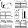
Figure 1.
Figure 1.
CCR5 regulates p53 transcriptional activity. (A) Time course induction of p53 (dotted line, ▴), p21WAF1 (solid line, ▪), and Mdm2 (dashed line, •) in CCL5-stimulated MCF-7 cells. Western blots from four independent experiments were quantified by densitometry and the values were normalized using the tubulin loading control. Data points are plotted relative to mean values obtained before chemokine addition (n = 5). (B) CCL5-induced p53 targets in MCF-7 cells expressing the p53175H mutant. One representative experiment of three is shown. (C and D) MCF-7 cells transfected with control or p53-specific siRNA oligonucleotides were stimulated with CCL5 (C) and incubated at 37°C for the times indicated or γ irradiated (30 Gy; D) and incubated for 3 h at 37°C. Cell lysates were analyzed by Western blot. One representative experiment of three is shown. (E) CCR5 was detected in live mock- or KDELΔ32-expressing MCF-7 cells by FACS®. Surface CCR5 expression in mock- and KDELΔ32-expressing cells was estimated by multiplying average fluorescence intensity by the percentage of CCR5+ cells (n = 3). (F and G) Mock- and KDELΔ32-expressing cells were stimulated with CCL5 (F) or γ irradiated (30 Gy; G) and incubated at 37°C for the times indicated. Cell lysates were analyzed by Western blot. One representative experiment of three is shown.
Figure 2.
Figure 2.
CCR5 regulates p53 activity through in a Gi-, JAK2-, and p38 MAPK–dependent manner. (A) Measurement of p21WAF1 and Mdm2 induction analyzed by Western blot after CCL5 stimulation of MCF-7 cells pretreated with the indicated chemical inhibitors. Data points represent the mean ± SD of densitometric values obtained in two independent experiments for each inhibitor. Data are plotted relative to those obtained in DMSO-treated cells before CCL5 addition. (B and C) Mock- and KDELΔ32-expressing cells were stimulated with CCL5 (B) or UV irradiated (30 J/m2, C), and then incubated at 37°C for the times indicated (for CCL5) or for 30 min (for UV irradiation). Cell lysates were analyzed by Western blot with anti–phospho-p38 (pp38) or anti-p38–specific antibodies. One representative experiment of three is shown. (D and E) Mock-, dnMKK3-, and dnMKK6-expressing cells and cells coexpressing dnMKK3 and dnMKK6 were stimulated with CCL5 (D) or UV irradiated (30 J/m2, E), and then incubated at 37°C as above. CCL5-induced p21WAF1 mdm2 up-regulation was visualized by Western blot (D). In the case of UV irradiation (E), cell lysates were analyzed by Western blot with anti–phospho-p38 (pp38) or anti-p38–specific antibodies. In all cases, dnMKK3 and dnMKK6 expression was detected using an anti-Flag antibody. One representative experiment of two is shown.
Figure 3.
Figure 3.
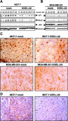
Cell surface levels of CCR5 specifically affect the proliferation of breast tumor xenografts with functional p53. BrdU (A and B) and TUNEL (C and D) staining of mock- and KDELΔ32-expressing MCF-7 (A and C) or MDA-MB-231 (B and D) xenografts. Counterstaining was done with hematoxylin and eosin for BrdU and nuclei were DAPI stained for TUNEL. ×40. Data points at the right represent the percentage of BrdU+ or TUNEL+ nuclei determined in four random fields for each tumor analyzed (two-tailed Mann-Whitney test). *, P < 0.05.
Figure 4.
Figure 4.
p21WAF1 and phospho-p38-MAPK detection in xenografts from mock- and KDELΔ32-expressing tumor cells. (A and B) Lysates from mock- and KDELΔ32-expressing MCF-7 (A) or MDA-MB-231 (B) xenografts were analyzed sequentially with anti-p21WAF1, anti-Mdm2, anti-p53, and anti-tubulin antibodies by Western blot. (C) Cryosections of xenografts derived from mock- and KDELΔ32-expressing MCF-7 and MDA-MB-231 cells, as indicated, were stained with anti-p21WAF1 antibody followed by a peroxidase-labeled second antibody. Counterstaining was performed with hematoxylin and eosin. (D) Paraffin sections from mock- and KDELΔ32-expressing MCF-7 xenografts were stained with an anti–phospho-p38 antibody followed by a peroxidase-labeled second antibody, and were hematoxylin and eosin counterstained. For C and D, the background staining with the second antibody was also analyzed (not depicted). ×40.
Figure 5.
Figure 5.
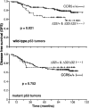
The Δ32 polymorphism affects human breast cancer progression. Kaplan-Meier DFS curves for the 547 breast cancer patients evaluated according to p53 expression and the Δ32 polymorphism, as indicated. CCR5 wild-type (+/+) patients, solid line; Δ32/+ and Δ32/Δ32 patients, dashed line.

References

    1. Murphy, P. 2001. Chemokines and molecular basis of cancer metastasis. N. Engl. J. Med. 354:833–835. - PubMed
    1. Balkwill, F., and A. Mantovani. 2001. Inflammation and cancer: back to Virchow? Lancet. 357:539–545. - PubMed
    1. Homey, B., A. Muller, and A. Zlotnik. 2002. Chemokines: agents for the immunotherapy of cancer? Nat. Rev. Immunol. 2:175–184. - PubMed
    1. Mashino, K., N. Sadanaga, H. Yamaguchi, F. Tanaka, M. Ohta, K. Shibuta, H. Inoue, and M. Mori. 2002. Expression of chemokine receptor CCR7 is associated with lymph node metastasis of gastric carcinoma. Cancer Res. 62:2937–2941. - PubMed
    1. Muller, A., B. Homey, H. Soto, N. Ge, D. Catron, M. Buchanan, T. McClanahan, E. Murphy, W. Yuan, S. Wagner, et al. 2001. Involvement of chemokine receptors in breast cancer metastasis. Nature. 410:50–56. - PubMed

Publication types

Substances