. 2003 Nov 3;198(9):1403-14.
doi: 10.1084/jem.20030050.

Age-dependent requirement for gammadelta T cells in the primary but not secondary protective immune response against an intestinal parasite

Affiliations

Age-dependent requirement for gammadelta T cells in the primary but not secondary protective immune response against an intestinal parasite

Elizabeth Ramsburg et al. J Exp Med. .

Abstract

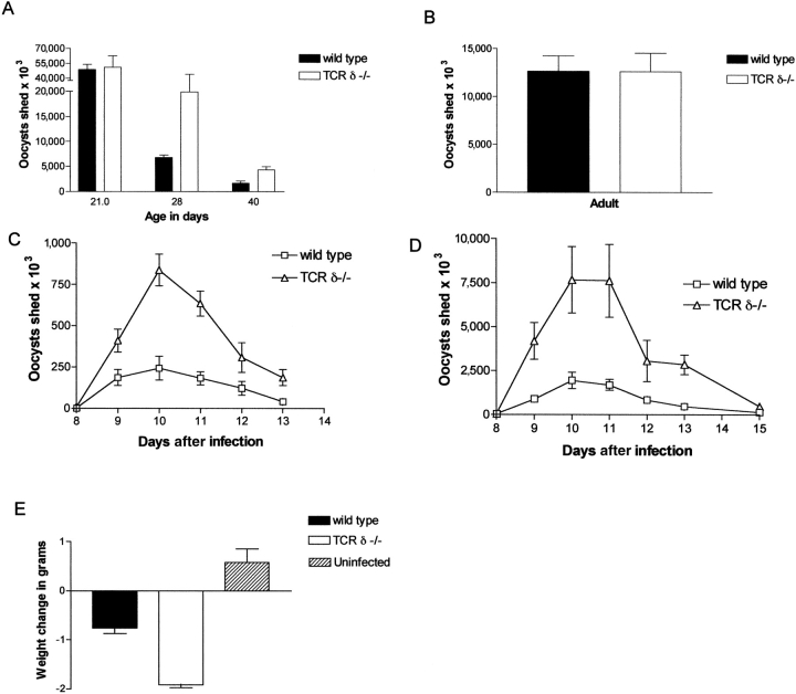
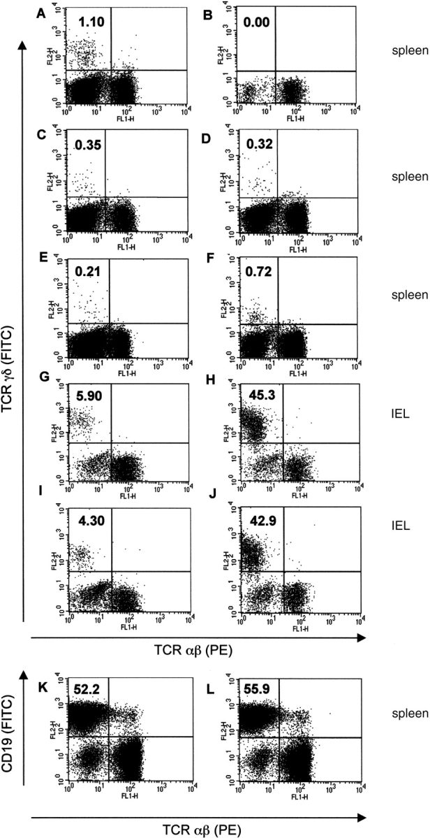
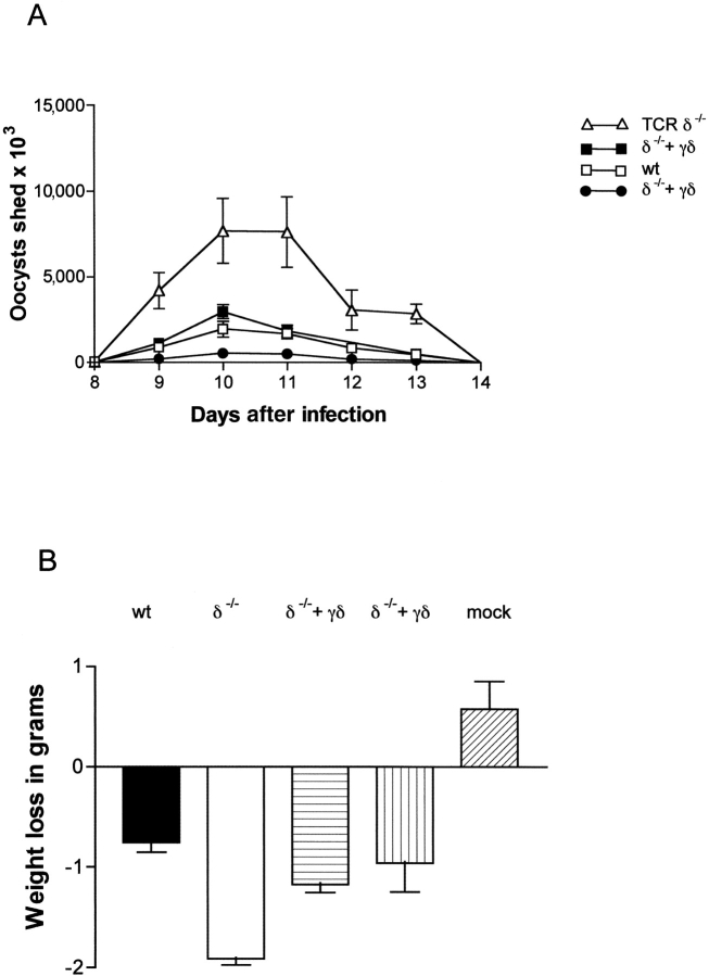
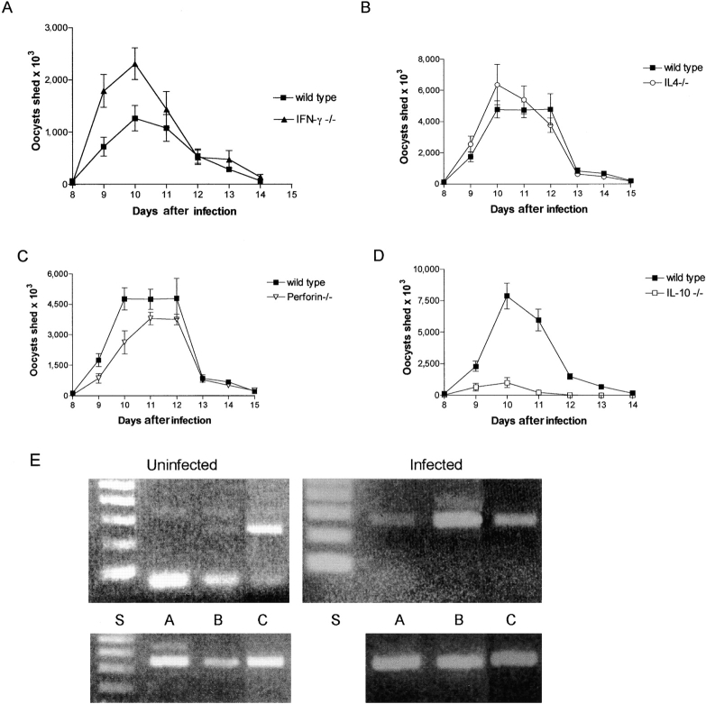
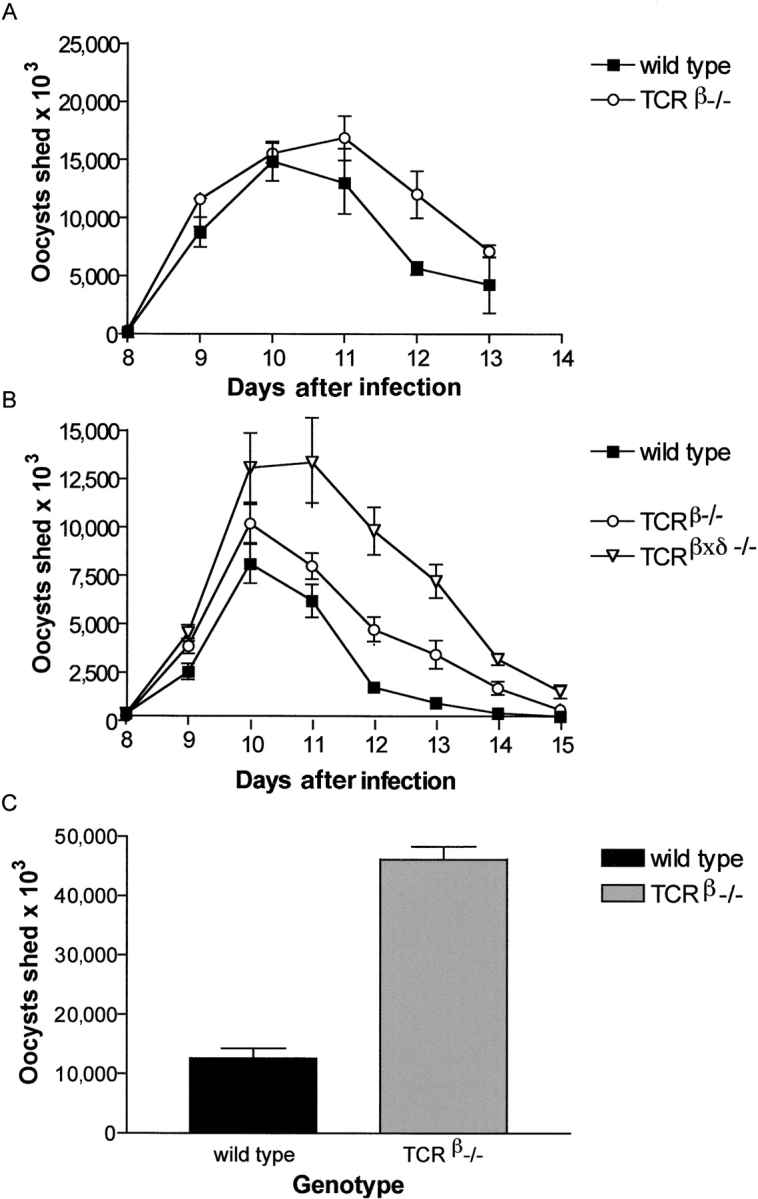
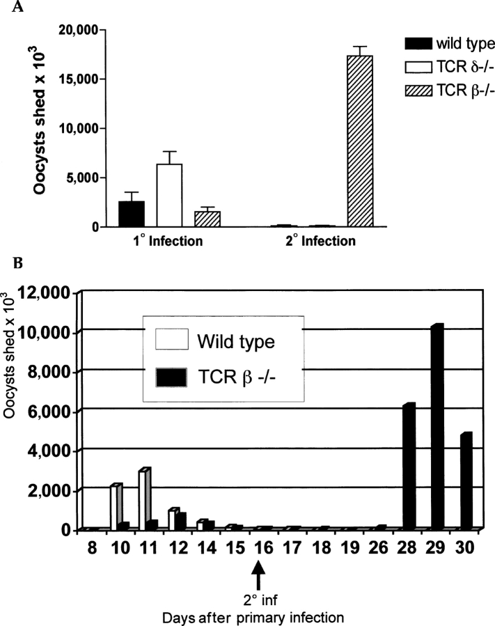
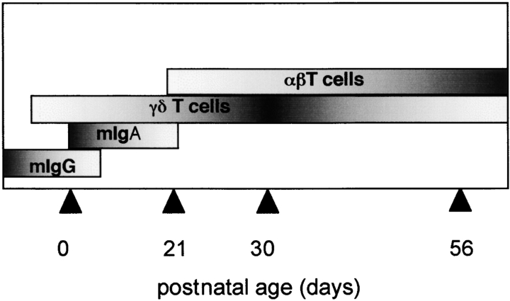
Between weaning (3 wk of age) and adulthood (7 wk of age), mice develop increased resistance to infection with Eimeria vermiformis, an abundant intestinal parasite that causes coccidiosis. This development of resistance was perturbed in T cell receptor (TCR)delta(-/-) mice, which at 4 wk of age remained largely susceptible to infection and prone to infection-associated dehydration. These phenotypes were rescued by the repopulation of gammadelta cells after adoptive transfer of lymphoid progenitors into newborn recipients. Because alphabeta T cells are necessary and sufficient for the protection of adult mice against E. vermiformis, the requirement for gammadelta cells in young mice shows a qualitative difference between the cellular immune responses operating at different ages. An important contribution toward primary immune protection in young hosts may have provided a strong selective pressure for the evolutionary conservation of gammadelta cells. This notwithstanding, the development of effective, pathogen-specific immunity in young mice requires alphabeta T cells, just as it does in adult mice.


Figures

Figure 1.
Figure 1.
Age-dependent gain of immunocompetence is delayed in mice lacking γδ T cells. (A) Infection of T cell–intact (shaded bars, wild type) and γδ T cell–deficient (white bars, TCRδ2/2) mice at 21 d old (wild type, n = 7; TCRδ2/2, n = 10); 28 d old (wild type, n = 4; TCRδ2/2, n = 3); and 40 d old (wild type, n = 4; TCRδ2/2, n = 3). (B) Infection of adult T cell–intact (shaded bar; wild type) and γδ T cell–deficient mice (white bar, TCRδ2/2). Mice were gavage infected with 1,000 (A) or 2,000 (B) sporulated E. vermiformis oocysts. Bars reflect average total oocysts shed over the patent period ±SEM. (C and D) 30-d-old T cell–intact (wild type, open squares) and γδ T cell–deficient (TCRδ−/−, open triangles) mice were gavage infected with 100 (C, wild type, n = 8 and TCRδ−/−, n = 11) or 1,000 (D, wild type, n = 7 and TCRδ−/−, n = 3) sporulated E. vermiformis oocysts. Plots depict average daily oocyst yields. (E) Weight change during days 7–10 after infection (1,000 oocysts) of infected T cell–intact (wild type, shaded bar, n = 7), γδ T cell–deficient (TCRδ−/−, white bar, n = 3), and mock-infected TCRδ−/− mice (diagonally striped bars).
Figure 2.
Figure 2.
γδ cells are present in the spleen and gut of reconstituted animals. Splenocytes analyzed by flow cytometry for TCRγδ (A–J, y axis) and TCRαβ (x axis), and C19 (K and L, y axis) and TCRαβ (x axis). All plots shown were pregated on live lymphocytes. (A) Unmanipulated C57.BL/6 mice; (B) mock-reconstituted TCRδ−/− mice; (C–F) TCRδ−/− mice reconstituted with E14 fetal liver cells from wild-type donors (C and D); and TCR β −/− donors (E and F). (G–J) IELs were isolated 12 wk after neonatal reconstitution of TCRδ−/− mice with E14 fetal liver cells from wild-type (G and H) and TCRβ−/− (I and J) donors; this harvest date was 8 wk after infection. (A–J) Percentage of total splenic/gut lymphocytes comprising γδ cells is indicated in the top left quadrant. (K and L) Analysis of B cell composition of splenocytes from TCRδ−/− mice and TCRδ−/− mice reconstituted with E14 fetal liver cells from wild-type donors. Percent of total splenic lymphocytes comprising CD19+ B cells is indicated in the top left quadrant.
Figure 3.
Figure 3.
Replacement of γδ cells restores resistance to infection among 30-d-old TCRδ−/− mice. Reconstituted and control mice were gavage infected with 1,000 E. vermiformis oocysts. (A) Oocysts shed by TCRδ−/− mice (open triangles, n = 3; same group as in Fig. 1 D), wild-type mice (open squares, n = 7; same group as in Fig. 1 D), TCRδ−/− mice reconstituted with fetal liver from wild-type donors (closed squares, n = 11), and TCRδ−/− mice reconstituted with fetal liver from αβ T cell–deficient donors (closed circles, n = 7). (B) 30-d-old wild-type mice (shaded bars, n = 7), TCRδ−/− mice mock reconstituted (white bar, n = 3), and TCRδ−/− mice reconstituted with E14 fetal liver cells from either wild-type mice (horizontally striped bar, n = 11) or αβ cell–deficient mice (vertically striped bar, n = 7) were weighed daily at the peak of infection. Bars represent average weight loss in grams ± SD among members of a group during days 7–10 after infection. Age-matched control mice were mock infected with sterile water (diagonally striped bar, n = 7).
Figure 4.
Figure 4.
IFNγ−/− mice partially phenocopy susceptibility of TCRδ−/−mice. The susceptibility of 30-d-old wild-type and various cytokine deficient mice was compared as follows: (A) 30-d-old IFNγ−/− mice (closed triangles, n = 4) and wild type (closed squares, n = 4); (B) 30-d-old IL-4−/− mice (open circles, n = 4) and wild type (closed squares, n = 3); (C) perforin−/− mice (inverted open triangles, n = 4) and wild type (closed squares, n = 3); and (D) IL-10−/− mice (open squares, n = 4) and wild type (closed squares, n = 6). (E) RT-PCR analysis of IFNγ (top) and HPRT (bottom) RNA expression in splenic and intestinal αβ T (lane A) and γδ cells (lane B) harvested from mice that 6 d previously, at the age of 30 d, were either uninfected (left) or E. vermiformis infected (right). S, 100 bp size marker. IFNγ, bottom marker = 100 bp, product = 267 bp. HPRT, bottom marker = 200 bp, product = 351 bp. C, positive control (activated adult mouse spleen cells). The bona fide band is in each case the lower of a doublet; the fast running band in the top panel for the uninfected samples corresponds to primer dimers. The γδ cell sample from infected mice shows an IFNγ signal comparable to the HPRT signal, whereas the signal in uninfected mice was much weaker than the HPRT signal.
Figure 5.
Figure 5.
Young αβ T cell–deficient mice display variable susceptibility to infection. (A) Wild-type (closed squares, n = 7) and TCRβ−/− mice (open circles, n = 7) were infected (1,000 oocytes) at 21–24 d of age. (B) Wild-type mice (closed squares, n = 6), TCRβ−/− mice (open circles, n = 7), and TCRβxδ−/− mice (inverted triangles, n = 6) were infected (1,000 oocysts) at 30 d of age. (C) Wild-type (shaded bar) and TCRβ−/− mice (gray bar, n = 7) were infected (2,000 oocysts) as adults. Bars reflect total oocyst production ±SEM over the patent period.
Figure 6.
Figure 6.
αβ cells are required for the acquisition of memory. (A) 30-d-old wild-type mice (shaded bars, n = 5), TCRδ−/− mice (white bars, n = 3), and TCRβ−/− mice (striped bars, n = 2) mice were infected with 1,000 E. vermiformis oocysts (1° infection), and rechallenged at 8 wk of age with 10,000 oocysts (2° infection). (B) 11-d-old wild-type (white bars, n = 6) and TCRβ−/− mice (shaded bars, n = 4) were infected with 100 oocysts and rechallenged at 28 d of age with 1,000 oocysts. In both cases, only αβ T cell–intact mice were immune to reinfection.
Figure 7.
Figure 7.
Development of murine immune competence proceeds in overlapping stages. The model depicts the proposed relative contributions of maternal antibody (mIgG and mIgA), γδ T cells, αβ T cells, and innate factors to immune protection among young mice as age increases. Darker shading indicates higher dependence on a particular factor. Age increases from left to right with 0, 21, 30, and 56 d of age (arrowheads). Weaning typically occurs at 21 d, and animals are considered adults by 56 d of age.

References

    1. Adkins, B. 2000. Development of neonatal Th1/Th2 function. Int. Rev. Immunol. 19:157–171. - PubMed
    1. Marshall-Clarke, S., D. Reen, L. Tasker, and J. Hassan. 2000. Neonatal immunity: how well has it grown up? Immunol. Today. 21:35–41. - PubMed
    1. Haynes, B.F., and C.S. Heinly. 1995. Early human T cell development: analysis of the human thymus at the time of initial entry of hematopoietic stem cells into the fetal thymic microenvironment. J. Exp. Med. 181:1445–1458. - PMC - PubMed
    1. Cuisinier, A.M., F. Fumoux, D. Moinier, L. Boubli, V. Guigou, M. Milili, C. Schiff, M. Fougereau, and C. Tonnelle. 1990. Rapid expansion of human immunoglobulin repertoire (VH, V kappa, V lambda) expressed in early fetal bone marrow. New Biol. 2:689–699. - PubMed
    1. Billingham, R., L. Brent, and P.B. Medawar. 1953. Actively acquired tolerance of foreign cells. Nature. 172:603–606. - PubMed

Publication types

Substances