. 2003 Nov;112(9):1351-60.
doi: 10.1172/JCI19435.

Mitochondrial DNA mutations in human colonic crypt stem cells

Affiliations

Mitochondrial DNA mutations in human colonic crypt stem cells

Robert W Taylor et al. J Clin Invest. 2003 Nov.

Abstract

The mitochondrial genome encodes 13 essential subunits of the respiratory chain and has remarkable genetics based on uniparental inheritance. Within human populations, the mitochondrial genome has a high rate of sequence divergence with multiple polymorphic variants and thus has played a major role in examining the evolutionary history of our species. In recent years it has also become apparent that pathogenic mitochondrial DNA (mtDNA) mutations play an important role in neurological and other diseases. Patients harbor many different mtDNA mutations, some of which are mtDNA mutations, some of which are inherited, but others that seem to be sporadic. It has also been suggested that mtDNA mutations play a role in aging and cancer, but the evidence for a causative role in these conditions is less clear. The accumulated data would suggest, however, that mtDNA mutations occur on a frequent basis. In this article we describe a new phenomenon: the accumulation of mtDNA mutations in human colonic crypt stem cells that result in a significant biochemical defect in their progeny. These studies have important consequences not only for understanding of the finding of mtDNA mutations in aging tissues and tumors, but also for determining the frequency of mtDNA mutations within a cell.


Figures

Figure 1
Figure 1
Respiratory chain deficiency in normal human colonic mucosa. (a) H&E preparations showing normal mucosal structure. Scale bar: 100 μm. (b) Higher magnification of a, showing normal crypt structure. Scale bar: 50 μm. (c) Normal cytochrome c oxidase activity (brown) in colonic crypts following dual cytochrome c oxidase and succinate dehydrogenase histochemistry. Scale bar: 100 μm. (d) Absence of histochemically detectable cytochrome c oxidase activity in colonic crypts (blue). Note delineation of crypt territories at the luminal surface. Scale bar: 100 μm. (e) Single cytochrome c oxidase–deficient colonic crypt. Scale bar: 100 μm. (f) Higher magnification of e, showing crypt base. Scale bar: 20 μm. (g) Presence of multiple, adjacent cytochrome c oxidase–deficient crypts. Scale bar: 100 μm. (h) Transverse section showing a similar cluster of cytochrome c oxidase–deficient crypts. Scale bar: 100 μm. The specificity of the dual cytochrome c oxidase and succinate dehydrogenase histochemical assay was established. (i) Colonic mucosa incubated in standard cytochrome c oxidase medium. (j) Colonic mucosa incubated as i, but in the presence of 2.5 mM sodium azide, a specific inhibitor of cytochrome c oxidase. (k) Colonic mucosa incubated in standard succinate dehydrogenase medium. (l) Colonic mucosa incubated as k, but in the presence of 50 mM sodium malonate, a competitive inhibitor of succinate dehydrogenase. Scale bars: 100 μm (ik).
Figure 2
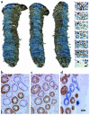
Figure 2
Partial respiratory chain deficiency in some crypts and immunocytochemistry of cytochrome c oxidase deficiency. (a) Reconstructed images were produced from 59 transverse sections (8 μm) covering the full thickness of the colonic lamina propria. Representative images of the sections reacted for cytochrome c oxidase and succinate dehydrogenase are shown in the panel on the extreme right, with the reconstructed crypt arrowed. Scale bar: 50 μm. The position of each image corresponds to the approximate position in the adjacent reconstructed images. The images of the reconstructed colonic crypt are each rotated clockwise through 45°C to show a continuous ribbon of cytochrome c oxidase–deficient cells originating at the crypt base. (bd) Representative serial transverse sections of colonic crypts showing immunoreactivity of cytochrome c oxidase subunit I (b), immunoreactivity of cytochrome c oxidase subunit IV (c), and dual cytochrome c oxidase/succinate dehydrogenase histochemistry (d). Scale bar: 50 μm (bd).
Figure 3
Figure 3
Examples of frameshift mutations within cytochrome c oxidase structural genes associated with cytochrome c oxidase deficiency. (a) Sequencing electropherogram detailing a region of COIII containing a short, poly-C tract of six residues (nucleotides 9532–9537) amplified from a cytochrome c oxidase positive–reacting colonic crypt. (b) Sequencing of this region in an alternative cytochrome c oxidase–deficient crypt revealed a homoplasmic single nucleotide insertion (C9537ins). The frameshift induced by this insertion creates a premature stop codon predicting the synthesis of a truncated cytochrome c oxidase subunit III polypeptide of 110 amino acids and as such is likely to be the cause of the enzyme defect (20). (c) In another cytochrome c oxidase–deficient colonic crypt, we identified a single C nucleotide deletion within this poly-C tract (C9537del) present at near homoplasmic levels. Similar to the C9537ins mutation, this change also leads to the premature truncation of the cytochrome c oxidase III subunit, resulting in cytochrome c oxidase deficiency. Abbreviations for the amino acid residues are as follows:Asn, asparagine; Gln, glutamine; Gly, glycine; Ile, isoleucine; Leu, leucine; Pro, proline; Thr, threonine. Ter represents termination codon.
Figure 4
Figure 4
Mathematical modeling of mitochondrial DNA mutation and replication in colonic crypt stem cells. Simulated cells contained approximately 10,000 mtDNA and divided every 24 hours. Each symbol is the result of 300 (at high mutation rates) to 3,000 (at low mutation rates) independent simulations. (a) The percentage of simulated crypt stem cells containing detectable (>30%) mutant mtDNA during a human lifespan. Each curve corresponds to a different mutation rate indicated on the figure. The model predicts that the random partitioning of mitochondrial genomes during crypt cell division will cause random genetic drift and lead to the clonal expansion of somatic mtDNA mutations during human life. (b) The percentage of simulated crypt stem cells containing a detectable amount of mtDNA mutations as a function of the mtDNA mutation rate (circles, no mutations, > 30%; triangles, one mutation, >30%; inverted triangles, two mutations, >30%; asterisk, three or more mutations. The curves represent the theoretical simple probability functions). The model predicts that the mutation rate must be approximately 5 × 10⁻⁵ per genome per day in order to simultaneously generate crypt stem cells that contain no mutations, and some that have two or more detectable mutations, at age 80 years. Previous simulations demonstrate that the random partitioning of individual genomes during stem cell division will cause random genetic drift and clonal expansion of somatic mtDNA mutations, and the speed of the random drift is dependent upon the frequency of the crypt stem cell divisions. Changing the rate of stem cell division to once per 48 hours did not alter the simulation results.
Figure 5
Figure 5
Incidence of cytochrome c oxidase–deficient colonic crypts per decade. Only crypts in which at least 50% of all cells were cytochrome c oxidase deficient were included. The crypts were counted on at least ten different transverse sections for 28 individual patients. The average value for each patient was included in the mean ± SEM value given for each decade. The number of patients per decade was 35–44 years, four patients; 45–54 years, seven patients; 55–64 years, five patients; 65–74 years, six patients; 75–84 years, six patients. (See also Table 2.) R2 is a measure of the conformation to an exponential increase where 0 represents no association and 1 is a perfect fit.

Comment in

  • Tales from the crypt.
    Schon EA. Schon EA. J Clin Invest. 2003 Nov;112(9):1312-6. doi: 10.1172/JCI20249. J Clin Invest. 2003. PMID: 14597757 Free PMC article.

References

    1. Chinnery PF, et al. The epidemiology of pathogenic mitochondrial DNA mutations. Ann. Neurol. 2000;48:188–193. - PubMed
    1. Lightowlers RN, Chinnery PF, Turnbull DM, Howell N. Mammalian mitochondrial genetics: heredity, heteroplasmy and disease. Trends Genet. 1997;13:450–455. - PubMed
    1. Sciacco M, Bonilla E, Schon EA, DiMauro S, Moraes CT. Distribution of wild-type and common deletion forms of mtDNA in normal and respiration-deficient muscle fibers from patients with mitochondrial myopathy. Hum. Mol. Genet. 1994;3:13–19. - PubMed
    1. Brierley EJ, Johnson MA, Lightowlers RN, James OF, Turnbull DM. Role of mitochondrial DNA mutations in human aging: implications for the central nervous system and muscle. Ann. Neurol. 1998;43:217–223. - PubMed
    1. Cortopassi G, Arnheim N. Detection of specific mitochondrial DNA deletion in tissues of older humans. Nucleic Acids Res. 1990;275:169–180. - PMC - PubMed

Publication types

Substances