. 2003 Nov;112(9):1395-406.
doi: 10.1172/JCI17700.

Inhibition of endogenous thioredoxin in the heart increases oxidative stress and cardiac hypertrophy

Affiliations

Inhibition of endogenous thioredoxin in the heart increases oxidative stress and cardiac hypertrophy

Mitsutaka Yamamoto et al. J Clin Invest. 2003 Nov.

Abstract

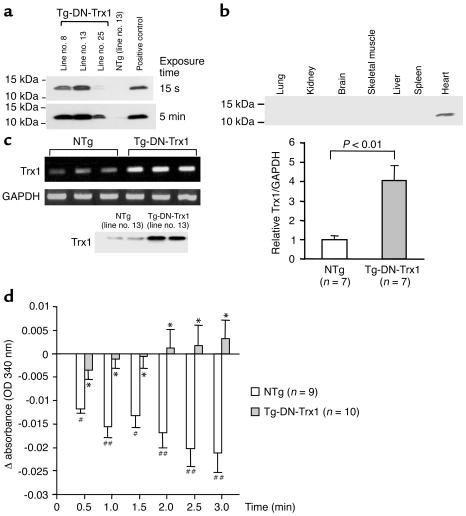
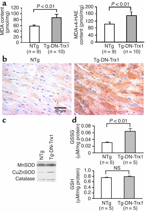
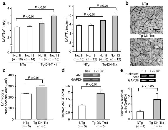
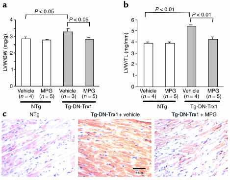
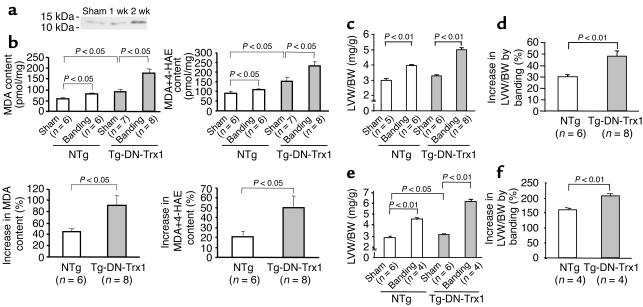
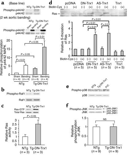
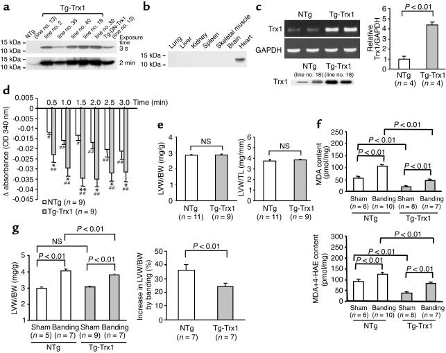
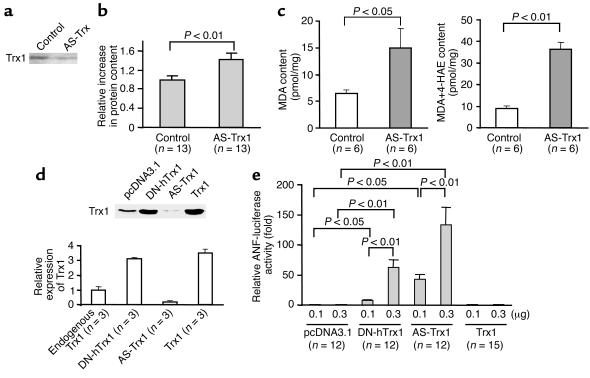
Thioredoxin 1 (Trx1) has redox-sensitive cysteine residues and acts as an antioxidant in cells. However, the extent of Trx1 contribution to overall antioxidant mechanisms is unknown in any organs. We generated transgenic mice with cardiac-specific overexpression of a dominant negative (DN) mutant (C32S/C35S) of Trx1 (Tg-DN-Trx1 mice), in which the activity of endogenous Trx was diminished. Markers of oxidative stress were significantly increased in hearts from Tg-DN-Trx1 mice compared with those from nontransgenic (NTg) mice. Tg-DN-Trx1 mice exhibited cardiac hypertrophy with maintained cardiac function at baseline. Intraperitoneal injection of N-2-mercaptopropionyl glycine, an antioxidant, normalized cardiac hypertrophy in Tg-DN-Trx1 mice. Thoracic aortic banding caused greater increases in myocardial oxidative stress and enhanced hypertrophy in Tg-DN-Trx1 compared with NTg mice. In contrast, transgenic mice with cardiac-specific overexpression of wild-type Trx1 did not show cardiac hypertrophy at baseline but exhibited reduced levels of hypertrophy and oxidative stress in response to pressure overload. These results demonstrate that endogenous Trx1 is an essential component of the cellular antioxidant mechanisms and plays a critical role in regulating oxidative stress in the heart in vivo. Furthermore, inhibition of endogenous Trx1 in the heart primarily stimulates hypertrophy, both under basal conditions and in response to pressure overload through redox-sensitive mechanisms.


Figures

Figure 1
Figure 1
(a) Heart homogenates were prepared from Tg-DN-Trx1 and NTg mice. Immunoblot analyses were conducted using anti-hTrx1 Ab. Short (15-second) and long (5-minute) exposures of the immunoblot are shown. After long exposure, endogenous mouse Trx1 was detected. Note that the anti-hTrx1 Ab (clone 2G11) does not detect mouse Trx1 as efficiently as it detects hTrx1. (b) Tissue homogenates were prepared from various organs. Immunoblot analyses were conducted using anti-hTrx1 Ab. (c) RT-PCR analyses of Trx1 and GAPDH. Total RNA was extracted from Tg-DN-Trx1 (line no. 13) and NTg mice. The lower left panel indicates protein expression of total Trx1, determined using anti-hTrx1 Ab (clone 4H9), which detects both mouse Trx1 and hTrx1. (d) The disulfide oxidoreductase activity of Trx was determined by the insulin reduction assay. Time-dependent reduction of NADPH, determined by spectrophotometry, is shown. *P < 0.01 compared with NTg. #P < 0.05, ##P < 0.01 compared with 0 min.
Figure 2
Figure 2
(a) Heart homogenates were prepared from Tg-DN-Trx1 mice and NTg littermates. Tissue levels of MDA alone and MDA plus 4-HAE, markers of lipid peroxidation, were found to be increased in Tg-DN-Trx1 mice. (b) LV myocardial sections were subjected to immunostaining with 8-OHdG, a marker of oxidative DNA damage, which was increased in Tg-DN-Trx1 mice. The result is representative of three experiments. (c) Heart homogenates were prepared from Tg-DN-Trx1 mice and NTg littermates. Immunoblot analyses of MnSOD, CuZnSOD, and catalase are shown. Note that neither the level of MnSOD nor the level of catalase differed between Tg-DN-Trx1 and NTg mice, while that of CuZnSOD was higher (about 2.5-fold) in Tg-DN-Trx1 mice. n = 3. (d) Tissue levels of GSSG and GSH were determined using fresh samples.
Figure 3
Figure 3
(a) LVW/BW (mg/g; left) and LVW/TL (mg/mm; right) were determined in Tg-DN-Trx1 mice (line nos. 8 and 13) and NTg littermates. The age of the animals was 85 ± 3 days in Tg-DN-Trx1 line no. 8, 81 ± 4 days in Tg-DN-Trx1 line no. 13, 84 ± 4 days in NTg line no. 8, and 79 ± 5 days in NTg line no. 13. (b) Silver staining of LV myocardial sections, showing LV myocyte cross-sectional area in Tg-DN-Trx1 mice and NTg littermates. (c) Mean LV myocyte cross-sectional area was determined from 100–200 myocytes in each animal. The experiments were conducted using five NTg and six Tg-DN-Trx1 mice. (d and e) mRNA expression of ANF (d), α-skeletal actin (e), and GAPDH was determined by RT-PCR. The intensity of the ethidium bromide bands was quantitated. The intensity of ANF or α-skeletal actin bands was normalized by that of GAPDH bands.
Figure 4
Figure 4
The effect of antioxidant treatment on base-line cardiac hypertrophy in Tg-DN-Trx1 mice. Tg-DN-Trx1 mice and NTg littermates were treated with an intraperitoneal injection of MPG (100 mg/kg/d) or saline (vehicle) for 4 weeks. LVW/BW (a) and LVW/TL (b) were determined. (c) LV myocardial sections obtained from NTg mice (left) and Tg-DN-Trx1 mice treated with saline (center) or MPG (right) were subjected to immunostaining with 8-OHdG.
Figure 5
Figure 5
Tg-DN-Trx1 mice and NTg littermates were subjected to either transverse aortic banding or sham operation. (a) The effect of pressure overload on expression of endogenous mouse Trx1 in NTg mice. The result is representative of four experiments. (bd) Aortic banding was applied for 2 weeks. (b) The effect of pressure overload on oxidative stress in the heart. The heart content of MDA and of MDA plus 4-HAE, the percentage increase in MDA, and the percentage increase in MDA plus 4-HAE are shown. (c and d) LVW/BW (c) and percentage increase in LVW/BW in response to aortic banding (d) were determined. Similar results were obtained for LVW/TL. The percentage increase in LVW/BW and LVW/TL (not shown) was significantly greater in Tg-DN-Trx1 than in NTg mice in response to pressure overload. (e and f) Aortic banding was applied for 4 weeks. LVW/BW (e) and percentage increase in LVW/BW in response to aortic banding (f) were determined. The percentage increase in LVW/BW was significantly greater in Tg-DN-Trx1 than in NTg mice.
Figure 6
Figure 6
The activities of the Ras–Raf-1–ERK pathway, p38-MAPK, and p46/p54–JNK1 were examined in Tg-DN-Trx1 mice and NTg littermates. (a) Some mice were subjected to aortic banding for 2 weeks. Activities of p42/p44–ERK (a), Raf-1 (b), p38-MAPK (e), and p46/p54–JNK1 (f) were determined using anti-phosphospecific Ab’s. The same filter was reprobed with respective non-phosphospecific Ab. (e) Cell extracts prepared from cardiac myocytes overexpressing mammalian sterile 20–like kinase 1 (Mst1) were used as positive control (P/C). The gel picture is representative of three to ten experiments in each immunoblot analysis. (c) The activity of Ras was determined using the Ras-binding domain in Raf-1 coupled with agarose. (d) S-thiolation of Ras was determined as described in Methods. COS-7 cells grown in 60-mm dishes were transfected with the indicated expression plasmids (2 μg). Cells were incubated with biotinylated cysteine (0.5 mM), and S-thiolated proteins were isolated by streptavidin-Sepharose. Samples were subjected to immunoblot analyses with anti-Ras Ab. AS-Trx1, antisense Trx1.
Figure 7
Figure 7
Characterization of transgenic mice with cardiac-specific overexpression of wild-type Trx1 (Tg-Trx1 mice). (a) Heart homogenates were prepared from Tg-Trx1 mice and NTg littermates. Immunoblot analyses were conducted using anti-hTrx1 Ab. Short (3-second) and long (2-minute) exposures of the immunoblot are shown. After long exposure, endogenous mouse Trx1 was detected. (b) Tissue homogenates were prepared from various organs. Immunoblot analyses were conducted using anti-hTrx1 Ab. (c) RT-PCR analyses of Trx1 and GAPDH. Total RNA was extracted from Tg-Trx1 (line no. 18) and NTg mice. The lower left panel indicates protein expression of total Trx1, determined using anti-hTrx1 Ab (clone 4H9). (d) The disulfide oxidoreductase activity of Trx was determined by the insulin reduction assay. Time-dependent reduction of NADPH, determined by spectrophotometry, is shown. *P < 0.05; **P < 0.01 compared with NTg. #P < 0.05, ##P < 0.01 compared with 0 min. (e) LVW/BW (mg/g; left) and LVW/TL (mg/mm; right) were determined in Tg-Trx1 mice (line no. 18) and NTg littermates (line no. 18). The age of the animals was 94 ± 9 days in Tg-Trx1 and 97 ± 7 days in NTg mice. Base-line hypertrophy was not observed in any other lines of Tg-Trx1 mice (not shown). (f and g) Tg-Trx1 mice and NTg littermates were subjected to either transverse aortic banding or sham operation. Aortic banding was applied for 2 weeks. (f) The effect of pressure overload on oxidative stress in the heart. The heart contents of MDA and MDA plus 4-HAE are shown. (g) LVW/BW (left) and percentage increase in LVW/BW in response to aortic banding (right) were determined. Similar results were obtained for LVW/TL (not shown).
Figure 8
Figure 8
(ac) Neonatal rat cardiac myocytes were treated with either control Morpholino oligo or Morpholino antisense oligo for rat Trx1 (2 μM) using osmotic delivery. Myocytes were harvested 48 hours after application of the oligo. (a) Expression of Trx1 was determined by immunoblotting with anti-Trx1 Ab (4H9). (b) Total cardiac myocyte protein content was determined. The mean value in control Morpholino oligo–treated myocytes was designated as 1. (c) Cellular content of MDA and MDA plus 4-HAE was determined as described in Methods. (d) COS-7 cells grown in 60-mm dishes were transfected with either empty vector plasmid or pcDNA3.1 harboring DN-hTrx1, antisense rat Trx1 (AS-Trx1), or Trx1. Forty-eight hours after transfection, cells were harvested, and expression of Trx1 was determined by immunoblot analysis. The level of Trx1 expression quantitated by densitometric analyses is shown. (e) Cardiac myocytes were transfected with ANF-luciferase (-638) (ANF-luciferase reporter gene containing 638 base pairs upstream of the rat ANF gene transcription start site) and SV40-β-galactosidase, together with pcDNA3.1 alone (empty vector) or with pcDNA3.1 harboring DN-hTrx1, antisense rat Trx1, or Trx1. Forty-eight hours after transfection, activities of luciferase and β-galactosidase were determined. The activity of luciferase was normalized by that of β-galactosidase.

References

    1. Sawyer DB, et al. Role of oxidative stress in myocardial hypertrophy and failure. J. Mol. Cell. Cardiol. 2002;34:379–388. - PubMed
    1. Sorescu D, Griendling KK. Reactive oxygen species, mitochondria, and NAD(P)H oxidases in the development and progression of heart failure. Congest. Heart Fail. 2002;8:132–140. - PubMed
    1. Nakamura H, Nakamura K, Yodoi J. Redox regulation of cellular activation. Annu. Rev. Immunol. 1997;15:351–369. - PubMed
    1. Masutani H, Yodoi J. Thioredoxin. Overview. Methods Enzymol. 2002;347:279–286. - PubMed
    1. Kishimoto C, et al. Serum thioredoxin (TRX) levels in patients with heart failure. Jpn. Circ. J. 2001;65:491–494. - PubMed

Publication types