

Accuracy and applications of group MEG studies using cortical source locations estimated from participants' scalp surfaces
- PMID: 14601140
- PMCID: PMC6872117
- DOI: 10.1002/hbm.10133
Accuracy and applications of group MEG studies using cortical source locations estimated from participants' scalp surfaces
Abstract
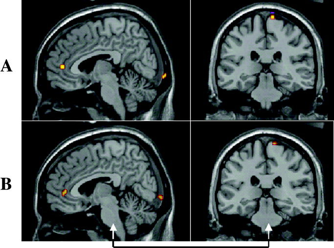
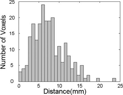
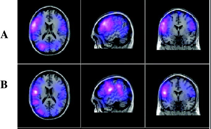
We contend that powerful group studies can be conducted using magnetoencephalography (MEG), which can provide useful insights into the approximate distribution of the neural activity detected with MEG without requiring magnetic resonance imaging (MRI) for each participant. Instead, a participant's MRI is approximated with one chosen as a best match on the basis of the scalp surface from a database of available MRIs. Because large inter-individual variability in sulcal and gyral patterns is an inherent source of blurring in studies using grouped functional activity, the additional error introduced by this approximation procedure has little effect on the group results, and offers a sufficiently close approximation to that of the participants to yield a good indication of the true distribution of the grouped neural activity. T1-weighted MRIs of 28 adults were acquired in a variety of MR systems. An artificial functional image was prepared for each person in which eight 5 x 5 x 5 mm regions of brain activation were simulated. Spatial normalisation was applied to each image using transformations calculated using SPM99 with (1) the participant's actual MRI, and (2) the best matched MRI substituted from those of the other 27 participants. The distribution of distances between the locations of points using real and substituted MRIs had a modal value of 6 mm with 90% of cases falling below 12.5 mm. The effects of this approach on real grouped SAM source imaging of MEG data in a verbal fluency task are also shown. The distribution of MEG activity in the estimated average response is very similar to that produced when using the real MRIs.
Copyright 2003 Wiley-Liss, Inc.
Figures
Similar articles
-
Similarities between simulated spatial spectra of scalp EEG, MEG and structural MRI.Brain Topogr. 2009 Nov;22(3):191-6. doi: 10.1007/s10548-009-0104-7. Epub 2009 Jun 26. Brain Topogr. 2009. PMID: 19557510 Free PMC article.
-
Pseudo-MRI Engine for MRI-Free Electromagnetic Source Imaging.Hum Brain Mapp. 2025 Feb 1;46(2):e70148. doi: 10.1002/hbm.70148. Hum Brain Mapp. 2025. PMID: 39902833 Free PMC article.
-
Sharing individualised template MRI data for MEG source reconstruction: A solution for open data while keeping subject confidentiality.Neuroimage. 2022 Jul 1;254:119165. doi: 10.1016/j.neuroimage.2022.119165. Epub 2022 Apr 1. Neuroimage. 2022. PMID: 35378289
-
Mapping function in the human brain with magnetoencephalography, anatomical magnetic resonance imaging, and functional magnetic resonance imaging.J Clin Neurophysiol. 1995 Sep;12(5):406-31. doi: 10.1097/00004691-199509010-00002. J Clin Neurophysiol. 1995. PMID: 8576388 Review.
-
Magnetoencephalography (MEG) and magnetic source imaging (MSI).Neurologist. 2004 May;10(3):138-53. doi: 10.1097/01.nrl.0000126589.21840.a1. Neurologist. 2004. PMID: 15140274 Review.
Cited by
-
Frontoparietal network and neuropsychological measures in typically developing children.Neuropsychologia. 2021 Aug 20;159:107914. doi: 10.1016/j.neuropsychologia.2021.107914. Epub 2021 Jun 10. Neuropsychologia. 2021. PMID: 34119500 Free PMC article.
-
A tool for functional brain imaging with lifespan compliance.Nat Commun. 2019 Nov 5;10(1):4785. doi: 10.1038/s41467-019-12486-x. Nat Commun. 2019. PMID: 31690797 Free PMC article.
-
Alpha Frequency Entrainment Reduces the Effect of Visual Distractors.J Cogn Neurosci. 2019 Sep;31(9):1392-1403. doi: 10.1162/jocn_a_01422. Epub 2019 May 6. J Cogn Neurosci. 2019. PMID: 31059352 Free PMC article.
-
Beta and gamma oscillations index cognitive interference effects across a distributed motor network.Neuroimage. 2020 Jun;213:116747. doi: 10.1016/j.neuroimage.2020.116747. Epub 2020 Mar 14. Neuroimage. 2020. PMID: 32179103 Free PMC article.
-
Hippocampal and cortical oscillatory dynamics reflect semantic processing and predict behavioural performance.J Physiol. 2025 May;603(10):3089-3106. doi: 10.1113/JP287373. Epub 2025 May 5. J Physiol. 2025. PMID: 40320916
References
-
- Carducci F, Babiloni C, Babiloni F, Cincotti F, Moretti D, Rizzuto M, Rossini PM (2001): Localization of cortical sites in subjects not having MRIs. Neuroimage 13: S91.
-
- Friston K, Holmes A, Worsley K, Poline J, Frith C, Frackowiak R (1995): Statistical parametric mapping in functional imaging: a general linear approach. Hum Brain Mapp 2: 189–210.
-
- Grave de Peralta‐Menendez R, Gonzales‐Andino SL (1998): A critical analysis of linear inverse solutions to the neuroelectromagnetic inverse problem. IEEE Trans Biomed Eng 45: 440–448. - PubMed
-
- Hillebrand A, Barnes GR (2002): A quantitative assessment of the sensitivity of whole‐head MEG to activity in the adult human cortex. Neuroimage 16: 638–650. - PubMed
-
- Kozinska D, Tretiak OJ, Nissanov J, Ozturk C (1997): Multidimensional alignment using the Euclidean distance transform. Graphical Models Image Process 59: 373–387.
MeSH terms
LinkOut - more resources
Full Text Sources
Medical
