. 2003 Nov;69(11):6361-9.
doi: 10.1128/AEM.69.11.6361-6369.2003.

Cold shock response and major cold shock proteins of Vibrio cholerae

Affiliations

Cold shock response and major cold shock proteins of Vibrio cholerae

Partha Pratim Datta et al. Appl Environ Microbiol. 2003 Nov.

Abstract

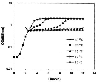
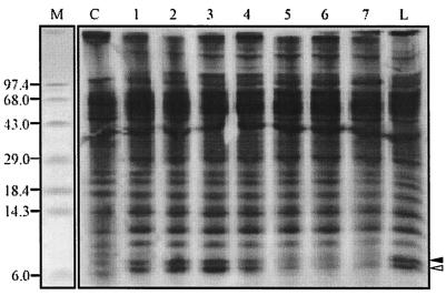
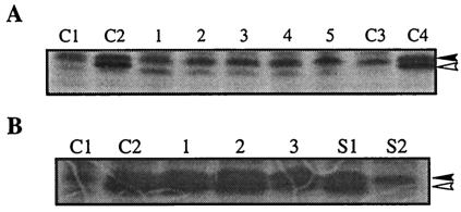
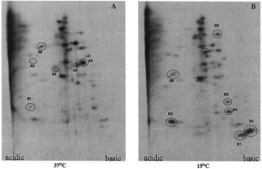
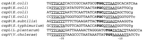
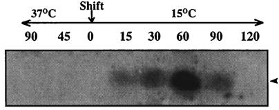
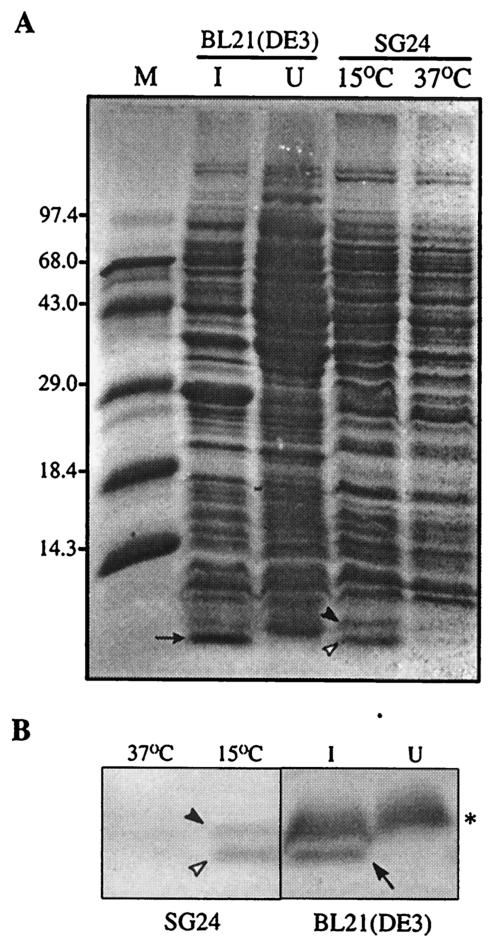
When exponentially growing Vibrio cholerae cells were shifted from 37 degrees C to various lower temperatures, it was found that the organism could adapt and grow at temperatures down to 15 degrees C, below which the growth was completely arrested. There was no difference between the patterns of the cold shock responses in toxinogenic and nontoxinogenic strains of V. cholerae. Gel electrophoretic analyses of proteins of cold-exposed cells revealed significant induction of two major cold shock proteins (Csps), whose molecular masses were 7.7 kDa (CspA(VC)) and 7.5 kDa (CspV), and six other Csps, most of which were much larger. We cloned, sequenced, and analyzed the cspV gene encoding the CspV protein of V. cholerae O139 strain SG24. Although CspA(VC) and CspV have similar kinetics of synthesis and down-regulation, the corresponding genes, cspA and cspV, which are located in the small chromosome, are not located in the same operon. A comparative analysis of the kinetics of synthesis revealed that the CspV protein was synthesized de novo only during cold shock. Although both CspA(VC) and CspV were stable for several hours in the cold, the CspV protein was degraded rapidly when the culture was shifted back to 37 degrees C, suggesting that this protein is probably necessary for adaptation at lower temperatures. Northern blot analysis confirmed that the cspV gene is cold shock inducible and is regulated tightly at the level of transcription. Interestingly, the cspV gene has a cold shock-inducible promoter which is only 12 nucleotides from the translational start site, and therefore, it appears that no unusually long 5' untranslated region is present in its mRNA transcript. Thus, this promoter is an exception compared to other promoters of cold shock-inducible genes of different organisms, including Escherichia coli. Our results suggest that V. cholerae may use an alternative pathway for regulation of gene expression during cold shock.


Figures

FIG. 1.
FIG. 1.
Growth kinetics of V. cholerae O139 strain SG24 at various temperatures. The arrow indicates the OD585 at which cultures were shifted from 37°C to low temperatures.
FIG. 2.
FIG. 2.
Autoradiogram showing the kinetics of induction of CspAVC and CspV. Pulse-labeling at 15°C was performed for the following periods: lane 1, 0 to 30 min; lane 2, 30 to 60 min; lane 3, 60 to 90 min; lane 4, 90 to 120 min; lane 5, 120 to 150 min; lane 6, 150 to 180 min; and lane 7, 270 to 300 min. Lane C contained cells that were labeled for 10 min at 37°C. Lane L contained cells that were labeled at 15°C for 5 h. The solid and open arrowheads indicate the positions of CspAVC and CspV, respectively. Lane M contained standard molecular mass markers, whose sizes (in kilodaltons) are indicated on the left.
FIG. 3.
FIG. 3.
Stability of CspAVC and CspV. (A) Cells were pulse-labeled for the following periods after they were shifted back from 15 to 37°C: lane 1, 0 to 15 min; lane 2, 15 to 30 min; lane 3, 30 to 45 min; lane 4, 45 to 60 min; and lane 5, 0 to 3.5 h. Lanes C1 and C2 contained cells that were labeled for 10 min at 37°C and for 60 to 90 min at 15°C, respectively. Lanes C3 and C4 contained cells that were labeled for 3.5 h at 37°C and at 15°C, respectively. The solid and open arrowheads indicate the positions of CspAVC and CspV, respectively. (B) Lanes C1 and C2 contained cells that were labeled for 10 min at 37°C and for 60 to 90 min at 15°C, respectively. Cells were labeled for 2 h at 15°C and then chased with nonradioactive methionine (see Materials and Methods) for the following periods: lane 1, 1 h; lane 2, 3 h; and lane 3, 12 h. Lanes S1 and S2 contained cells that were labeled at 15°C for 1 h, shifted back to 37°C, and chased with nonradioactive methionine for 15 and 30 min, respectively. The solid and open arrowheads indicate the positions of CspAVC and CspV, respectively.
FIG. 4.
FIG. 4.
Role of Csps in cold adaptation of V. cholerae and status of CspAVC and CspV in the presence of chloramphenicol. (A) Growth of V. cholerae SG24 at various temperatures was monitored as described in Materials and Methods. The solid and open arrowheads indicate the OD585 values at which V. cholerae cells were shifted from 37°C to low temperatures and from 15 to 10°C, respectively. The curved arrow indicates the enhanced growth rate at 10°C after preincubation at 15°C. (B) Lanes 1 and 2 contained V. cholerae cells grown at 37°C in absence and presence of chloramphenicol, respectively. [35S]methionine labeling of cellular proteins in the presence of chloramphenicol at 15°C was done for the following periods: lane 3, 0 to 30 min; lane 4, 30 to 60 min; lane 5, 60 to 90 min; lane 6, 150 to 180 min; and lane 7, 5 to 5.5 h. For lanes 8 and 9, chloramphenicol was added 4.5 h after the shift from 37 to 15°C and this was followed by labeling at 5 to 5.5 h and at 6 to 6.5 h, respectively. The solid and open arrowheads indicate the positions of CspAVC and CspV, respectively.
FIG. 5.
FIG. 5.
Autoradiograms of 2D-E gels showing the major Csps of V. cholerae. Proteins were labeled and separated as described in Materials and Methods. The spots for proteins that were down-regulated due to a shift from 37 to 15°C and the spots for proteins which were induced due to cold shock are circled in panels A and B, respectively, and are numbered on the basis of size in ascending order in the autoradiograms. The same amount of protein was loaded in each gel.
FIG. 6.
FIG. 6.
Comparison of the promoter regions of cspA homologues. The promoter regions of the E. coli cspA (35), cspB (17), cspG (23), and cspI (37) genes, the Bacillus subtilis cspB gene (38), the Salmonella enterica serovar Typhimurium cspB gene (9), and the Lactobacillus plantarum cspL gene (19) and P2 of the cspV gene of V. cholerae (this study) are compared. The putative transcriptional start sites are indicated by lowercase type; the proposed −10 and −35 regions are underlined; and the highly conserved TGn motifs just upstream of the −10 regions in the homologues are indicated by boldface type.
FIG. 7.
FIG. 7.
Northern blot analysis of the cspV mRNA transcripts after cold shock. A 210-bp DNA fragment of the cspV gene was used as a probe. The time (in minutes) at which an aliquot was removed from a V. cholerae culture growing either at 37°C or at 15°C is indicated above each lane. The zero indicates the time of the shift from 37 to 15°C. The same amount of RNA was loaded in each lane. The arrowhead indicates the position of the hybridized band of cspV transcripts.
FIG. 8.
FIG. 8.
(A) Overexpression of the CspV protein in E. coli. Lanes I and U contained cells grown under IPTG-induced and uninduced conditions, respectively. The arrow indicates the position of the overexpressed CspV protein. The solid and open arrowheads indicate the positions of CspAVC and CspV, respectively. Lane M contained protein molecular mass standards, whose sizes (in kilodaltons) are indicated on the left. (B) Immunoblot detection of cold shock-induced CspAVC (solid arrowhead), CspV (open arrowhead), and overexpressed CspV (arrow) by using rabbit polyclonal antiserum against the CspA protein of E. coli. Lanes I and U contained cells grown under IPTG-induced and uninduced conditions, respectively. The asterisk indicates the position of a nonspecific protein band of E. coli that cross-reacted with the antiserum.

References

    1. Ausubel, F. M., R. Brent, R. E. Kingston, D. D. Moore, J. G. Seidman, J. A. Smith, and K. Struhl. 1995. Current protocols in molecular biology. John Wiley & Sons, Inc., New York, N.Y.
    1. Bae, W., P. G. Jones, and M. Inouye. 1997. CspA, the major cold shock protein of Escherichia coli, negatively regulates its own gene expression. J. Bacteriol. 179:7081-7088. - PMC - PubMed
    1. Bhadra, R. K., S. Roychoudhury, and J. Das. 1994. Vibrio cholerae O139 biotype El Tor. Lancet 343:728. - PubMed
    1. Bik, E. M., A. E. Bunschoten, R. D. Gouw, and F. Mooi. 1995. Genesis of the novel epidemic Vibrio cholerae O139 strain: evidence for horizontal transfer of genes involved in polysaccharide synthesis. EMBO J. 14:209-216. - PMC - PubMed
    1. Carroll, W. J., M. C. Mateescu, K. Chava, R. R. Colwell, and A. K. Bej. 2001. Response and tolerance of toxigenic Vibrio cholerae O1 to cold temperatures. Antonie Leeuwenhoek 79:377-384. - PubMed

Publication types

MeSH terms

Substances

Associated data

LinkOut - more resources