

Cerebral perfusion in infants and neonates: preliminary results obtained using dynamic susceptibility contrast enhanced magnetic resonance imaging
- PMID: 14602704
- PMCID: PMC1763235
- DOI: 10.1136/fn.88.6.f525
Cerebral perfusion in infants and neonates: preliminary results obtained using dynamic susceptibility contrast enhanced magnetic resonance imaging
Abstract
Background: Previous studies have used the dynamic susceptibility contrast enhanced (DSCE) magnetic resonance (MR) imaging technique to measure cerebral perfusion in adults.
Objective: To assess the feasibility of the technique in a heterogeneous cohort of sick human infants and identify cerebral perfusion abnormalities.
Methods: Perfusion measurements were made by characterising the changing concentration of an injected bolus of contrast agent using a series of MR images acquired during the first pass of the contrast bolus. Qualitative values of relative cerebral blood flow (rCBF) were then calculated from these data on a pixel by pixel basis to generate parametric maps of perfusion.
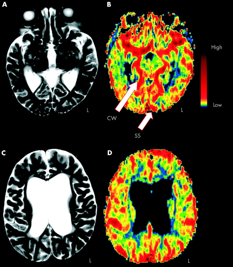
Results: Images of perfusion were successfully calculated from 12 out of 27 neonates and infants, all with established cerebral pathology. Normal vascular anatomical structures such as the circle of Willis were identified within all calculated images. Values of rCBF were generally larger in grey matter than in white matter. In several patients, perfusion abnormalities resulted in structural abnormalities which were detected in conventional MR imaging at follow up. The acquisition of perfusion data was most difficult when the least mature brains were examined because of motion artefacts and a smaller head size with a lower level of rCBF than adults.
Conclusions: This preliminary study shows that: (a) maps of rCBF can be acquired from neonates and infants; (b) characterisation of the bolus passage becomes progressively easier as the brain matures; (c) early abnormalities in cerebral perfusion may have negative prognostic implications; (d) the main difficulty when using the DSCE technique to study neonates relates to image artefacts resulting from bulk head motion.
Figures

References
Publication types
MeSH terms
Substances
LinkOut - more resources
Full Text Sources
Medical