. 2003 Nov 5;23(31):9961-7.
doi: 10.1523/JNEUROSCI.23-31-09961.2003.

Lmx1b, Pet-1, and Nkx2.2 coordinately specify serotonergic neurotransmitter phenotype

Affiliations

Lmx1b, Pet-1, and Nkx2.2 coordinately specify serotonergic neurotransmitter phenotype

Leping Cheng et al. J Neurosci. .

Abstract

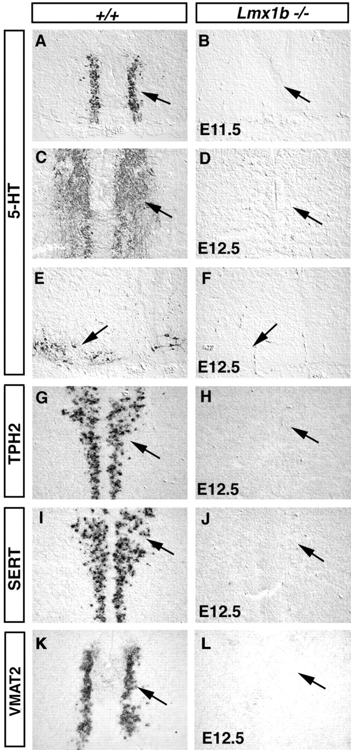
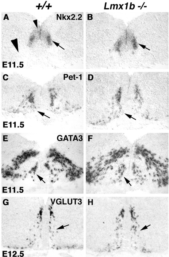
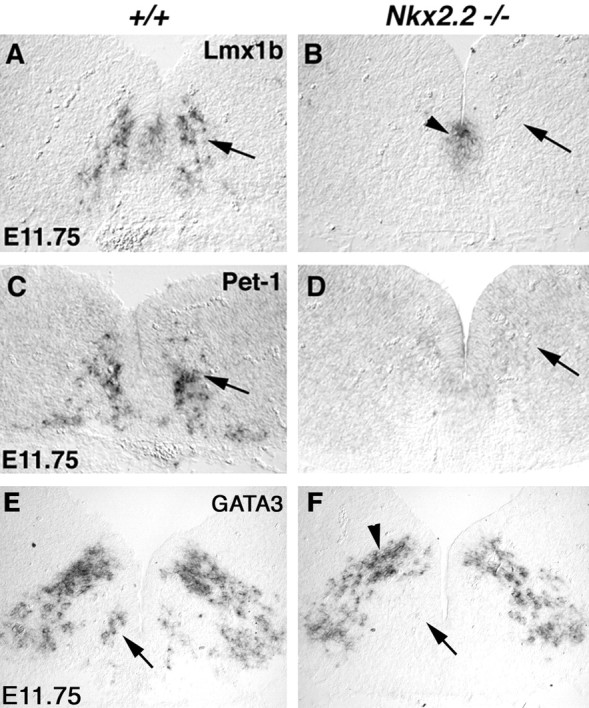
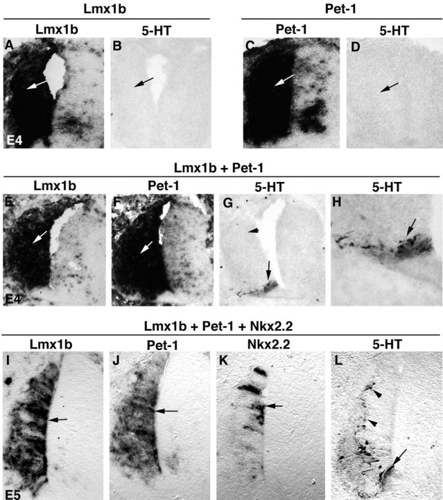
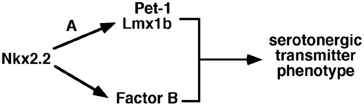
Serotonergic (5-HT) neurons in the brainstem modulate a wide range of physiological processes and behaviors. Two transcription factor genes, Pet-1 and Nkx2.2, are necessary but not sufficient to specify the 5-HT transmitter phenotype. Here we show that the Lim class homeobox gene Lmx1b is required for proper formation of the entire 5-HT system in the hindbrain, as indicated by the loss of expression of genes necessary for serotonin synthesis and transport in Lmx1b null mice. Lmx1b and Pet1 act downstream of Nkx2.2, and their expression is independently regulated at the time when 5-HT transmitter phenotype is specified. Ectopic expression of Lmx1b plus Pet-1 is able to induce formation of 5-HT cells in the most ventral spinal cord, where Nkx2.2 is normally expressed. Combined expression of all three genes, Lmx1b, Pet-1, and Nkx2.2, drives 5-HT differentiation in the dorsal spinal cord. Our studies therefore define a molecular pathway necessary and sufficient to specify the serotonergic neurotransmitter phenotype.


Figures

Figure 1.
Figure 1.
Lmx1b is expressed in serotonergic neurons. Transverse sections through E11.5 pons (A, B), E12.5 caudal pons (C, D), E12.5 medulla (E, F), and E14.5 caudal pons (G-I) are shown. In situ hybridization was performed with the indicated probes (A-F). Lmx1b and Pet-1 are expressed in neurons derived from the ventral midline (A-F, arrows) that correspond to the primordial raphe nuclei. Lmx1b is also expressed weakly in the floor plate (A, arrowhead) and other areas in the medulla (A, star; E, arrowhead). G-H, Coronal sections through the E14.5 pons. Double immunostaining was performed with Lmx1b antibody (G, red) and 5-HT antibody (H, green). I is the superimposed image showing that Lmx1b is expressed in all 5-HT cells (with yellow nuclei) (I, arrow). A small subset of Lmx1b-positive cells is 5-HT negative (I, arrowhead).
Figure 2.
Figure 2.
Development of 5-HT neurons is compromised in Lmx1b mutants. Transverse sections through the pons (A-D, G-J) and the medulla (E, F, K, L) of embryos with indicated stages and genotypes are shown. 5-HT immunostaining (A-F) and in situ hybridization (G-L) were performed with the indicated probes (G-L).
Figure 3.
Figure 3.
Prospective 5-HT neurons are formed in Lmx1b null embryos. Transverse sections through E11.5 medulla (A-D) and E12.5 pons (E-H) are shown. In situ hybridization was performed with the indicated probes.
Figure 4.
Figure 4.
Nkx2.2 is necessary for the expression of Lmx1b, Pet-1, and GATA3. Transverse sections through medulla of E11.75 wild-type (A, C, D) and Nkx2.2 mutants (B, D, F) are shown. In situ hybridization was performed with the indicated probes. Expression of Lmx1b, Pet-1, and GATA3 in prospective 5-HT neurons is lost in Nkx2.2 null medulla (B, D, F, arrows). Residual Lmx1b expression in the floor plate is still detected in Nkx2.2 mutants (B, arrowhead). Also, GATA3 expression in the more dorsally localized neurons is also unaffected in the mutants (F, arrowhead).
Figure 5.
Figure 5.
Lmx1b, Pet-1, and Nkx2.2 in combination are sufficient to induce 5-HT cell fate in the chick spinal cord. Transverse sections through the spinal cord are shown. Plasmids were electroporated at E2 and analyzed at E4 (A-H) or E5 (I-L). The plasmids used for electroporation are shown above the lines. Expression of the transgenes (the names of which are shown at the top of the panels and below the lines) was detected by in situ hybridization. Also, 5-HT immunostaining was performed (B, D, G, H, L). H is the high magnification of the positive area shown in G.
Figure 6.
Figure 6.
A molecular pathway controlling 5-HT cell fate specification. Nkx2.2 plus an unknown cofactor (A) directly or indirectly activate the expression of Pet-1 and Lmx1b in prospective 5-HT neurons. Nkx2.2 might need a different cofactor to activate another downstream factor (B), or it removes an unknown inhibitor (data not shown) to allow Lmx1b and Pet-1 to promote 5-HT cell fate.

References

    1. Adams KA, Maida JM, Golden JA, Riddle RD ( 2000) The transcription factor Lmx1b maintains Wnt1 expression within the isthmic organizer. Development 127: 1857-1867. - PubMed
    1. Asbreuk CH, Vogelaar CF, Hellemons A, Smidt MP, Burbach JP ( 2002) CNS expression pattern of Lmx1b and coexpression with ptx genes suggest functional cooperativity in the development of forebrain motor control systems. Mol Cell Neurosci 21: 410-420. - PubMed
    1. Blakely RD, Berson HE, Fremeau RTJ, Caron MG, Peek MM, Prince HK, Bradley CC ( 1991) Cloning and expression of a functional serotonin transporter from rat brain. Nature 354: 66-70. - PubMed
    1. Briscoe J, Sussel L, Serup P, Hartigan-O'Connor D, Jessell TM, Rubenstein J, Ericson J ( 1999) Homeobox gene Nkx2.2 and specification of neuronal identity by graded Sonic hedgehog signalling. Nature 398: 622-627. - PubMed
    1. Chang AS, Chang SM, Starnes DM, Schroeter S, Bauman AL, Blakely RD ( 1996) Cloning and expression of the mouse serotonin transporter. Brain Res Mol Brain Res 43: 185-192. - PubMed

Publication types

MeSH terms