. 2003 Nov 5;23(31):10013-20.
doi: 10.1523/JNEUROSCI.23-31-10013.2003.

The contraceptive agent Provera enhances GABA(A) receptor-mediated inhibitory neurotransmission in the rat hippocampus: evidence for endogenous neurosteroids?

Affiliations

The contraceptive agent Provera enhances GABA(A) receptor-mediated inhibitory neurotransmission in the rat hippocampus: evidence for endogenous neurosteroids?

Delia Belelli et al. J Neurosci. .

Abstract

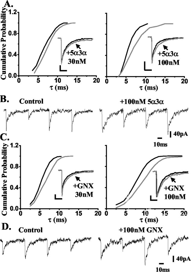
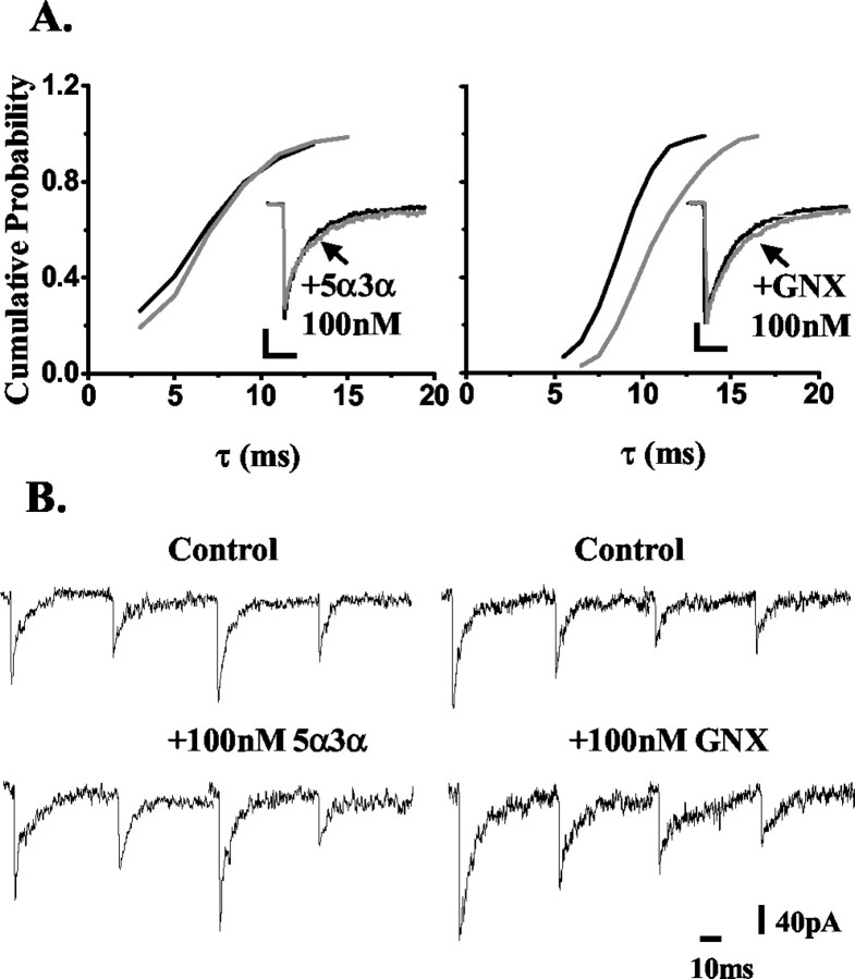
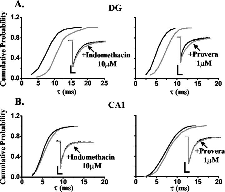
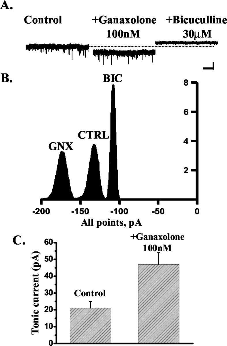
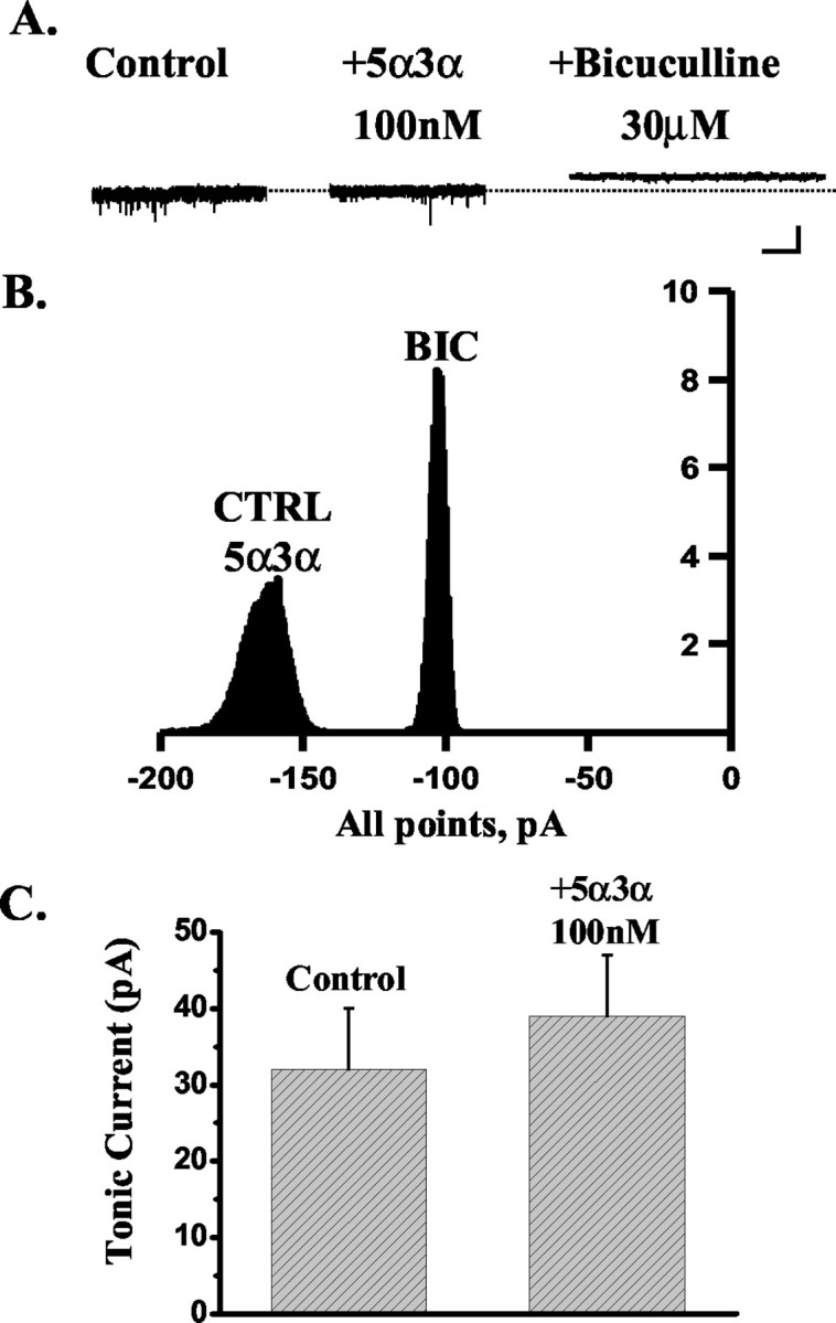
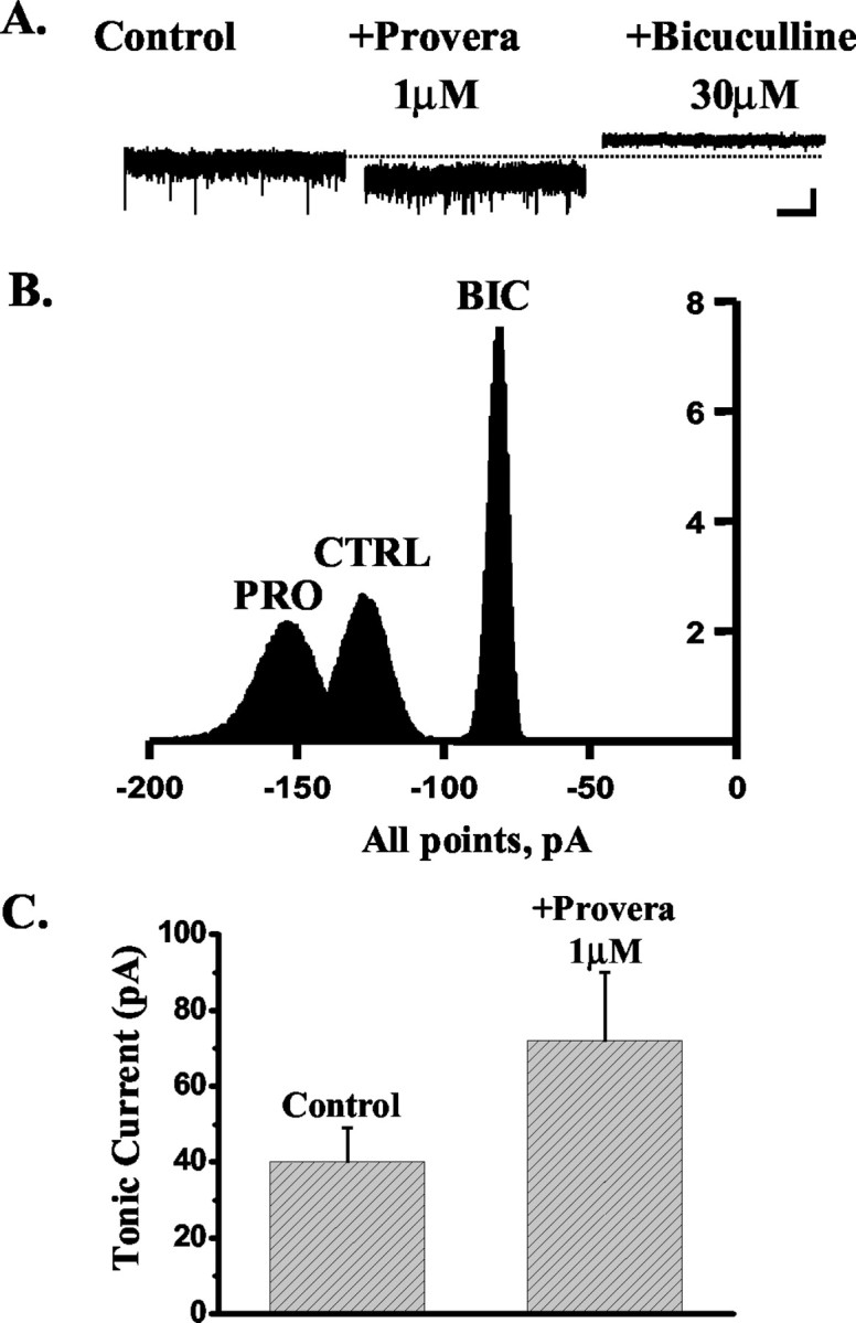
Neurosteroids typified by 5alpha-pregnan-3alpha-ol-20-one (5alpha3alpha) have emerged as the most potent endogenous positive modulators of the GABAA receptor, the principal mediator of fast inhibitory transmission within the CNS. Neurosteroids can be synthesized de novo in the brain in levels sufficient to modulate GABA(A) receptor function and, thus, might play an important physiological-pathophysiological role. Indirect support for this proposal comes from the observation that neurosteroid action is region and neuron selective. However, the mechanism(s) that imparts specificity of action remains primarily elusive. Although neurosteroids are relatively promiscuous toward different GABA(A) receptor isoforms, the contribution of local neurosteroid metabolism has been relatively unexplored. Here, we investigate the role of neurosteroid metabolism by using electrophysiological techniques to compare the actions of 5alpha3alpha and its metabolically stable synthetic analog ganaxolone on inhibitory neurotransmission in CA1 and dentate gyrus neurons. Furthermore, we evaluate the contribution of a key enzyme in neurosteroid metabolism [i.e., 3alpha-hydroxysteroidoxidoreductase (3alpha-HSOR)] to the inactivation of endogenous, or exogenously applied 5alpha3alpha. We show that low concentrations of ganaxolone, but not of 5alpha3alpha, enhance inhibitory transmission in dentate gyrus, whereas both steroids are similarly effective in CA1 neurons. Furthermore, inhibition of 3alpha-HSOR by the contraceptive agent Provera results in enhanced synaptic and extrasynaptic GABA(A) receptor-mediated inhibition in the dentate gyrus but not in the CA1 region. Collectively, these findings advocate a crucial role for local steroid metabolism in shaping GABA(A) receptor-mediated inhibition in a regionally dependent manner and suggest a novel action by the contraceptive agent on inhibitory centers in the CNS.


Figures

Figure 1.
Figure 1.
The effect of 5α3α and ganaxolone (GNX) on the decay of mIPSCs recorded from CA1 neurons. A, A cumulative probability plot of the decay of all mIPSCs, expressed as the cumulative time constant τ (see Materials and Methods), recorded from an exemplar CA1 neuron before and after the application of 30 (left) and 100 nm (right) 5α3α. The rightward shift of this relationship induced by either steroid concentration indicates that all mIPSCs recorded from this cell were sensitive to the neurosteroid. The insets illustrate the normalized ensemble average of all mIPSCs from the same CA1 neurons before and after application of 30 (left) and 100 nm (right) 5α3α. Calibration: 20 pA (left), 10 pA (right), 10 msec. B, Examples of individual mIPSC traces recorded from the same CA1 neuron before and after the application of 100 nm 5α3α. C, A cumulative probability plot of the decay of all mIPSCs, expressed as the cumulative time constant τ (see Materials and Methods), recorded from an exemplar CA1 neuron before and after the application of 30 (left) and 100 nm (right) ganaxolone. The rightward shift of this relationship induced by either steroid concentration indicates that all mIPSCs recorded from this cell were sensitive to this steroid. The insets illustrate the normalized ensemble average of all mIPSCs from the same CA1 neurons before and after application of 30 (left) and 100 nm (right) ganaxolone. Calibration: 10 pA, 10 msec. D, Examples of individual mIPSC traces recorded from the same CA1 neuron before and after the application of 100 nm ganaxolone.
Figure 2.
Figure 2.
The effect of 5α3α and ganaxolone (GNX) on the decay of mIPSCs recorded from DG neurons. A, A cumulative probability plot of the decay of all mIPSCs, expressed as the cumulative time constant τ (see Materials and Methods), recorded from an exemplar DG neuron before and after the application of 100 nm 5α3α (left) and 100 nm ganaxolone (right). The rightward shift of this relationship induced by ganaxolone indicates that all mIPSCs recorded from this cell were sensitive to this steroid. In contrast, the lack of the rightward shift induced by 100 nm 5α3α indicates that all mIPSCs recorded from this cell were insensitive to this neurosteroid. The insets illustrate the normalized ensemble average of all mIPSCs from the same DG neurons before and after application of 100 nm 5α3α (left) and 100 nm ganaxolone (right). Calibration: 20 pA, 10 msec. B, Examples of individual mIPSC traces recorded from the same DG neurons before and after the application of 100 nm 5α3α (left) and 100 nm ganaxolone (right).
Figure 3.
Figure 3.
The effect of indomethacin and Provera on the decay of mIPSCs recorded from DG and CA1 neurons. A, A cumulative probability plot of the decay of all mIPSCs, expressed as the cumulative time constant τ (see Materials and Methods), recorded from an exemplar DG neuron before and after the application of 10 μm indomethacin (left) and 1 μm Provera (right). The rightward shift of this relationship induced by either indomethacin or Provera indicates that all mIPSCs recorded from these cells were sensitive to the 3α-HSOR inhibitor. The insets illustrate the normalized ensemble average of all mIPSCs from the same DG neurons before and after application of 10 μm indomethacin (left) and 1 μm Provera (right). B, A cumulative probability plot of the decay of all mIPSCs, expressed as the cumulative time constant τ (see Materials and Methods), recorded from an exemplar CA1 neuron before and after the application of 10 μm indomethacin (left) and 1 μm Provera (right). The modest rightward shift of this relationship induced by either indomethacin or Provera indicates that all mIPSCs recorded from these cells were marginally sensitive to the 3α-HSOR inhibitor. Note that the magnitude of the shift induced by either 3α-HSOR inhibitor in the CA1 neuron is much smaller compared with that observed for a DG neuron. The insets illustrate the normalized ensemble average of all mIPSCs from the same CA1 neurons before and after application of 10 μm indomethacin (left) and 1 μm Provera (right). Calibration: 20 pA, 10 msec.
Figure 4.
Figure 4.
The effect of ganaxolone on the tonic current recorded from DG neurons. A, The tonic current, calculated as the difference between the holding current in the presence and absence of 30 μm bicuculline (see Materials and Methods), recorded from an exemplar DG neuron is enhanced by the application of 100 nm ganaxolone, and the steroid effect is blocked by the subsequent application of 30 μm bicuculline. The dashed line indicates the holding current (pA) under control conditions. Calibration: 50 pA, 5 sec. B, Corresponding all-point histograms illustrate the amplitude of the holding current under control conditions (CTRL) in the presence of 100 nm ganaxolone (GNX) and after the application of 30 μm bicuculline (BIC). C, Bar graph summarizing the enhancement of the tonic current induced by 100 nm ganaxolone in five DG neurons.
Figure 5.
Figure 5.
The effect of 5α3α on the tonic current recorded from DG neurons. A, The tonic current, calculated as the difference between the holding current (pA) in the presence and absence of 30 μm bicuculline (see Materials and Methods), recorded from an exemplar DG neuron is insensitive to the application of 100 nm 5α3α. The dashed line indicates the holding current under control conditions. Calibration: 50 pA, 5 sec. B, Corresponding all-point histograms illustrate the amplitude of the holding current under control conditions (CTRL) in the presence of 100 nm 5α3α and after the application of 30 μm bicuculline (BIC). Note that all-point histograms illustrating the amplitude of the holding current under control conditions and in the presence of 100 nm 5α3α overlap, thus indicating the lack of sensitivity of the tonic current recorded from this cell to 5α3α action. C, Bar graph summarizing the lack of a significant effect by 100 nm 5α3α on the tonic current recorded from five DG neurons.
Figure 6.
Figure 6.
The effect of Provera on the tonic current recorded from DG neurons. A, The tonic current, calculated as the difference between the holding current (pA) in the presence and absence of 30 μm bicuculline (see Materials and Methods), recorded from an exemplar DG neuron is enhanced by the application of 1 μm Provera, and this effect is blocked by the subsequent application of 30 μm bicuculline. The dashed line indicates the holding current under control conditions. Calibration: 50 pA, 5 sec. B, Corresponding all-point histograms illustrate the amplitude of the holding current under control conditions (CTRL) in the presence of 1 μm Provera (PRO) and after the application of 30 μm bicuculline (BIC) C, Bar graph summarizing the enhancement of the tonic current induced by 1 μm Provera in five DG neurons.

References

    1. Belelli D, Lan NC, Gee KW ( 1990) Anticonvulsant steroids and the GABA/benzodiazepine receptor-chloride ionophore complex. Neurosci Biobehav Rev 14: 315-322. - PubMed
    1. Belelli D, Callachan H, Hill-Venning C, Peters JA, Lambert JJ ( 1996a) Interaction of positive allosteric modulators with human and Drosophila recombinant GABA receptors expressed in Xenopus laevis oocytes. Br J Pharmacol 118: 563-576. - PMC - PubMed
    1. Belelli D, Lambert JJ, Peters JA, Gee KW, Lan NC ( 1996b) Modulation of human recombinant GABAA receptors by pregnanediols. Neuropharmacology 35: 1223-1231. - PubMed
    1. Belelli D, Casula A, Ling A, Lambert JJ ( 2002) The influence of subunit composition on the interaction of neurosteroids with GABAA receptors. Neuropharmacology 43: 651-661. - PubMed
    1. Brickley SG, Cull-Candy SG, Farrant M ( 1996) Development of a tonic form of synaptic inhibition in rat cerebellar granule cells resulting from persistent activation of GABAA receptors. J Physiol (Lond) 497: 753-759. - PMC - PubMed

Publication types

MeSH terms

LinkOut - more resources