

The complete genome sequence and analysis of Corynebacterium diphtheriae NCTC13129
- PMID: 14602910
- PMCID: PMC275568
- DOI: 10.1093/nar/gkg874
The complete genome sequence and analysis of Corynebacterium diphtheriae NCTC13129
Abstract
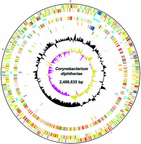
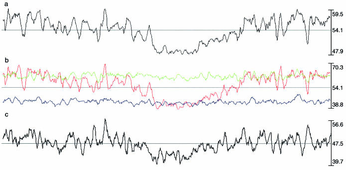
Corynebacterium diphtheriae is a Gram-positive, non-spore forming, non-motile, pleomorphic rod belonging to the genus Corynebacterium and the actinomycete group of organisms. The organism produces a potent bacteriophage-encoded protein exotoxin, diphtheria toxin (DT), which causes the symptoms of diphtheria. This potentially fatal infectious disease is controlled in many developed countries by an effective immunisation programme. However, the disease has made a dramatic return in recent years, in particular within the Eastern European region. The largest, and still on-going, outbreak since the advent of mass immunisation started within Russia and the newly independent states of the former Soviet Union in the 1990s. We have sequenced the genome of a UK clinical isolate (biotype gravis strain NCTC13129), representative of the clone responsible for this outbreak. The genome consists of a single circular chromosome of 2 488 635 bp, with no plasmids. It provides evidence that recent acquisition of pathogenicity factors goes beyond the toxin itself, and includes iron-uptake systems, adhesins and fimbrial proteins. This is in contrast to Corynebacterium's nearest sequenced pathogenic relative, Mycobacterium tuberculosis, where there is little evidence of recent horizontal DNA acquisition. The genome itself shows an unusually extreme large-scale compositional bias, being noticeably higher in G+C near the origin than at the terminus.
Figures


References
-
- Holmes R.K. (2000) Biology and molecular epidemiology of diphtheria toxin and the tox gene. J. Infect. Dis., 181 (Suppl. 1), S156–S167. - PubMed
-
- Hadfield T.L., McEvoy,P., Polotsky,Y., Tzinserling,V.A. and Yakovlev,A.A. (2000) The pathology of diphtheria. J. Infect. Dis., 181 (Suppl. 1), S116–S120. - PubMed
-
- Cha J.H., Chang,M.Y., Richardson,J.A. and Eidels,L. (2003) Transgenic mice expressing the diphtheria toxin receptor are sensitive to the toxin. Mol. Microbiol., 49, 235–240. - PubMed
-
- Public Health Laboratory Service (1997) Diptheria acquired during a cruise in the Baltic Sea. Commun. Dis. Rep. Weekly, 7, 137–138. - PubMed
Publication types
MeSH terms
Substances
Associated data
- Actions
Grants and funding
LinkOut - more resources
Full Text Sources
Other Literature Sources
Molecular Biology Databases
