. 2003 Nov 25;100(24):13839-44.
doi: 10.1073/pnas.1936150100. Epub 2003 Nov 6.

J protein cochaperone of the mitochondrial inner membrane required for protein import into the mitochondrial matrix

Affiliations

J protein cochaperone of the mitochondrial inner membrane required for protein import into the mitochondrial matrix

Patrick D D'Silva et al. Proc Natl Acad Sci U S A. .

Abstract

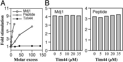
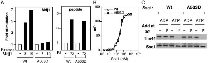
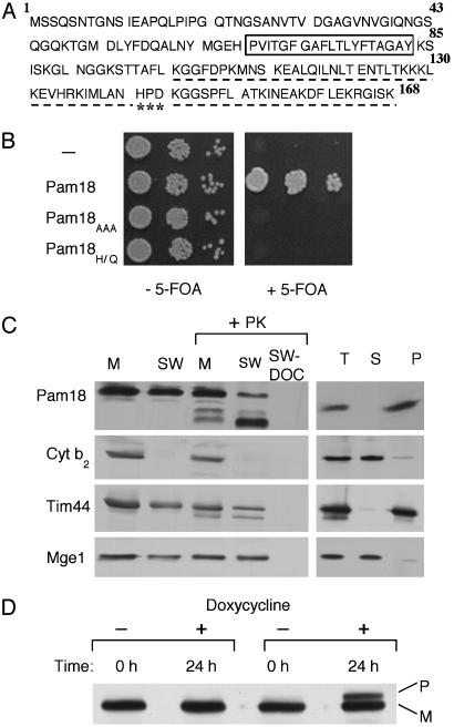
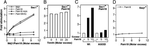
The major Hsp70 of the mitochondrial matrix (Ssc1 in yeast) is critically important for the translocation of proteins from the cytosol, across the mitochondrial inner membrane, and into the matrix. Tim44, a peripheral inner membrane protein with limited sequence similarity to the J domain of J-type cochaperones, tethers Ssc1 to the import channel. Here we report that, unlike a J protein, Tim44 does not stimulate the ATPase activity of Ssc1, nor does it affect the stimulation by either a known mitochondrial J protein or a peptide substrate. Thus, we conclude that Tim44 does not function as a J protein cochaperone of Ssc1; rather, it tethers Ssc1 to the import channel through interactions independent of those critical for J protein function. However, a previously unstudied essential gene, PAM18, encodes an 18-kDa protein that contains a J domain and is localized to the mitochondrial inner membrane. Pam18 stimulates the ATPase activity of Ssc1; depletion of Pam18 in vivo disrupts import of proteins into the mitochondrial matrix. We propose that Pam18 is the J protein partner for Ssc1 at the import channel and is critical for Ssc1's function in protein import.


Figures

Fig. 1.
Fig. 1.
Effect of Tim44 on the ATPase activity of Ssc1. Ssc1:ATP complex (≈0.2 μM) was incubated at 23°C in the presence of various concentrations of Mdj1 or peptide P5. The rate of conversion to ADP was measured and plotted as the fold stimulation over the basal rate determined in the absence of additional components. (A) Mdj1, peptide P5, or Tim44, as indicated. (B) Reactions with 1 μM Mdj1 (Left)or5 μM peptide P5 (Right) were carried out in the presence of indicated concentrations of Tim44.
Fig. 2.
Fig. 2.
Functional defect of Ssc1A503D. (A) ATPase activity. Ssc1:ATP and Ssc1A503D:ATP complexes (≈0.2 μM) were incubated at 23°C in the presence of Mdj1 or peptide P5. The basal rates of hydrolysis of the mutant and wild-type protein were indistinguishable. (B) Peptide binding measured by fluorescence anisotropy: Fluorescein-labeled F-P5 (10 nM) was incubated in the presence of the indicated concentrations of wild-type and A503D mutant protein and ADP. Anisotropy measurements were taken at 25°C; raw polarization values were fitted to a one-site binding (hyperbola) equation to determine the Kd. (C) Effect of peptide on Ssc1:Tim44 interaction. Tim44 (0.09 μM) was incubated with Ssc1 (0.6 μM) in the presence of either ATP or ADP, as indicated, for 30 min. In some cases, 10 μM peptide P5 was added, and incubation continued for an additional 30 min. The mixtures were immunoprecipitated by using Ssc1-specific antibodies as described in Materials and Methods. The presence of Tim44 and Ssc1 in the precipitate was assessed by immunoblot analysis by using antibodies specific for the respective proteins.
Fig. 3.
Fig. 3.
In vivo function of the J protein Pam18. (A) Predicted amino acid sequence of Pam18. A box is drawn around the putative membrane-spanning region. The region having sequence identity to a J domain is underlined; the highly conserved HPD is indicated by ***. (B) Requirement in vivo of J domain of Pam18. Strains carrying plasmids expressing no protein (–), wild-type Pam18, and mutant Pam18, Pam18AAA or Pam18H141Q (Pam18HI/Q) were spotted as a 1- fold dilution series on media lacking (Left) or containing (Right) 5-fluoroorotic acid. Plates were incubated for 3 days at 30°C. (C) Fractionation of mitochondria. Mitochondria were purified from cells expressing Pam18 having an HA tag at the C terminus. Mitochondria were separated into membrane and soluble components (Right). Mitochondrial protein (T) and fractional equivalents of membrane pellet (P) and supernatant (S) were analyzed. Mitochondria (M) were subjected to swelling to disrupt the outer membrane (SW) (Left). Equivalent amounts were treated with proteinase K and in some cases treated with detergent (sodium deoxycholate), as indicated. The fractions were subjected to immunoblot analysis by using antibodies specific for the HA tag on Pam18, as well as an antibody specific for known proteins of the intermembrane space (cytochrome b2), inner membrane (Tim44), and matrix (Mge1). (D) Δpam18 cells carrying Pam18 under control of the tetracycline regulatable promoter were grown overnight in rich media, at which point they were subcultured in the presence or absence of the tetracycline analogue doxycycline. After 24 h whole-cell lysates were prepared and separated by SDS/PAGE, followed by immunoblot analysis by using antibodies specific for Hsp60.
Fig. 4.
Fig. 4.
Stimulation of Ssc1 ATPase activity by Pam18. Hsp70:ATP complexes were prepared, and the rate of hydrolysis was measured at 23°C in the presence of various concentrations of Mdj1, wild-type Pam18, or mutant Pam18, in the presence or absence of Tim44. The rate of conversion to ADP was measured and is plotted as the fold stimulation over the basal rate. In each panel, the Hsp70 for which ATP hydrolysis was measured is indicated by an asterisk. (A) Ssc1:ATP (≈0.2 μM) was incubated in the presence of various concentrations of wild-type Pam18, Mdj1, or the Pam 18 mutant proteins Pam18AAA or Pam18H141Q. (B) Reactions with 1 μM Pam18 were carried out in the presence of indicated concentrations of Tim44. (C) Ssc1:ATP and Ssc1A503D:ATP complexes (≈0.2 μM) were incubated in the presence of excess Pam18. (D) Ssq1:ATP complexes (0.1 μM) in the presence of various concentrations of Pam18.

References

    1. Neupert, W. & Brunner, M. (2002) Nat. Rev. Mol. Cell Biol. 8, 555–565. - PubMed
    1. Pfanner, N. & Geissler, A. (2001) Nat. Rev. Mol. Cell Biol. 5, 339–349. - PubMed
    1. Matouschek, A., Pfanner, N. & Voos, W. (2000) EMBO Rep. 5, 404–410. - PMC - PubMed
    1. Bukau, B. & Horwich, A. L. (1998) Cell 92, 351–366. - PubMed
    1. Hartl, F. & Hayer-Hartl, M. (2002) Science 295, 1852–1858. - PubMed

Publication types

MeSH terms