. 2003 Nov 15;171(10):5537-46.
doi: 10.4049/jimmunol.171.10.5537.

Inhibition of fibrocyte differentiation by serum amyloid P

Affiliations

Inhibition of fibrocyte differentiation by serum amyloid P

Darrell Pilling et al. J Immunol. .

Abstract

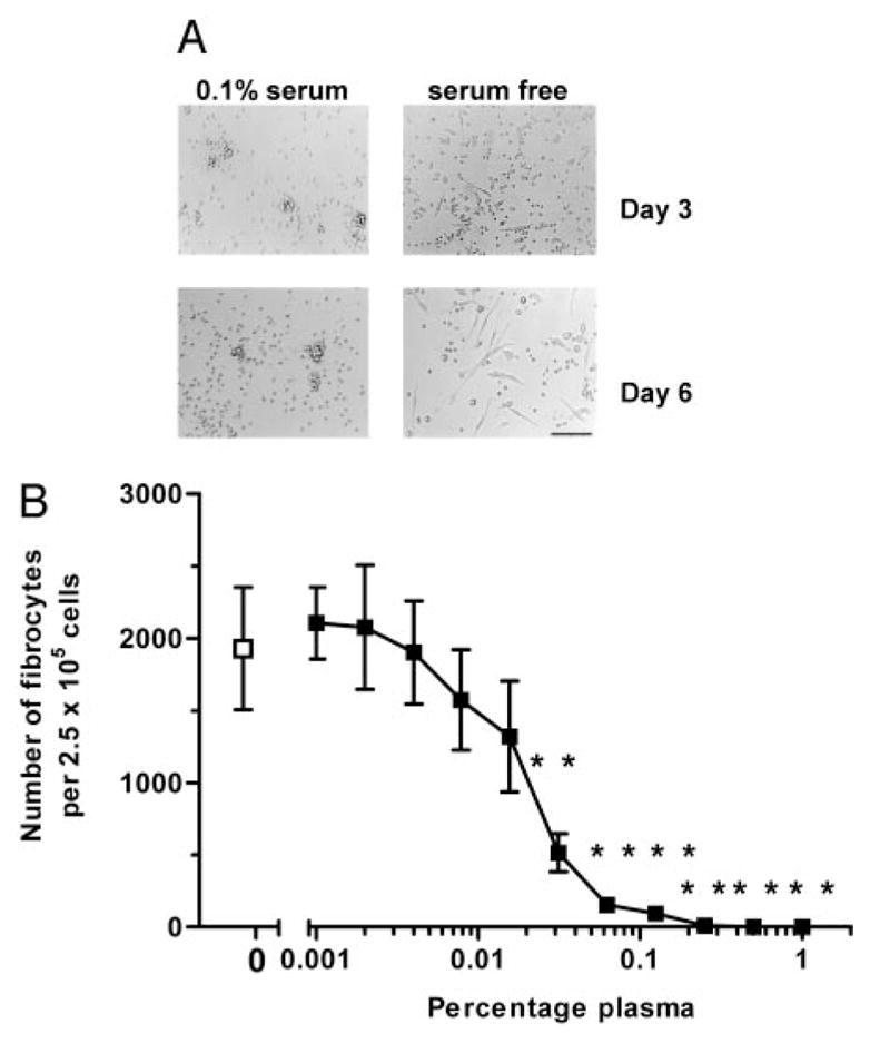
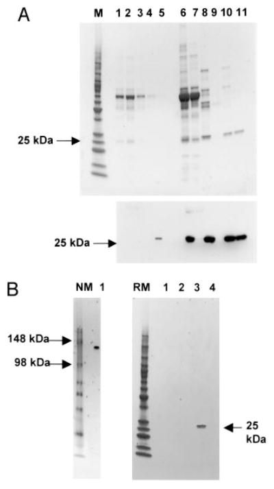
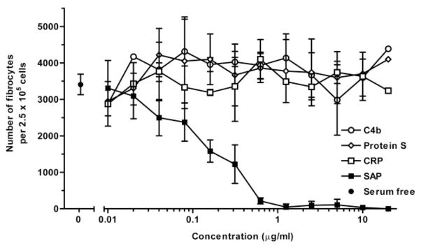
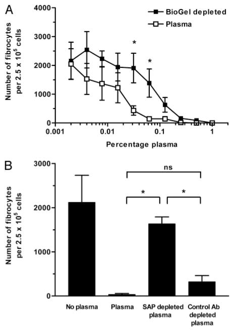
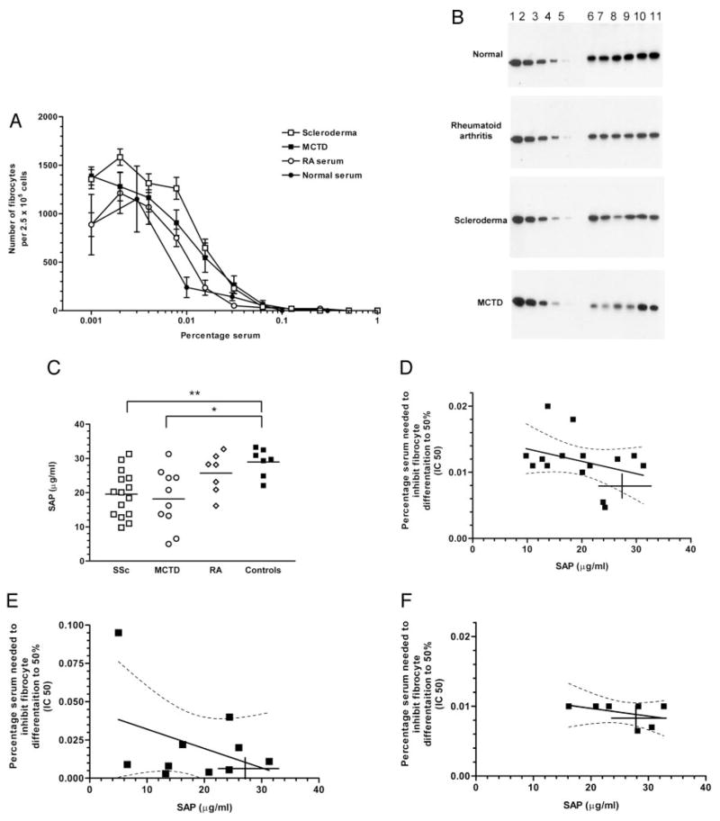
Wound healing and the dysregulated events leading to fibrosis both involve the proliferation and differentiation of fibroblasts and the deposition of extracellular matrix. Whether these fibroblasts are locally derived or from a circulating precursor population is unclear. Fibrocytes are a distinct population of fibroblast-like cells derived from peripheral blood monocytes that enter sites of tissue injury to promote angiogenesis and wound healing. We have found that CD14(+) peripheral blood monocytes cultured in the absence of serum or plasma differentiate into fibrocytes within 72 h. We purified the factor in serum and plasma that prevents the rapid appearance of fibrocytes, and identified it as serum amyloid P (SAP). Purified SAP inhibits fibrocyte differentiation at levels similar to those found in plasma, while depleting SAP reduces the ability of plasma to inhibit fibrocyte differentiation. Compared with sera from healthy individuals and patients with rheumatoid arthritis, sera from patients with scleroderma and mixed connective tissue disease, two systemic fibrotic diseases, were less able to inhibit fibrocyte differentiation in vitro and had correspondingly lower serum levels of SAP. These results suggest that low levels of SAP may thus augment pathological processes leading to fibrosis. These data also suggest mechanisms to inhibit fibrosis in chronic inflammatory conditions, or conversely to promote wound healing.


Figures

FIGURE 1
FIGURE 1
Serum and plasma inhibit the rapid differentiation of fibroblast-like cells. A, PBMC at 2.5 × 10⁵/ml were cultured in serum-free medium for 3 or 6 days in the presence or absence of 0.1% human serum, and then examined by microscopy for the appearance of fibroblast-like cells. Bar is 100 μm. B, PBMC at 2.5 × 10⁵/ml were cultured in flat-bottom 96-well plates in serum-free medium for 6 days in dilutions of human plasma. Cells were then air dried, fixed, and stained, and fibrocytes were enumerated by morphology. Results are expressed as mean ± SD of the number of fibrocytes per 2.5 × 10⁵ PBMCs (n = 7 separate experiments). Plasma significantly inhibited fibrocyte differentiation above 0.031% (p < 0.005), as determined by a Mann-Whitney test.
FIGURE 2
FIGURE 2
Expression of surface molecules on fibroblast-like cells. PBMC were cultured on glass slides in serum-free medium for 6 days. Cells were air dried and analyzed by immunohistochemistry. mAbs used are as indicated, and identified by biotin-conjugated goat anti-mouse Ig, followed by ExtrAvidin peroxidase. Cells were counterstained with Mayer’s hematoxylin to identify nuclei. Positive staining was identified by brown staining; nuclei are counterstained blue. Insert for CD83 shows positive staining on a dendritic cell.
FIGURE 3
FIGURE 3
Characterization of the inhibitory molecule present in plasma that inhibits fibrocyte differentiation. Citrated plasma was treated with BaCl2, and the precipitated material was collected by centrifugation and dialyzed against 20 mM sodium phosphate containing 10 mM EDTA and protease inhibitors. This material was then fractionated by heparin and ion exchange chromatography. A, Fractions were analyzed by PAGE on a 4–20% reducing gel. M, m.w. markers. Lane 1, plasma; lane 2, BaCl2 supernatant; lane 3, wash 1; lane 4, wash 2; lane 5, BaCl2 precipitate (lanes 1–5, diluted 1/500 in sodium phosphate buffer; lanes 6–11, undiluted); lane 6, BaCl2 precipitate; lane 7, heparin flow through; lane 8, heparin fraction; lane 9, High Q flow through; lane 10, High Q fraction; lane 11, gel-purified fraction. Upper panel is a gel stained with Coomassie blue; lower panel is a duplicate gel assessed by Western blotting, using a rabbit anti-SAP Ab. B, Active fractions from the High Q ion exchange column and eluted from gel slices were analyzed by 4–20% PAGE on native or reducing gels. NM, native gel markers; lane 1, active fraction. RM, reduced gel markers; lanes 1–3, control gel slice samples, lane 4, active fraction.
FIGURE 4
FIGURE 4
Inhibition of fibrocyte differentiation by SAP, but not CRP or other plasma proteins. PBMC at 2.5 × 10⁵/ml were cultured in serum-free medium for 6 days in the presence of commercially available purified SAP, CRP, protein S, or C4b, and then examined for the appearance of fibroblast-like cells. Cells were then air dried, fixed, and stained, and fibrocytes were enumerated by morphology. Results are mean ± SD of fibrocytes per 2.5 × 10⁵ PBMC (n = 3 separate experiments). Compared with the other proteins, SAP at concentrations greater than 1 μg/ml significantly inhibited fibrocyte differentiation (p < 0.001), as determined by ANOVA.
FIGURE 5
FIGURE 5
Depletion of SAP from plasma reduces the inhibition of fibrocyte differentiation. A, SAP was depleted from plasma using Bio-Gel A agarose beads. Control plasma was diluted in 10 mM Tris, pH 8, 150 mM NaCl, and 5 mM CaCl2 buffer alone. PBMC at 2.5 × 10⁵/ml were cultured in serum-free medium for 6 days in the presence of dilutions of plasma. Cells were then air dried, fixed, and stained, and fibrocytes were enumerated by morphology. Results are mean ± SD of fibrocytes per 2.5 × 10⁵ PBMC (n = 3 separate experiments). At two dilutions of plasma, 0.3 and 0.6%, the depletion had a significant effect on fibrocyte differentiation (p < 0.05). B, Anti-SAP Ab-coated protein G beads were used to deplete plasma. Twenty percent plasma in sodium phosphate buffer was incubated with protein G coupled to anti-SAP or rabbit IgG control Ab. After depletion, plasma was diluted to 0.25% and cultured with PBMC at 2.5 × 10⁵/ml for 6 days. Fibrocytes were enumerated by morphology. Results are mean ± SD of fibrocytes per 2.5 × 10⁵ PBMC (n = 4 separate experiments). Depletion of SAP from plasma significantly impaired the inhibition of fibrocyte differentiation (p < 0.05), as determined by Mann-Whitney test. ns, Indicates a statistically insignificant difference.
FIGURE 6
FIGURE 6
Sera from scleroderma and MCTD patients have a reduced ability to inhibit fibrocyte differentiation and a corresponding low level of SAP. A, PBMC at 2.5 × 10⁵/ml were cultured in serum-free medium for 6 days in the presence of dilutions of sera from scleroderma, MCTD, RA patients, and normal controls. Cells were then air dried, fixed, and stained, and fibrocytes were enumerated by morphology. Results are mean ± SD of fibrocytes per 2.5 × 10⁵ PBMC. Sera were from patients with RA (n = 7 patients), systemic sclerosis (n = 15 patients), MCTD (n = 10 patients), or from normal controls (n = 7). B, Commercial SAP preparations were used as standards at 2, 1, 0.5, 0.25, and 0.125 μg/ml. SAP standards (lanes 1–5) and six randomly selected normal or patient serum samples (lanes 6–11) were analyzed by PAGE on a 4–20% reducing gel. Western blots of the gels were then stained with anti-SAP Abs. C, Concentration of SAP in serum samples was assessed by ELISA. Bar indicates the mean value. There were significantly lower levels of SAP in patients with SSc (p < 0.01) and MCTD (p < 0.05) compared with normal sera, as determined by ANOVA. There was no significant difference in the SAP levels between RA patients and normal sera. D, Correlation of SAP concentration and IC50 inhibitory activity in sera from SSc, MCTD, and RA patients, respectively. The solid lines in D, E and F represent the regression line with the dotted line the 95% confidence limits. Crosshairs represent the range (maximum and minimum) of SAP and IC50 values observed in the controls.

References

    1. Clark RA. Fibrin and wound healing. Ann NY Acad Sci. 2001;936:355. - PubMed
    1. Butcher EC, Picker LJ. Lymphocyte homing and homeostasis. Science. 1996;272:60. - PubMed
    1. Akbar AN, Salmon M. Cellular environments and apoptosis: tissue microenvironments control activated T cell death. Immunol Today. 1997;18:72. - PubMed
    1. Buckley CD, Pilling D, Lord JM, Akbar AN, Scheel-Toellner D, Salmon M. Fibroblasts regulate the switch from acute resolving to chronic persistent inflammation. Trends Immunol. 2001;22:199. - PubMed
    1. Akbar AN, Salmon M. Chronic inflammatory states maintained by abnormal stromal microenvironments. Immunology. 1996;89:51.

Publication types

MeSH terms

Substances