

Focal aggregation of voltage-gated, Kv2.1 subunit-containing, potassium channels at synaptic sites in rat spinal motoneurones
- PMID: 14608003
- PMCID: PMC1664801
- DOI: 10.1113/jphysiol.2003.056192
Focal aggregation of voltage-gated, Kv2.1 subunit-containing, potassium channels at synaptic sites in rat spinal motoneurones
Abstract
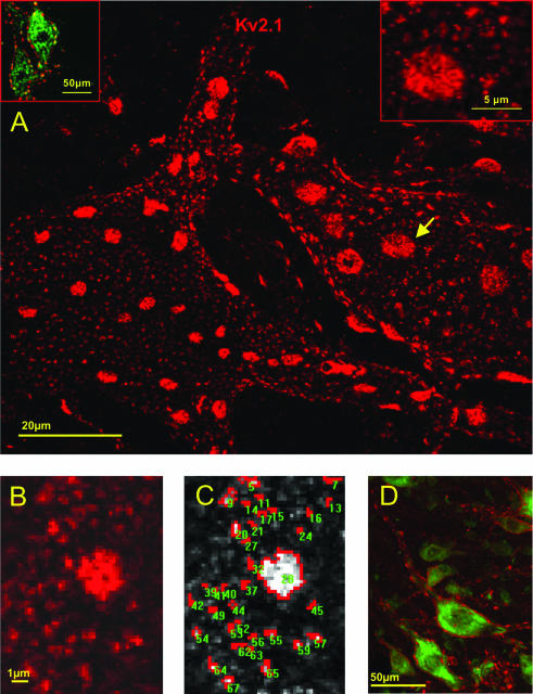
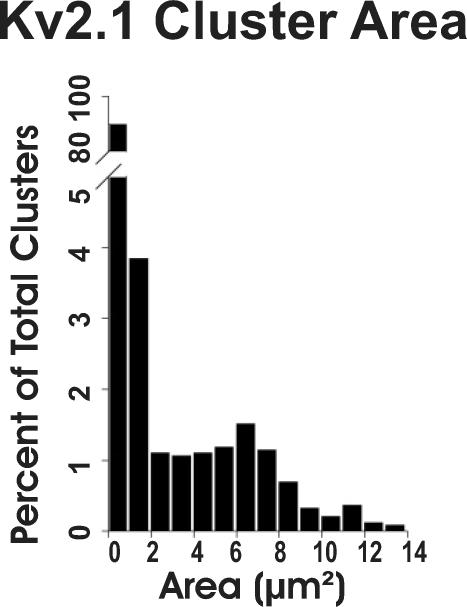
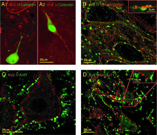
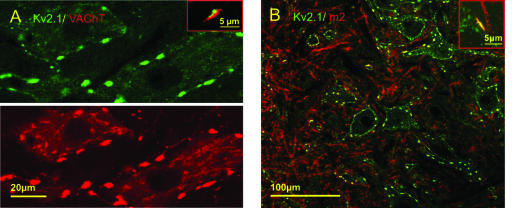
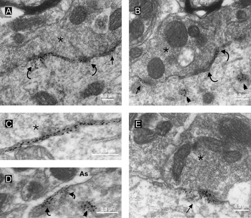
Delayed rectifier K+ currents are involved in the control of alpha-motoneurone excitability, but the precise spatial distribution and organization of the membrane ion channels that contribute to these currents have not been defined. Voltage-activated Kv2.1 channels have properties commensurate with a contribution to delayed rectifier currents and are expressed in neurones throughout the mammalian central nervous system. A specific antibody against Kv2.1 channel subunits was used to determine the surface distribution and clustering of Kv2.1 subunit-containing channels in the cell membrane of alpha-motoneurones and other spinal cord neurones. In alpha-motoneurones, Kv2.1 immunoreactivity (-IR) was abundant in the surface membrane of the soma and large proximal dendrites, and was present also in smaller diameter distal dendrites. Plasma membrane-associated Kv2.1-IR in alpha-motoneurones was distributed in a mosaic of small irregularly shaped, and large disc-like, clusters. However, only small to medium clusters of Kv2.1-IR were observed in spinal interneurones and projection neurones, and some interneurones, including Renshaw cells, lacked demonstrable Kv2.1-IR. In alpha-motoneurones, dual immunostaining procedures revealed that the prominent disc-like domains of Kv2.1-IR are invariably apposed to presynaptic cholinergic C-terminals. Further, Kv2.1-IR colocalizes with immunoreactivity against postsynaptic muscarinic (m2) receptors at these locations. Ultrastructural examination confirmed the postsynaptic localization of Kv2.1-IR at C-terminal synapses, and revealed clusters of Kv2.1-IR at a majority of S-type, presumed excitatory, synapses. Kv2.1-IR in alpha-motoneurones is not directly associated with presumed inhibitory (F-type) synapses, nor is it present in presynaptic structures apposed to the motoneurone. Occasionally, small patches of extrasynaptic Kv2.1-IR labelling were observed in surface membrane apposed by glial processes. Voltage-gated potassium channels responsible for the delayed rectifier current, including Kv2.1, are usually assigned roles in the repolarization of the action potential. However, the strategic localization of Kv2.1 subunit-containing channels at specific postsynaptic sites suggests that this family of voltage-activated K+ channels may have additional roles and/or regulatory components.
Figures
Similar articles
-
The K+ channel, Kv2.1, is apposed to astrocytic processes and is associated with inhibitory postsynaptic membranes in hippocampal and cortical principal neurons and inhibitory interneurons.Neuroscience. 1998 May;84(1):37-48. doi: 10.1016/s0306-4522(97)00519-8. Neuroscience. 1998. PMID: 9522360
-
Frequency-dependent regulation of rat hippocampal somato-dendritic excitability by the K+ channel subunit Kv2.1.J Physiol. 2000 Jan 1;522 Pt 1(Pt 1):19-31. doi: 10.1111/j.1469-7793.2000.t01-2-00019.xm. J Physiol. 2000. PMID: 10618149 Free PMC article.
-
Association of potassium channel Kv3.4 subunits with pre- and post-synaptic structures in brainstem and spinal cord.Neuroscience. 2004;126(4):1001-10. doi: 10.1016/j.neuroscience.2004.03.051. Neuroscience. 2004. PMID: 15207333
-
Potassium currents in motoneurones.Prog Neurobiol. 1995 Dec;47(6):513-31. doi: 10.1016/0301-0082(95)00032-1. Prog Neurobiol. 1995. PMID: 8787033 Review.
-
Kv2 channels create endoplasmic reticulum / plasma membrane junctions: a brief history of Kv2 channel subcellular localization.Channels (Austin). 2019 Dec;13(1):88-101. doi: 10.1080/19336950.2019.1568824. Channels (Austin). 2019. PMID: 30712450 Free PMC article. Review.
Cited by
-
Control of single channel conductance in the outer vestibule of the Kv2.1 potassium channel.J Gen Physiol. 2006 Aug;128(2):231-46. doi: 10.1085/jgp.200509465. J Gen Physiol. 2006. PMID: 16880266 Free PMC article.
-
Calcium- and metabolic state-dependent modulation of the voltage-dependent Kv2.1 channel regulates neuronal excitability in response to ischemia.J Neurosci. 2005 Nov 30;25(48):11184-93. doi: 10.1523/JNEUROSCI.3370-05.2005. J Neurosci. 2005. PMID: 16319318 Free PMC article.
-
Motoneuron excitability: the importance of neuromodulatory inputs.Clin Neurophysiol. 2009 Dec;120(12):2040-2054. doi: 10.1016/j.clinph.2009.08.009. Epub 2009 Sep 27. Clin Neurophysiol. 2009. PMID: 19783207 Free PMC article. Review.
-
Synaptic mechanisms underlying modulation of locomotor-related motoneuron output by premotor cholinergic interneurons.Elife. 2020 Feb 21;9:e54170. doi: 10.7554/eLife.54170. Elife. 2020. PMID: 32081133 Free PMC article.
-
Deletion of the Kv2.1 delayed rectifier potassium channel leads to neuronal and behavioral hyperexcitability.Genes Brain Behav. 2014 Apr;13(4):394-408. doi: 10.1111/gbb.12120. Epub 2014 Mar 7. Genes Brain Behav. 2014. PMID: 24494598 Free PMC article.
References
-
- Alvarez FJ, Dewey DE, Harrington DA, Fyffe REW. Cell-type specific organization of glycine receptor clusters in the mammalian spinal cord. J Comp Neurol. 1997;379:150–170. - PubMed
-
- Antonucci DE, Lim ST, Vassanelli S, Trimmer JS. Dynamic localization and clustering of dendritic Kv2.1 voltage-dependent potassium channels in developing hippocampal neurons. Neuroscience. 2001;108:69–81. - PubMed
Publication types
MeSH terms
Substances
Grants and funding
LinkOut - more resources
Full Text Sources
Molecular Biology Databases