. 2003 Nov;133(11):3614-8.
doi: 10.1093/jn/133.11.3614.

Choline availability during embryonic development alters progenitor cell mitosis in developing mouse hippocampus

Affiliations

Choline availability during embryonic development alters progenitor cell mitosis in developing mouse hippocampus

Corneliu N Craciunescu et al. J Nutr. 2003 Nov.

Abstract

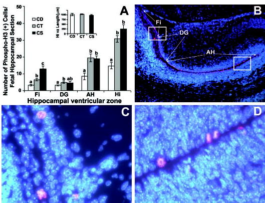
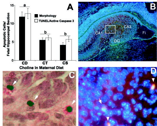
Previously, we reported that dietary choline influences development of the hippocampus in fetal rat brain. It is important to know whether similar effects of choline occur in developing fetal mouse brain because interesting new experimental approaches are now available using several transgenic mouse models. Timed-pregnant mice were fed choline-supplemented (CS), control (CT) or choline-deficient (CD) AIN-76 diet from embryonic day 12 to 17 (E12-17). Fetuses from CD dams had diminished concentrations of phosphocholine and phosphatidylcholine in their brains compared with CT or CS fetuses (P < 0.05). When we analyzed fetal hippocampus on day E17 for cells with mitotic phase-specific expression of phosphorylated histone H3, we detected fewer labeled cells at the ventricular surface of the ventricular zone in the CD group (14.8 +/- 1.9) compared with the CT (30.7 +/- 1.9) or CS (36.6 +/- 2.6) group (P < 0.05). At the same time, we detected more apoptotic cells in E17 hippocampus using morphology in the CD group (11.8 +/- 1.4) than in CT (5.6 +/- 0.6) or CS (4.2 +/- 0.7) group (P < 0.05). This was confirmed using terminal deoxynucleotidyl transferase (TdT)-mediated dUTP-digoxigenin anti-digoxigenin fluorescein conjugate antibody nick end-labeling (TUNEL) and activated caspase-3 immunoreactivity. We conclude that the dietary availability of choline to the mouse dam influences progenitor cell proliferation and apoptosis in the fetal brain.


Figures

FIGURE 1
FIGURE 1
Maternal dietary choline deficiency in timed-pregnant mice fed choline-supplemented (CS), control (CT) or choline-deficient (CD) AIN-76 diet from embryonic day 12 to 17 (E12–17) decreases mitosis in embryonic mice on day E17 at the ventricular surface of the ventricular zone of the hippocampus. All mice were killed on E17 and coronal sections were prepared from the brains of fetuses from each group for the analysis of mitosis using the mitosis-specific marker anti-phospho-histone H3 as described in Materials and Methods. The DAPI nuclear DNA counterstaining is blue, whereas the Cy3 conjugated secondary antibody bound to the anti-phospho-histone H3 (Ser10) primary antibody stains red. Panel A: In CD fetal hippocampus compared with CT, there were fewer phospho-histone H3 positive cells at the ventricular surface of the ventricular zone adjacent to fimbria (Fi), dentate gyrus (DG) and Ammon’s Horn (AH), and this was reflected in the calculated values for the whole hippocampal section length of ventricular zone (Hi). Compared with the CT group, the CS group had a higher incidence of phospho-histone H3-labeled cells at the ventricular surface of the ventricular zone only in the fimbria. The graph insert shows the equivalence of the sections in terms of total hippocampal ventricular zone length. Values are means ± SEM of at least 6 pups per group from 6 dams. Means without a common letter differ, P < 0.05 (for each pair using Student’s t test, for all pairs using Tukey-Kramer HSD test, and comparison with control using Dunnett’s Method). Panel B shows a representative fetal hippocampus at a magnification of 50X with the regions of interest marked. Panels C and D are 400X magnifications of the boxed regions in panel B, and show representative labeled cells in the hippocampal regions.
FIGURE 2
FIGURE 2
Maternal dietary choline deficiency in timed-pregnant mice fed choline-supplemented (CS), control (CT) or choline-deficient (CD) AIN-76 diet from embryonic day 12 to 17 (E12–17) increases apoptosis in day E17 mouse hippocampus. Coronal sections were prepared from the brains of day E17 fetuses from each diet group and were analyzed for apoptosis using a combination of classical apoptotic morphology, active caspase-3 immunoreactivity and TUNEL (terminal deoxynucleotidyl transferase (TdT)-mediated dUTP-digoxigenin anti-digoxigenin fluorescein conjugate antibody nick end-labeling). Consecutive serial sections were used for TUNEL and active (cleaved) caspase-3 staining as described in Materials and Methods. Panel A: The graph shows apoptotic cell counts using morphological criteria (means ± SEM, n = 10–12 pups/group from at least 5 dams), and TUNEL-cleaved caspase-3 immunostaining (means ± SEM, n = 6 pups/group from 3 different dams). Means without common letters differ, P < 0.05 (for each pair using Student’s t test, for all pairs using Tukey-Kramer HSD test, and comparison with control using Dunnett’s Method). Panel B: A 50X magnification image of the right hippocampal hemisphere showing green TUNEL-positive cells distributed in the cortical plate (CA1, CA2,3), dentate gyrus (DG) and fimbria (Fi). Panel C: A representative image at 400X magnification is shown; the arrows point to cells showing apoptotic morphology. Panel D: Higher power view of the boxed area in panel B obtained by screening the fluorescent images for observing DAPI (blue, nuclear counterstaining), fluorescein isothiocyanate (green, positive TUNEL), and rhodamine (red, activated caspase-3); spherical TUNEL and activated caspase-3 positive nuclei are indicated by arrowheads. Normal nuclei stain blue.

References

    1. Jones JP, Meck W, Williams CL, Wilson WA, Swartzwelder HS. Choline availability to the developing rat fetus alters adult hippocampal long-term potentiation. Brain Res Dev Brain Res. 1999;118:159–167. - PubMed
    1. Meck WH, Smith RA, Williams CL. Organizational changes in cholinergic activity and enhanced visuospatial memory as a function of choline administered prenatally or postnatally or both. Behav Neurosci. 1989;103:1234–1241. - PubMed
    1. Meck W, Williams C. Perinatal choline supplementation increases the threshold for chunking in spatial memory. Neuroreport. 1997;8:3053–3059. - PubMed
    1. Meck W, Williams C. Characterization of the facilitative effects of perinatal choline supplementation on timing and temporal memory. Neuroreport. 1997;8:2831–2835. - PubMed
    1. Tees RC. The influences of rearing environment and neonatal choline dietary supplementation on spatial learning and memory in adult rats. Behav Brain Res. 1999;105:173–188. - PubMed

Publication types