. 2003 Aug;224(2):114-21.
doi: 10.1016/j.cellimm.2003.08.008.

Effector CD4 cell tolerization is mediated through functional inactivation and involves preferential impairment of TNF-alpha and IFN-gamma expression potentials

Affiliations

Effector CD4 cell tolerization is mediated through functional inactivation and involves preferential impairment of TNF-alpha and IFN-gamma expression potentials

Meixiao Long et al. Cell Immunol. 2003 Aug.

Abstract

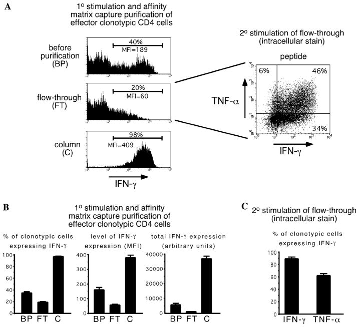
It has recently been shown that effector/memory T cells can undergo peripheral tolerization in response to self-antigen. In the present study, we found that within 24h self-antigen profoundly impairs the ability of CD4 effectors to express TNF-alpha (and to a lesser extent IFN-gamma); however, several days of self-antigen exposure is required to impair non-effector functions such as IL-2 expression and proliferation. Since only half of the initial effector CD4 cell population expresses effector cytokines following brief antigenic stimulation, tolerization might have been mediated either through functional inactivation of effector-competent cells, or alternatively by the selective deletion of competent and expansion of non-competent cells. When briefly stimulated effectors were fractionated based on their expression of IFN-gamma, the IFN-gamma(-) sub-population was able to express IFN-gamma following secondary stimulation, indicating that all effector CD4 cells are functionally competent. Furthermore, both IFN-gamma(+) and IFN-gamma(-) sub-populations underwent tolerization in response to self-HA (although the former was slightly more prone to deletion at later time points). Thus, effector CD4 cell tolerization is mediated primarily through the functional inactivation of effector-competent cells.


Figures

Fig. 1
Fig. 1
Cytokine expression potentials of effector CD4 cells tolerized in response to self-antigen. Resting Th1 effector clonotypic CD4 cells (identified as Thy1.1+) were recovered from vacc-HA-infected NT primary adoptive transfer recipients, relabeled with CFSE, retransferred into secondary recipients expressing either viral-HA (NT + vacc-HA, V) or self-HA (C3-HAhigh transgenics, S), and recovered from spleens either one or eight days later for analysis by performing intracellular staining for IFN-γ, TNF-α and IL-2 following in vitro restimulation with HA peptide. (A) Representative dot plots are shown for the day 8 analysis, with the percentages of cytokine-expressing clonotypic CD4 cells (and levels of IFN-γ and TNF-α expression (mean fluorescence intensity, MFI) in parentheses) shown for each quadrant (which were set based on staining with isotype control Abs, data not shown). Note that cytokine staining was never detected in the absence of peptide stimulation (data not shown). (B) Quantitative analysis of the day 1 time point. Total cytokine expression is calculated as the product of the % of cytokine positive clonotypic CD4 cells × the MFI per positively expressing cell, and expressed in arbitrary units as previously described [14]. For comparison, cytokine expression is also shown for time-matched primary resting clonotypic effector CD4 cells that had not been re-exposed to HA (1°). Additionally, the ratios of total cytokine expression for viral-HA vs. self-HA and 1° effectors vs. self-HA are shown, as well as the frequencies of recovered clonotypic CD4 cells. N = 3 for both viral-HA and self-HA groups, and n = 6 for 1° effectors (collected from two separate experiments). (C) Quantitative analysis of the day 8 time point was performed as in panel B. N = 9 for viral-HA, and 10 for self-HA (each collected from three separate experiments), and n = 3 for 1° effectors. Data is expressed as the mean ± the standard error of the mean (SEM).
Fig. 2
Fig. 2
Resting effector CD4 cells require greater than 24 h to undergo division following exposure to tolerogen. Day 6 resting effector clonotypic CD4 cells were recovered from spleens of vacc-HA-infected NT primary adoptive transfer recipients, relabeled with CFSE, and their time course of blastogenesis (FSC) and division (CFSE-dilution) assessed following retransfer into vacc-HA and self-HA recipients and recovery from spleens. The plots shown are representative of 3–10 replicates for each group.
Fig. 3
Fig. 3
Separation and restimulation of IFN-γ+ and IFN-γ effector CD4 cell sub-populations. (A) Day 6 resting effector clonotypic CD4 cells were restimulated in vitro with HA peptide, stained and fractionated for IFN-γ expression using affinity matrix capture technology (the percentage and level of IFN-γ expression (MFI) are shown, with the cutoff for IFN-γ expression determined by a parallel staining reaction that lacked capture antibody (data not shown)). The IFN-γ fraction (flow-through, FT) was subsequently restimulated with HA peptide and stained for intracellular IFN-γ vs. TNF-α. Data shown is representative of three separate experiments. Quantitative analyses (n = 3) of affinity matrix capture staining and separation (B), and IFN-γ and TNF-α intracellular staining of the IFN-γ fraction following restimulation with HA peptide (C), are presented as in previous figures.
Fig. 4
Fig. 4
Response of retransferred IFN-γ+ and IFN-γ effector CD4 cell sub-populations. (A) Representative experiment (from a total of 3) in which IFN-γ+ and IFN-γ effector CD4 cell sub-populations fractionated in Fig. 3 were relabeled with CFSE, retransferred in equal numbers into C3-HAhigh (self-HA, S) or NT + vacc-HA (viral-HA, V) secondary recipients, and recovered from the spleen and lungs following perfusion 5 days later. Histogram plots show the percentage of clonotypic CD4 cells (Thy1.1+) within total lymphocytes, as well as CFSE fluorescence of gated Thy1.1+ cells (a dashed line is placed directly to the left of the location of undivided cells). (B) Table showing the frequency of IFN-γ+ and IFN-γ clonotypic CD4 cells recovered from the spleen and lungs of self-HA and viral-HA secondary recipients for three separate experiments. (C) IFN-γ vs. TNF-α intracellular staining dot plots of recovered IFN-γ+ and IFN-γ clonotypic effector CD4 cells corresponding to panel A following in vitro restimulation with HA peptide. (D) Quantitative analysis of total intracellular IFN-γ and TNF-α staining following in vitro restimulation with HA peptide is presented as in previous figures (n = 3 for each group).

Similar articles

Cited by

References

    1. Higgins AD, Mihalyo MA, Adler AJ. Effector CD4 cells are tolerized upon exposure to parenchymal self-antigen. J Immunol. 2002;169:3622–3629. - PMC - PubMed
    1. Kreuwel HT, Aung S, Silao C, Sherman LA. Memory CD8(+) T cells undergo peripheral tolerance. Immunity. 2002;17:73–81. - PubMed
    1. Wucherpfennig KW, Strominger JL. Molecular mimicry in T cell-mediated autoimmunity: viral peptides activate human T cell clones specific for myelin basic protein. Cell. 1995;80:695–705. - PMC - PubMed
    1. Gross DM, Forsthuber T, Tary-Lehmann M, Etling C, Ito K, Nagy ZA, Field JA, Steere AC, Huber BT. Identification of LFA-1 as a candidate autoantigen in treatment-resistant Lyme arthritis. Science. 1998;281:703–706. - PubMed
    1. Bachmaier K, Neu N, de la Maza LM, Pal S, Hessel A, Penninger JM. Chlamydia infections and heart disease linked through antigenic mimicry. Science. 1999;283:1335–1339. - PubMed

Publication types

Substances