. 2003 Nov 17;22(22):5983-93.
doi: 10.1093/emboj/cdg581.

On the role of the two extracytoplasmic substrate-binding domains in the ABC transporter OpuA

Affiliations

On the role of the two extracytoplasmic substrate-binding domains in the ABC transporter OpuA

Esther Biemans-Oldehinkel et al. EMBO J. .

Abstract

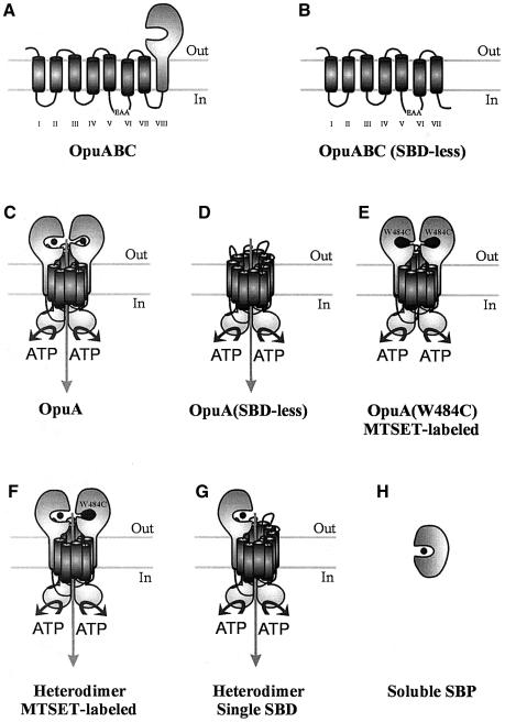
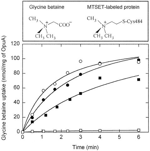
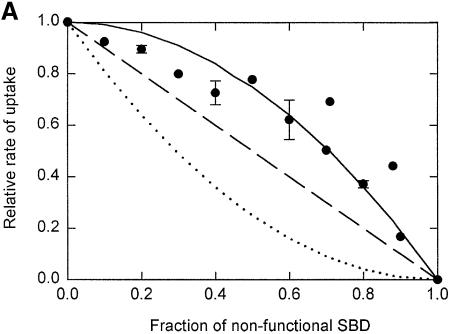
Members of two transporter families of the ATP-binding cassette (ABC) superfamily use two or even four extracytoplasmic substrate-binding domains (SBDs) for transport. We report on the role of the two SBDs in the translocation cycle of the ABC transporter OpuA from Lactococcus lactis. Heterooligomeric OpuA complexes with only one SBD or one functional and one non-functional SBD (inactivated by covalent linkage of a substrate mimic) have been constructed, and the substrate binding and transport kinetics of the purified transporters, reconstituted in liposomes, have been determined. The data indicate that the two SBDs of OpuA interact in a cooperative manner in the translocation process by stimulating either the docking of the SBDs onto the translocator or the delivery of glycine betaine to the translocator. It appears that one of these initial steps, but not the later steps in translocation or resetting of the system to the initial state, is rate determining for transport. These new insights on the functional role of the extracytoplasmic SBDs are discussed in the light of the current knowledge of substrate-binding-protein-dependent ABC transporters.


Figures

None
Fig. 1. Schematic representation of the different protein(s) (complexes) used in this study. (A) OpuABC, substrate-binding/translocator protein; (B) OpuABC(SBD-less), OpuABC lacking the signal anchor sequence [transmembrane segment VIII in (A)] and the SBD; (C) OpuA, the wild-type transporter, which is composed of two copies of OpuABC and two copies of OpuAA (the ATPase subunit); (D) OpuA(SBD-less), transporter complex formed from two copies of OpuABC(SBD-less) and two copies of OpuAA; (E) MTSET-labeled OpuA(W484C), OpuA complex with Trp to Cys substitution at position 484 of OpuABC and labeled with a thiol-specific glycine betaine mimic; (F) Heterodimer composed of one OpuABC, one MTSET-labeled OpuABC(W484C) and two OpuAA subunits; (G) heterodimer composed of one OpuABC, one OpuABC(SBD-less) and two OpuAA subunits; (H) soluble SBP, SBD of OpuA expressed as soluble protein.
None
Fig. 2. Visualization of overexpression and purification of the proteins. (A) Coomassie Brilliant Blue stained SDS–PAGE gel (12.5% polyacrylamide) of membrane vesicles (6.5 µg of total protein was loaded per lane) containing OpuA (lane 1), OpuA(W484C) (lane 2) and SBD-less OpuA (lane 3). In lanes 4–7, 2.5 µg of purified protein was loaded per lane; OpuA (lane 4), OpuA(W484C) (lane 5), SBD-less OpuA (lane 6) and soluble SBP (lane 7). The OpuAA component of wild-type OpuA runs at a lower molecular weight than that of OpuA(W484C) or SBD-less OpuA, because it does not contain a his tag; the OpuAA subunits of the other complexes carry a N-terminal 10-his tag. Importantly, OpuAA without a his-tag copurifies with his-tagged OpuABC. (B) Size-exclusion chromatography of DDM-solubilized and Ni2+-NTA-purified OpuA. (C) Size-exclusion chromatography of Ni2+-NTA purified soluble SBP. (D) SDS–PAGE gel (12.5% polyacrylamide) showing the effect of glycerol on the stability of the OpuA complex (OpuAA: 47 kDa, OpuABC: 63 kDa). Purified OpuA (lane 1) was allowed to slowly dissociate by decreasing the glycerol concentration to 5% and repurified on Ni-NTA (lane 2). In the control sample (lane 3), the glycerol concentration was lowered to 5% (30 min at 4°C to allow the complex to dissociate) then increased to 20% (30 min at 4°C to allow the complex to reassemble), after which OpuA was repurified on Ni-NTA. Per lane, 2.5 μg of protein was loaded.
None
Fig. 3. Glycine betaine transport in hybrid membranes. (A) Uptake (filled circles) and efflux (open circles) of [14C]glycine betaine (final concentration 34 µM) in hybrid membranes (final protein concentration 0.4 mg/ml) was assayed in 400 mM KPi pH 7.0. Efflux of glycine betaine was initiated by the addition of 9 mM Mg-ATP after 20 min of uptake (indicated by arrow). (B) Kinetics of [3H]glycine betaine uptake. The data were analyzed with the Michaelis–Menten (dashed line) and the Hill equation (solid line). The error bars indicate the standard deviation from the mean of three measurements. The inset shows the high concentration range. The residuals, i.e. the difference between the experimental data and fitted line, are plotted for the Michaelis–Menten (open circles) and for the Hill (filled circles) equations.
None
Fig. 4. Binding of glycine betaine to membranes with amplified levels of OpuA and to soluble SBP. Binding of [3H]glycine betaine to hybrid membranes (filled circles) was measured in 400 mM KPi pH 7.0, 5 mM EDTA, using the ammonium sulfate precipitation method. The final OpuA concentration was ∼15 µg/ml. The error bars indicate the standard deviation from the mean of three measurements. After correction for background binding, the data were fitted to a hyperbola of the form B = Bmax * S/(KD + S); the residuals are presented in the top panel. Binding of glycine betaine to soluble SBP (open circles) was measured in 50 mM KPi pH, 7.0, 200 mM KCl plus 10% (v/v) glycerol at a SBP concentration of 20 µg/ml by monitoring the change in protein fluorescence (ΔF). The data are from three independent measurements and the standard deviation is indicated. Since the KD value was at least five times larger than the protein concentration, the binding curve could be analyzed according to the equation ΔF = ΔFmax * S/(KD + S) (Lanfermeijer et al., 1999); S, glycine betaine concentration. The residuals are also presented in the upper panel.
None
Fig. 5. The effect of MTSET on transport activity in proteoliposomes. Wild-type OpuA and OpuA(W484C) were incubated with freshly prepared 100 µM MTSET for 1 h at room temperature while bound to the Ni-NTA resin, which was followed by an additional 20 column volumes of washing with 50 mM KPi pH 8.0, 200 mM KCl, 20% glycerol, 0.05% DDM and 15 mM imidazole; the purification was otherwise the same as described in Materials and methods. Uptake of [14C]glycine betaine (final concentration 34 µM) in both untreated wild-type (filled circles) and OpuA(W484C) (filled squares) and MTSET-treated wild-type (open circles) and OpuA(W484C) (open squares) was assayed in 400 mM KPi pH 7.0.
None
Fig. 6. Glycine betaine transport by heterooligomeric complexes formed at different ratios of wild-type OpuA and MTSET-labeled OpuA(W484C). (A) The initial rate of uptake of [14C]glycine betaine (final concentration 34 µM) was measured in 400 mM KPi pH 7.0, from the linear increase in uptake over a period of 60 s. Some points show the average plus standard deviation of the data from duplicate measurements on three independent proteoliposome batches. Different dependences of the transport activity (= y) on the fraction of substrate-binding defective protein (= b) are indicated: y = 1 – 2b + b2 (dotted line); y = 1 – b (dashed line); and y = 1 – b2 (solid line). (B) Wild-type OpuA and MTSET-labeled OpuA(W484C) were mixed in different ratios prior to membrane reconstitution, but the proteins were not allowed to form heterodimeric complexes. Uptake was performed as described in (A).
None
Fig. 7. Glycine betaine transport by heterooligomeric complexes formed at different ratios of wild-type OpuA and SBD-less OpuA. Glycine betaine transport experiments were performed as described in the legend of Figure 6. Each point represents the average plus standard deviation of three independent experiments and measurements carried out in duplicate. The data points follow the line corresponding to y = 1 – b (see legend of Figure 6 for details).
None
Fig. 8. Kinetics of glycine betaine transport in heterodimeric complexes. Proteoliposomes containing wild-type OpuA homodimers (filled circles), wild-type/MTSET-labeled W484C heterodimers (filled squares) and wild-type/SBD-less heterodimers (filled triangles) were used to assay the initial rate of uptake of [3H]glycine betaine in 400 mM KPi pH 7.0. The heterodimeric complexes were formed by mixing wild-type OpuA and OpuA(W484C) or SBD-less OpuA at ratios of 1:9 as described in Materials and methods. The activities of the heterodimeric complexes were normalized for protein content by ignoring the fraction (0.81) of homodimeric OpuA(W484C) or SBD-less OpuA complex, as these species have no detectable activity. In these experiments, the activity is dominated by the fraction (0.18, which is 95% of all active species) of heterodimeric complexes. Each point represents the average plus standard deviation of two measurements. The best fit of the data was obtained with the Hill (filled circles, wild-type OpuA) or Michaelis–Menten (filled squares and triangles, heterodimeric complexes) equations, which yielded random distribution of the residuals (not shown).

References

    1. Albers S.V., Elferink,M.G., Charlebois,R.L., Sensen,C.W., Driessen,A.J. and Konings,W.N. (1999) Glucose transport in the extremely thermoacidophilic Sulfolobus solfataricus involves a high-affinity membrane-integrated binding protein. J. Bacteriol., 181, 4285–4291. - PMC - PubMed
    1. Ames G.F., Liu,C.E., Joshi,A.K. and Nikaido,K. (1996) Liganded and unliganded receptors interact with equal affinity with the membrane complex of periplasmic permeases, a subfamily of traffic ATPases. J. Biol. Chem., 271, 14264–14270. - PubMed
    1. Borths E.L., Locher,K.P., Lee,A.T. and Rees,D.C. (2002) The structure of Escherichia coli BtuF and binding to its cognate ATP binding cassette transporter. Proc. Natl Acad. Sci. USA, 99, 16642–16647. - PMC - PubMed
    1. Chen J., Sharma,S., Quiocho,F.A. and Davidson,A.L. (2001) Trapping the transition state of an ATP-binding cassette transporter: evidence for a concerted mechanism of maltose transport. Proc. Natl Acad. Sci. USA, 98, 1525–1530. - PMC - PubMed
    1. Davidson A.L. (2002) Mechanism of coupling of transport to hydrolysis in bacterial ATP-binding cassette transporters. J. Bacteriol., 184, 1225–1233. - PMC - PubMed

Publication types

MeSH terms