. 2003 Nov 17;22(22):6068-77.
doi: 10.1093/emboj/cdg579.

Disruption of the nucleolus mediates stabilization of p53 in response to DNA damage and other stresses

Affiliations

Disruption of the nucleolus mediates stabilization of p53 in response to DNA damage and other stresses

Carlos P Rubbi et al. EMBO J. .

Abstract

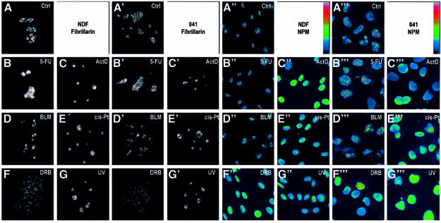
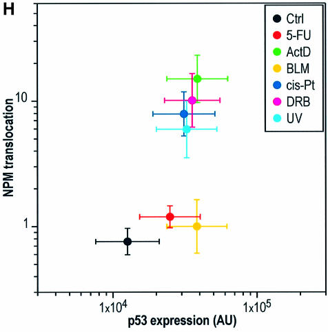
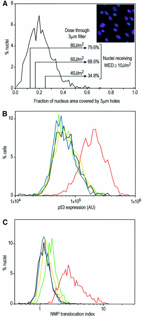
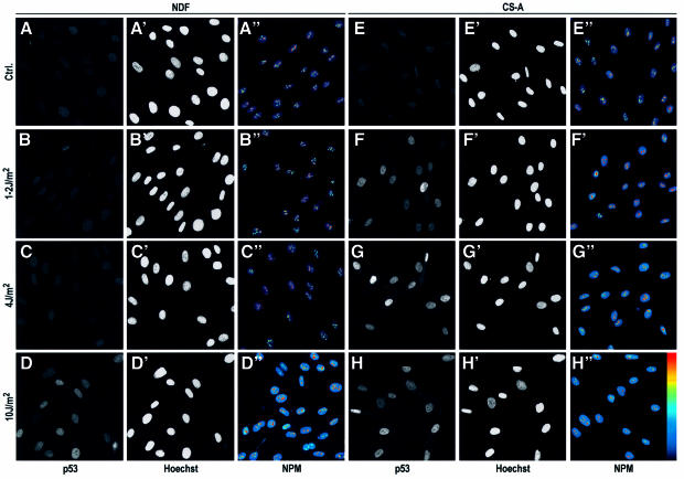
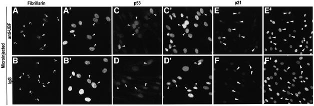
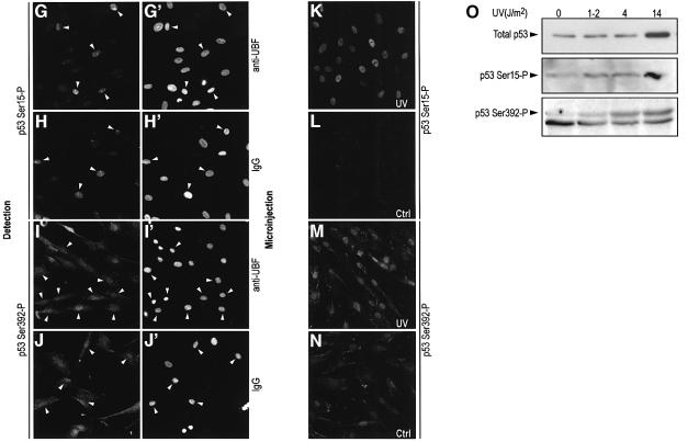
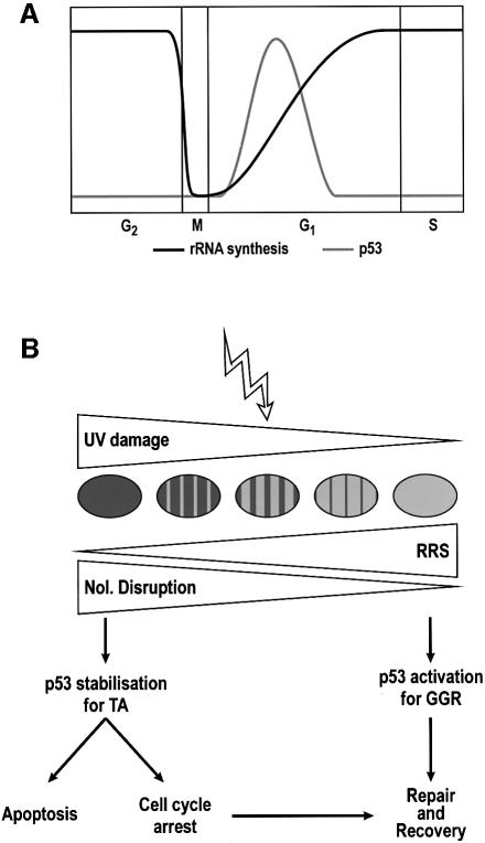
p53 protects against cancer through its capacity to induce cell cycle arrest or apoptosis under a large variety of cellular stresses. It is not known how such diversity of signals can be integrated by a single molecule. However, the literature reveals that a common denominator in all p53-inducing stresses is nucleolar disruption. We thus postulated that the impairment of nucleolar function might stabilize p53 by preventing its degradation. Using micropore irradiation, we demonstrate that large amounts of nuclear DNA damage fail to stabilize p53 unless the nucleolus is also disrupted. Forcing nucleolar disruption by anti-upstream binding factor (UBF) microinjection (in the absence of DNA damage) also causes p53 stabilization. We propose that the nucleolus is a stress sensor responsible for maintenance of low levels of p53, which are automatically elevated as soon as nucleolar function is impaired in response to stress. Our model integrates all known p53-inducing agents and also explains cell cycle-related variations in p53 levels which correlate with established phases of nucleolar assembly/disassembly through the cell cycle.


Figures

None
Fig. 1. Nucleolar disruption is independent of p53. (A–G′) Single confocal sections of nuclei of NDFs (AG) and 041 cells (A′–G′) stained for fibrillarin, 6 h after treatment with each indicated agent. Each image corresponds to a single nucleus. (A′′–G′′′) wide field images of NDFs (A′′–G′′) and 041 cells (A′′′–G′′′) stained for NPM after the same treatments. A pseudo-colour scale (indicated) was applied to each image to highlight all intensity ranges. (H) Dual plot of nuclear p53 expression level (DO-1 staining) and NPM translocation index for NDFs treated for 6 h with the indicated agents.
None
Fig. 2. Effects of micropore irradiation on p53 expression and NPM translocation in NDFs. (A) Distribution of the fraction of irradiated areas on NDF nuclei (n = 1699) observed by the ratio between the area exposed to UV irradiation through 3 µm Isopore filters (irradiated area detected by antibody labelling of photolesions) and the total nuclear projected area (Hoechst 33324). The insert shows an example field of micropore-irradiated nuclei, with CPDs labelled red and nuclei blue (Hoechst). (B) p53 expression levels in NDFs whole-nucleus irradiated at 10 J/m2 (red), micropore irradiated at 40 (green), 60 (yellow) and 80 J/m2 (blue), and non-irradiated (black). The fractions of nuclei receiving WED ≥10 J/m2 (see text) under each micropore irradiation condition are indicated in (A). (C) NPM translocation indext for NDFs irradiated in the same conditions as in (B). Cells in (A) were fixed immediately after irradiation, while cells in (B) and (C) were fixed 6 h after irradiation.
None
Fig. 3. Correlation of MRD for p53 expression and NPM translocation in NDFs and Cockayne syndrome complementation group A (CS-A) fibroblasts. NDFs (AD) and CS-A cells (EH) were irradiated at the indicated UV densities, fixed 6 h later and stained for p53 expression (A–D and E–H, with positions of nuclei indicated by Hoechst staining in A′–D′ and E′–H′) or NPM (A′′–D′′ and E′′–H′′). All images are wide-field. Pseudo-colour is used in NPM images.
None
Fig. 4. Effect of nucleolar disruption by microinjection of an anti-UBF antibody in NDFs. (A, C and E) Microinjection of anti-UBF (IgG1) with fibrillarin distribution (A) and p53 expression (C) assayed 6 h post-injection, and p21 expression (E) 18 h post-injection. (B, D and F) Microinjection of control purified mouse IgG1. (A′–F′) Reference Hoechst images. Arrows indicate microinjected cells. Lower magnification was used in p21 images in order to incorporate more cells into the field of view. (G–J) p53 phosphorylation in response to microinjection of anti-UBF (G and I) and non-specific IgG (H and J). p53 phosphorylation was detected for Ser15 (G and H) and Ser392 (I and J). (G′–J′) reference Hoechst images. (KN) Phosphorylation at Ser15 and Ser392 in control and UV-irradiated NDFs (14 J/m2) as indicated. (O) Western blot analysis of p53 expression and phosphorylation (Ser15 and Ser392) in control and UV-irradiated NDFs (1–2, 4 and 14 J/m2). Cells were harvested 6 h post-irradiation.
None
Fig. 5. (A) Schematic representation of the variations of rRNA synthesis rates and p53 levels along the normal cell cycle. (B) Scheme of the p53 response to high and low level DNA damage following UV irradiation. High level DNA damage implies compromise of the majority of the nucleolar population, impairing the total nucleolar function of the nucleus; low level refers to either localized high density damage or low whole area damage, such that the overall nucleolar function is not compromised. Recovery of RNA synthesis (RRS) will be faster the lower the DNA damage is. High level DNA damage stabilizes p53 and induces transactivation of downstream effector genes. Low level damage elicits a p53 response, with low (resting) levels consisting of chromatin relaxation for global NER.

References

    1. Abrahams P.J. et al. (1998) Impaired DNA repair capacity in skin fibroblasts from various hereditary cancer-prone syndromes. Mutat. Res., 407, 189–201. - PubMed
    1. Andera L. and Wasylyk,B. (1997) Transcription abnormalities potentiate apoptosis of normal human fibroblasts. Mol. Med., 3, 852–863. - PMC - PubMed
    1. Andersen J.S., Lyon,C.E., Fox,A.H., Leung,A.K., Lam,Y.W., Steen,H., Mann,M. and Lamond,A.I. (2002) Directed proteomic analysis of the human nucleolus. Curr. Biol., 12, 1–11. - PubMed
    1. Blattner C., Tobiasch,E., Litfen,M., Rahmsdorf,H.J. and Herrlich,P. (1999) DNA damage induced p53 stabilization: no indication for an involvement of p53 phosphorylation. Oncogene, 18, 1723–1732. - PubMed
    1. Blaydes J.P., Gire,V., Rowson,J.M. and Wynford-Thomas,D. (1997) Tolerance of high levels of wild-type p53 in transformed epithelial cells dependent on auto-regulation by mdm-2. Oncogene, 14, 1859–1868. - PubMed

Publication types

MeSH terms

Substances