. 2003 Nov 17;22(22):6137-47.
doi: 10.1093/emboj/cdg580.

Eme1 is involved in DNA damage processing and maintenance of genomic stability in mammalian cells

Affiliations

Eme1 is involved in DNA damage processing and maintenance of genomic stability in mammalian cells

Jacinth Abraham et al. EMBO J. .

Abstract

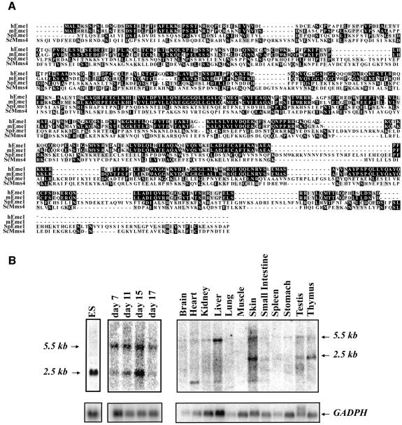
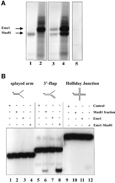
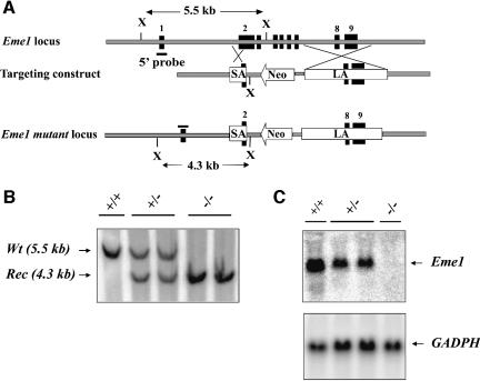
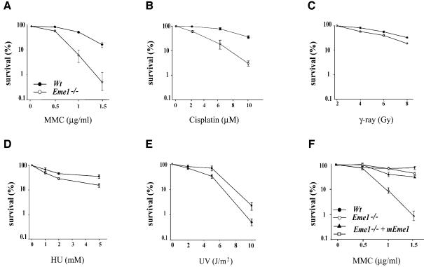
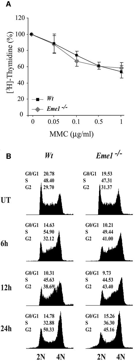
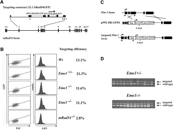
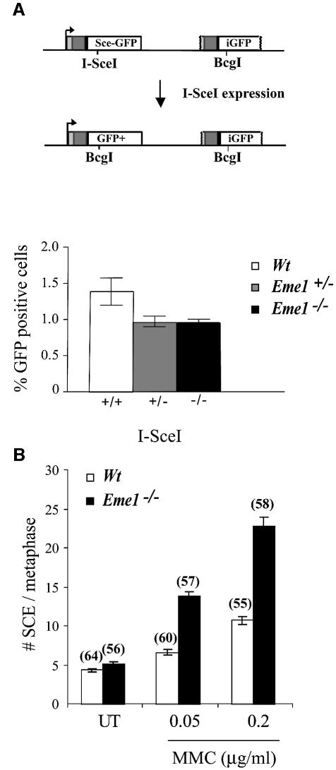
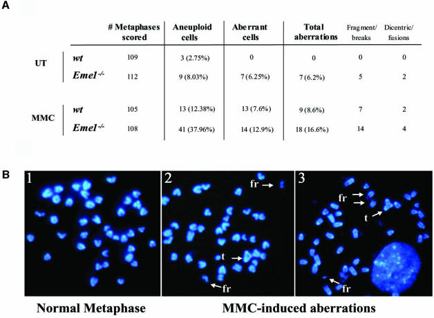
Yeast and human Eme1 protein, in complex with Mus81, constitute an endonuclease that cleaves branched DNA structures, especially those arising during stalled DNA replication. We identified mouse Eme1, and show that it interacts with Mus81 to form a complex that preferentially cleaves 3'-flap structures and replication forks rather than Holliday junctions in vitro. We demonstrate that Eme1-/- embryonic stem (ES) cells are hypersensitive to the DNA cross-linking agents mitomycin C and cisplatin, but only mildly sensitive to ionizing radiation, UV radiation and hydroxyurea treatment. Mammalian Eme1 is not required for the resolution of DNA intermediates that arise during homologous recombination processes such as gene targeting, gene conversion and sister chromatid exchange (SCE). Unlike Blm-deficient ES cells, increased SCE was seen only following induced DNA damage in Eme1-deficient cells. Most importantly, Eme1 deficiency led to spontaneous genomic instability. These results reveal that mammalian Eme1 plays a key role in DNA repair and the maintenance of genome integrity.


Figures

None
Fig. 1. Identification of mammalian Eme1. (A) Alignment of mouse, human and S.pombe Eme1 proteins with S.cerevisae Mms4. Amino acid identities and similarities are highlighted in black and gray, respectively. (B) Mouse Eme1 is mainly expressed in proliferative tissues. Radiolabeled Eme1 full-length cDNA was used to probe northern blots of mouse ES cells (left panel), mouse embryos at day 7, 11, 15 and 17 of gestation (middle panel) and mouse adult tissues (right panel). Northern blots were subsequently probed with a GADPH cDNA to assess loading.
None
Fig. 2. Endonuclease activity of the mouse Mus81–Eme1 complex. (A) Interaction of mouse Eme1 with Mus81. In vitro translation reactions containing the templates Mus81-Flag (lane 1), Eme1-HA (lane 2), both Mus81-Flag and Eme1-HA (lanes 3 and 4) or no template (lane 5) were subject to immunoprecipitation (IP). The IP antibodies were anti-Flag (lanes 1, 3 and 5) and anti-HA (lanes 2 and 4). In co-translation reactions, both Mus81 and Eme1 were immunoprecipitated by anti-Flag and anti-HA antibodies (lanes 3 and 4), demonstrating the physical interaction of these proteins. (B) Mus81–Eme1 protein complex has DNA structure-specific endonuclease activity. Three branched DNA substrates, splayed-arm (lanes 1–4), 3′-flap (lanes 5–8) and HJ (lanes 9–12), were assessed for cleavage by immunocomplex negative control (lanes 1, 5 and 9), human Mus81 fraction (lanes 2, 6 and 10), mouse Eme1 immunocomplex (lanes 3, 7 and 11) and mouse Mus81–Eme1 immunocomplex (lanes 4, 8 and 12). Cleavage reactions were resolved by 10% neutral PAGE and visualized by phosphor imager analysis. Mouse Mus81–Eme1 cleaved the 3′-flap structure well (lane 8), but had only faint endonuclease activity against the HJ substrate tested (lane 12).
None
Fig. 3. Generation of Eme1+/– and Eme1–/– ES cells. (A) Schematic representations of the Eme1 locus, the gene-targeting construct and the targeted Eme1 allele. Exons are denoted by solid black boxes. SA, short arm; LA, long arm; Neo, neomycin resistance gene; X, XbaI site. (B) Southern blot analysis of wild-type, Eme1+/– and Eme1–/– ES cells. Probing Southern blots of XbaI-digested ES cell DNA with a 5′-flanking Eme1 probe differentiates the wild-type allele (5.5 kb) from the recombined allele (4.3 kb). (C) Northern blot analysis showing loss of Eme1 mRNA in Eme1–/– ES cells. A 20 µg aliquot of total RNA from the indicated Eme1 ES genotypes was northern blotted and probed with a 32P-radiolabeled full-length Eme1 cDNA. The northern blot was subsequently probed with a GAPDH cDNA to assess loading of mRNA.
None
Fig. 4. Loss of Eme1 sensitizes ES cells to DNA damage. (AE) Wild-type (solid circles) and Eme1–/– (open circles) ES colony survival following DNA damage by MMC, cisplatin, IR, HU and UV treatment. Doses of the individual treatments are plotted on the x-axis, while the y-axis denotes the corresponding fold sensitivity of colonies over untreated controls. Eme1–/– ES cells were extremely sensitive to MMC and cisplatin treatments, and only mildly sensitive to the other agents. (F) MMC sensitivity of Eme1–/– ES clones complemented with HA-tagged Eme1 cDNA (closed triangle and open square). Stable transfection of Eme1-HA cDNA in Eme1-deficient ES cells restores MMC sensitivity to near wild-type levels.
None
Fig. 5. Intact intra-S-phase checkpoint and G2–M cell cycle arrest in the absence of Eme1. (A) The MMC-induced intra S-phase checkpoint is not affected in Eme1–/– ES cells. The proportion of [3H]thymidine incorporation in untreated wild-type (black square) and Eme1–/– (gray diamond) cells was set as 100%. The decreased proportion of [3H]thymidine incorporation following increasing doses of MMC treatments was similar for both wild-type and Eme1–/– ES cells. (B) G2–M checkpoint activation of wild-type and Eme1–/– ES cells following MMC treatment. Wild-type (left panel) and Eme1–/– (right panel) ES cells were subjected to 1 µg/ml of MMC for 1 h and returned to regular medium for 6, 12 and 24 h, following which time they were stained with propidium iodide and analyzed by FACS. Both genotypes displayed similar cell cycle profiles, with cells progressively accumulating in the G2/M phase.
None
Fig. 6. Eme1 does not impair homologous recombination processes such as gene targeting. (A) Schematic of the gene targeting strategy at the Rad54 locus. The Rad54 targeting construct (11.1hRAD54-GFP) and gene locus are depicted. Expression of Rad54–GFP is dependent on proper gene targeting at the Rad54 locus. (B) FACS analysis of the percentage of GFP+ cells after puromycin selection. Whereas gene targeting is clearly impaired in Rad54–/– ES cells, levels of GFP expression in Eme1+/– and Eme1–/– were similar to the wild-type control. (C) Gene targeting strategy at the Pim1 locus. (D) PCR analysis of gene-targeted clones. Eme1+/– and Eme1–/– ES cells were electroporated with the linear p59XDR-GFP6 targeting construct that favors gene targeting by selecting predominantly for survival of colonies that had integration at the Pim-1 locus. Homologous recombination-mediated gene targeting was tested by PCR, and showed no difference between the two genotypes.
None
Fig. 7. Induced DSB repair and sister chromatid exchange in the absence of Eme1. (A) Upper panel: schematic representation of the generation of a functional GFP gene by HR-mediated gene conversion. Transient I-SceI expression induces a DSB in a stably integrated construct bearing two non-functional GFP genes in tandem. HR- mediated gene conversion without crossing-over replaces the I-SceI with the BcgI site, and also restores GFP expression. Lower panel: quantification of GFP+ cells following I-SceI-induced DSB repair. Wild-type, Eme1+/– and Eme1–/– ES clones were analyzed by FACS to detect the proportion of GFP+ cells, indicative of successful gene conversion at the non-functional GFP locus. Wild-type, Eme1+/– and Eme1–/– ES clones showed no difference in the number of GFP+ cells, indicating equivalent HR-mediated gene conversion following an induced DSB. (B) Increased MMC-induced SCE in the absence of Eme1. The frequency of spontaneous SCE was similar between wild-type and Eme1–/– ES cells. However, a 2-fold increase of MMC- induced SCE was observed in Eme1-deficient ES cells by comparison with wild-type cells. The number of metaphases analyzed is given in parentheses above the corresponding histogram.
None
Fig. 8. Increased genomic instability in Eme1-deficient ES cells. (A) Spontaneous and MMC-induced chromosomal instability in Eme1-deficient ES cells. Metaphase spreads of untreated and MMC-treated wild-type and Eme1–/– ES cells were analyzed for chromosomal instability. Increased spontaneous aneuploidy is observed in Eme1–/– ES cells, with other aberrations represented by breaks, triradials and chromosomal fusions. (B) MMC treatment aggravates chromosomal aberrations in Eme1–/– ES cells. A normal metaphase spread from untreated Eme1–/– ES cells (1) and two metaphase spreads from MMC-treated Eme1–/– ES cells (2 and 3) showing MMC-induced accumulation of fragments (fr) and triradials (t).

References

    1. Bassing C.H., Swat,W. and Alt,F.W. (2002) The mechanism and regulation of chromosomal V(D)J recombination. Cell, 109, S45–S55. - PubMed
    1. Bastin-Shanower S.A., Fricke,W.M., Mullen,J.R. and Brill,S.J. (2003) The mechanism of Mus81–Mms4 cleavage site selection distinguishes it from the homologous endonuclease Rad1–Rad10. Mol. Cell. Biol., 23, 3487–3496. - PMC - PubMed
    1. Benson F.E. and West,S.C. (1994) Substrate specificity of the Escherichia coli RuvC protein. Resolution of three- and four-stranded recombination intermediates. J. Biol. Chem., 269, 5195–5201. - PubMed
    1. Boddy M.N., Lopez-Girona,A., Shanahan,P., Interthal,H., Heyer,W.D. and Russell,P. (2000) Damage tolerance protein Mus81 associates with the FHA1 domain of checkpoint kinase Cds1. Mol. Cell. Biol., 20, 8758–8766. - PMC - PubMed
    1. Boddy M.N., Gaillard,P.H., McDonald,W.H., Shanahan,P., Yates,J.R.,3rd and Russell,P. (2001) Mus81–Eme1 are essential components of a Holliday junction resolvase. Cell, 107, 537–548. - PubMed

Publication types