. 2003 Nov 17;198(10):1463-73.
doi: 10.1084/jem.20030232. Epub 2003 Nov 10.

LIME, a novel transmembrane adaptor protein, associates with p56lck and mediates T cell activation

Affiliations

LIME, a novel transmembrane adaptor protein, associates with p56lck and mediates T cell activation

Eun Mi Hur et al. J Exp Med. .

Abstract

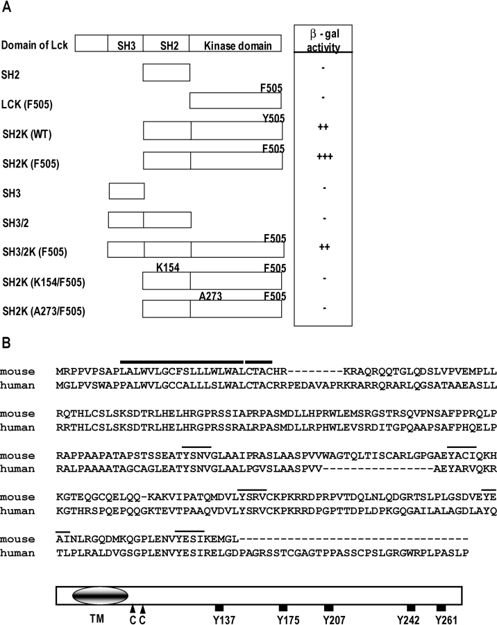
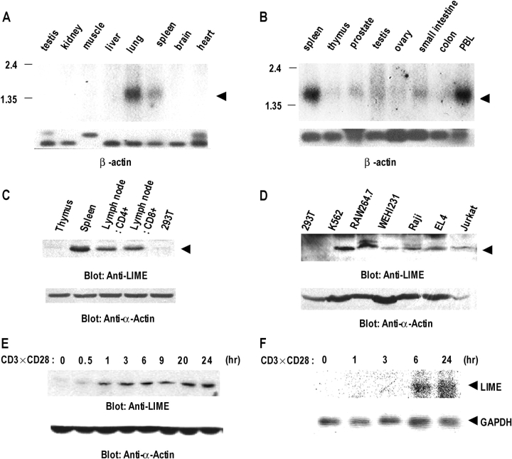
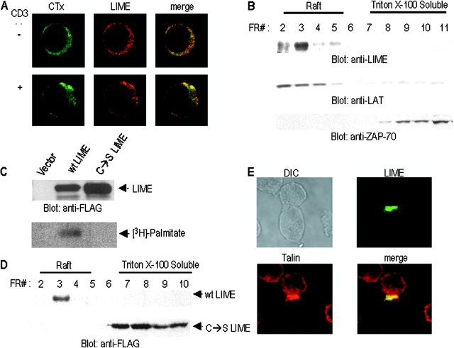
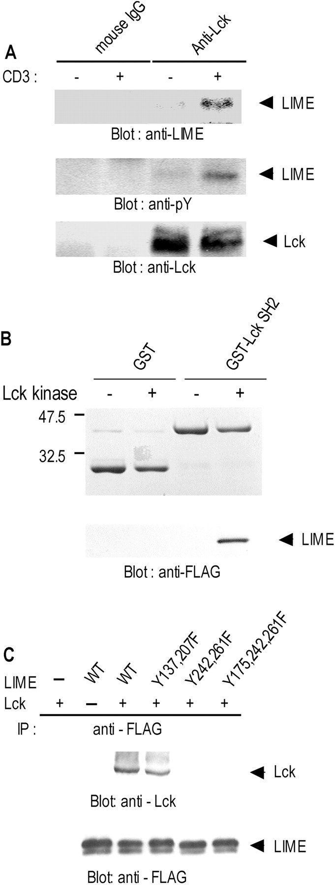
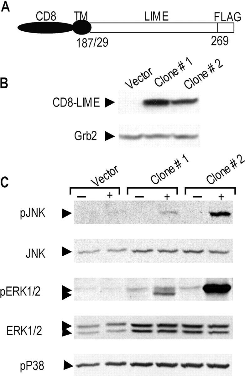
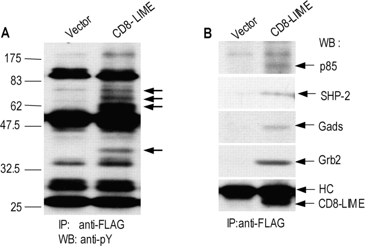
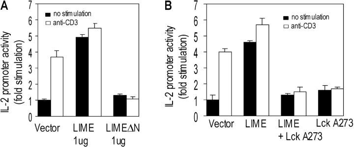
In this study, we identify and characterize a novel transmembrane adaptor protein, designated Lck-interacting membrane protein (LIME), as a binding partner of the Lck Src homology (SH)2 domain. LIME possesses a short extracellular domain, a transmembrane domain, and a cytoplasmic tail containing five tyrosine-based motifs. The protein is primarily expressed in hematopoietic cells and lung. Interestingly, LIME expression is up-regulated by TCR stimulation and sustained up to 24 h, suggesting that LIME acts throughout the early to late stages of T cell activation. LIME is localized to membrane rafts and distributed within the T cell-APC contact site. Upon TCR stimulation of Jurkat T cells, LIME associates with Lck as a tyrosine-phosphorylated protein. Experiments using Jurkat T cells expressing CD8-LIME chimera reveal that the protein associates with phosphatidylinositol 3-kinase, Grb2, Gads, and SHP2, and activates ERK1/2 and JNK but not p38. Moreover, overexpression of LIME in Jurkat T cells induces transcriptional activation of the IL-2 promoter. Our data collectively show that LIME is a raft-associated transmembrane adaptor protein linking TCR stimuli to downstream signaling pathways via associations with Lck.


Figures

Figure 1.
Figure 1.
cDNA cloning and the deduced amino acid sequence of LIME. (A) Binding specificity of LIME to Lck in a yeast two-hybrid system. Indicated regions of Lck were employed as bait and tested for binding to LIME. A part of LIME encompassing aa D52–270 was employed as prey. The binding affinity was scored as +++ (deep blue), ++ (intermediate blue), + (pale blue), and − (white) upon X-Gal (5-bromo-4-chloro-3-indoyl β-d-galactoside) staining. (B, top) The deduced amino acid sequences of mouse and human LIME and structural characteristics of the proteins. The transmembrane domain (TM) and CXXC motif are marked by bold lines. Putative tyrosine-based motifs are marked by thin lines on top of the sequence. Sequence data available from GenBank/EMBL/DDBJ under accession no. AF115339. (B, bottom) Schematic diagram of the predicted structure of murine LIME.
Figure 2.
Figure 2.
Expression profiles of LIME. (A and B) Northern blot analyses on mouse (A) and human (B) tissues. Multiple Tissue Northern blots were performed using LIME cDNA as a probe. The positions of the LIME transcript are indicated by arrowheads. A β-actin cDNA probe was used as control (bottom). (C) Expression of LIME in murine lymphoid tissues. Western blot analyses were performed on cell lysates isolated from indicated tissues (2 × 10⁶ cells/lane) using polyclonal mouse anti-LIME antibody. (D) Western blot analysis of LIME expression in various cell lines. Whole cell lysates (2 × 10⁶ cells/lane) from indicated cell lines were immunoblotted with mouse anti-LIME antiserum. 293T, human embryonic kidney; K562, human erythroleukemic cells; Raw 264.7, macrophage; WEHI 231, murine B cell; Raji, human B cell; EL-4, murine thymoma; Jurkat, human T cell leukemia. The same membrane was immunoblotted with anti–α-actin antibody. (E) Western blot analysis of LIME expression upon T cell activation. Resting lymphoid T cells were stimulated in vitro with anti-CD3ɛ and anti-CD28 mAb for the indicated times. LIME expression was determined by Western blotting. (F) Northern blot analysis of LIME expression upon TCR activation. Resting lymphoid T cells were stimulated and processed as described in E. GAPDH cDNA probe was used as control (bottom).
Figure 3.
Figure 3.
LIME localizes to membrane raft and is distributed within the immunological synapse upon contact with APC. (A) LIME cocaps with GM1. Jurkat T cells were either left unstimulated (−, top) or stimulated (+, bottom) with anti-CD3 (UCHT1). Subsequently, cells were stained with FITC-labeled cholera toxin B subunit (CTx) or anti-LIME antibody and Texas red–conjugated anti–rabbit IgG. (B) LIME localizes to raft. Jurkat T cells were stimulated with anti-CD3 for 10 min and solubilized in 1% Triton X-100. Lysates were subjected to sucrose gradient ultracentrifugation, and fractions (numbered 2–11 from top to bottom) were analyzed by immunoblotting with anti-LIME, anti-LAT, or anti-ZAP70 antibodies. LAT was employed as an established control marker of raft fraction. (C) LIME is palmitoylated. 293T cells were transfected with the expression plasmids encoding FALG-tagged WT LIME or mutant LIME in which Cys28 and Cys31 were mutated to serine (C→S LIME) and metabolically labeled with 3H-palmitate for 4 h. LIME proteins were immunoprecipitated with anti-FLAG antibody, and the immunoprecipitates were analyzed by immunoblot (top) and autoradiography (bottom). (D) Palmitoylation of membrane-proximal cysteines is required for localization of LIME to membrane raft. Cell lysates were prepared as described in C and subjected to sucrose gradient centrifugation. The fractions were analyzed by immunoblotting with anti-FLAG antibody. (E) Localization of LIME to the APC–T cell contact site. Jurkat T cells transfected with expression plasmids encoding LIME–GFP were mixed at a ratio of 1:1 with SEE-pulsed Raji B cells. Cells were fixed and examined by confocal microscopy. Recruitment of LIME to T cells was characterized by a bright band at the cell interface.
Figure 4.
Figure 4.
Binding of LIME to Lck. (A) In vivo association between LIME and Lck in T cells. Jurkat T cells were either left unstimulated or stimulated for 5 min by CD3 cross-linking. Digitonin lysates (1%) were subjected to precipitation with anti-Lck mAb or mouse IgG. Precipitates were analyzed by immunoblotting with anti-LIME and anti-phosphotyrosine mAbs. The amount of immunoprecipitated Lck is shown in the bottom panel. (B) LIME binds the Lck SH2 domain. Purified GST and GST–Lck SH2 fusion proteins were separated by SDS-PAGE and subjected to Coomassie staining (top). Subsequently, in vitro–translated FLAG–LIME was subjected to in vitro phosphorylation by purified Lck kinase and incubated with GST or GST–Lck SH2–coupled beads. Eluted proteins were separated by SDS-PAGE and immunoblotted with anti-FLAG antibody (bottom). (C) Mapping of sites in LIME responsible for binding to Lck. Tyrosine mutants of LIME were coexpressed with Lck in 293T cells by transient transfection. LIME mutants were immunoprecipitated with anti-FLAG mAb and analyzed by Western blotting with anti-Lck polyclonal antiserum (top). To control the amounts of precipitated LIME derivatives, the same membrane was blotted with anti-FLAG mAb after stripping (bottom).
Figure 5.
Figure 5.
LIME mediates activation of ERK and JNK. (A) Structure of CD8–LIME chimera used in this study. The COOH termini of chimera were tagged with FLAG. (B) Jurkat T cell lines stably expressing CD8–LIME were established, and expression levels of chimeric proteins in two independent clones (clone #1, #2) were determined by Western blotting using anti-FLAG mAb. The blot was stripped and reincubated with anti-Grb2 mAb as a control. (C) LIME activates ERK1/2 and JNK but not p38. Jurkat T cells stably expressing the CD8–LIME chimera were left unstimulated or stimulated by CD8 cross-linking. Phosphorylation of MAP kinases was determined by blotting with antibodies specific for phosphorylated JNK, ERK1/2, and p38. The same blot was stripped and reprobed with anti-ERK1/2 or JNK.
Figure 6.
Figure 6.
LIME recruits various SH2 domain–containing signaling proteins. (A) Jurkat T cells expressing CD8–LIME chimera were analyzed by immunoprecipitation with anti-FLAG antibody and subsequent Western blotting with anti-pY 4G10 mAb. Tyrosine-phosphorylated bands detected only in immunoprecipitates of CD8–LIME chimera–expressing cells are marked by arrows. (B) The same blot was stripped and reprobed with anti-PI3K p85 subunit, anti-SHP2, anti-Gads, anti-Grb2, and anti-FLAG antibodies. HC, IgG heavy chain.
Figure 7.
Figure 7.
LIME mediates TCR cross-linking–induced IL-2 promoter activation. (A and B) Jurkat T cells were cotransfected with 0.75 μg each of IL-2–luc reporter and empty vector or the indicated amounts of plasmids. Cells were either left unstimulated or stimulated with anti-CD3 antibody and subsequently assayed for luciferase activity. Results are depicted as fold stimulation compared with activity in unstimulated cells transfected with empty vector. Each experiment was performed three times in duplicates and normalized by β-galactosidase activity from cotransfected pCMV–β-gal.

Similar articles

Cited by

References

    1. Wange, R.L., and L.E. Samelson. 1996. Complex complexes: signaling at the TCR. Immunity. 5:197–205. - PubMed
    1. Weiss, A., and D.R. Littman. 1994. Signal transduction by lymphocyte antigen receptors. Cell. 76:263–274. - PubMed
    1. Cherukuri, A., M. Dykstra, and S.K. Pierce. 2001. Floating the raft hypothesis: lipid rafts play a role in immune cell activation. Immunity. 14(6):657–660. - PubMed
    1. Zhang, W., J. Sloan-Lancaster, J. Kitchen, R.P. Trible, and L.E. Samelson. 1998. LAT: the ZAP-70 tyrosine kinase substrate that links T cell receptor to cellular activation. Cell. 92:83–92. - PubMed
    1. Wange R.L. 2000. LAT, the linker for activation of T cells: a bridge between T cell-specific and general signaling pathways. Sci. STKE. 2000:RE1. - PubMed

Publication types

MeSH terms

Substances