. 2003 Nov 10;163(3):463-8.
doi: 10.1083/jcb.200305129.

Cloning of the first sn1-DAG lipases points to the spatial and temporal regulation of endocannabinoid signaling in the brain

Affiliations

Cloning of the first sn1-DAG lipases points to the spatial and temporal regulation of endocannabinoid signaling in the brain

Tiziana Bisogno et al. J Cell Biol. .

Abstract

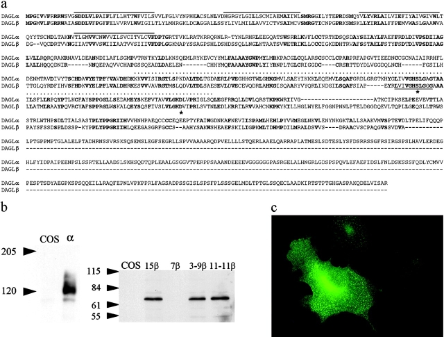
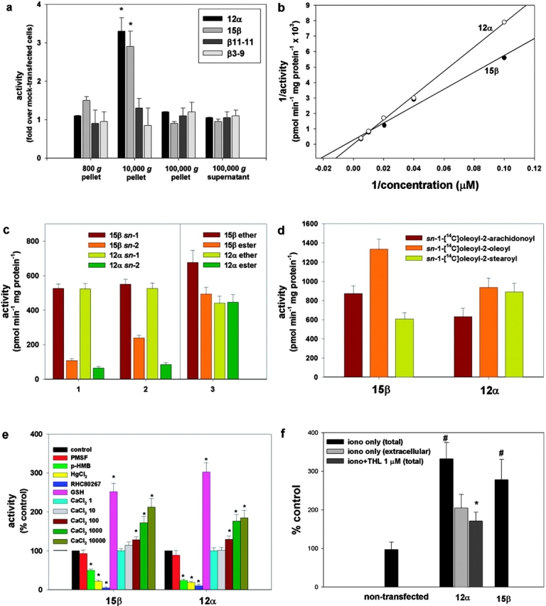
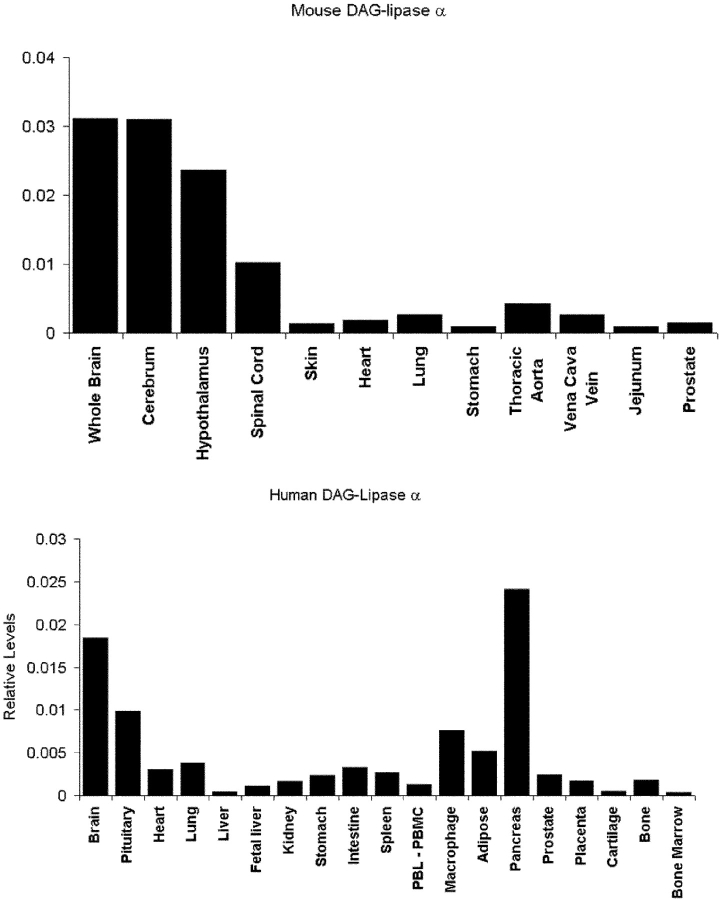
Diacylglycerol (DAG) lipase activity is required for axonal growth during development and for retrograde synaptic signaling at mature synapses. This enzyme synthesizes the endocannabinoid 2-arachidonoyl-glycerol (2-AG), and the CB1 cannabinoid receptor is also required for the above responses. We now report on the cloning and enzymatic characterization of the first specific sn-1 DAG lipases. Two closely related genes have been identified and their expression in cells correlated with 2-AG biosynthesis and release. The expression of both enzymes changes from axonal tracts in the embryo to dendritic fields in the adult, and this correlates with the developmental change in requirement for 2-AG synthesis from the pre- to the postsynaptic compartment. This switch provides a possible explanation for a fundamental change in endocannabinoid function during brain development. Identification of these enzymes may offer new therapeutic opportunities for a wide range of disorders.


Figures

Figure 1.
Figure 1.
Characterization and expression of the novel genes. An alignment between the human α (gi|20521123) and β (gi|21040277) gene products is shown in panel a. The four putative transmembrane domains, the serine lipase motif, and the lipase 3 motif are highlighted by lines above the sequence, a dotted line, and a line below the sequence, respectively. Mutated amino acids are denoted with an asterisk. A Western blot analysis (with molecular mass markers in kD) of control and transfected COS cells expressing the α and β gene products, including the mutated clones, is shown in panel b. The localization of the α transgene in transfected COS cells is shown in panel c.
Figure 2.
Figure 2.
Enzymatic characterization of the new lipase gene products expressed in COS cells (clones 12α and 15β). (a) Subcellular fractionation of the enzymes using sn-1-stearoyl-2-[14C]arachidonoyl-glycerol as a substrate. Controls included two clones expressing the highest levels of the β gene product with single point mutations in Asp 494 to Ala (clone 3-9β) and Ser 443 to Ala (clone 11-11β). Activity is expressed as fold over the activity in fractions from a clone transfected with the β construct that expressed no detectable transgene (clone 7β, control cells). (b) Lineweaver–Burk profiles of the activities in the 10,000 g pellets from clones 12α and 15β. Data points were subtracted from the activity found in control cells (clone 7β). Vmax values were 33.3 ± 4.5 and 3.45 ± 0.16 nmol min−1 mg protein−1 for the α and β form, respectively. (c) Selectivity of the two DAGLs for the sn-1 position of DAGs. The rate of formation is shown for: (1) [14C]oleic acid (sn-1 bars) and sn-1-[14C]oleoyl-glycerol (sn-2 bars) from sn-1-[14C]oleoyl-2-oleoyl-glycerol, this indicates sn-1 and sn-2 selectivity, respectively; (2) mono[14C]oleoyl-glycerol from either sn-1-[14C]oleoyl-2-oleoyl-glycerol (sn-2 bars) or sn-1-oleoyl-2-[14C]oleoyl-glycerol (sn-1 bars), this indicates sn-2 and sn-1 selectivity, respectively; and (3) [14C]oleic acid from either sn-1-[14C]oleoyl-2-arachidonyl glyceryl ether (ether bars), or sn-1-[14C]oleoyl-2-arachidonoyl glycerol (ester bars), this indicates sn-1 selectivity, without and with possible interference from sn-2 selectivity, respectively. (d) Rate of formation of [14C]oleic acid using DAG substrates with different fatty acids on the 2-position. (e) Effect of inhibitors, glutathione (GSH), and calcium on enzyme activity assessed using sn-1-[14C]oleoyl-2-arachidonoyl-glycerol as substrate. 1 mM PMSF; 1 mM p-hydroxy-mercuri-benzoate (p-HMB); 5 mM HgCl2; and 100 μM RHC80267. The concentrations of CaCl2 in the assay buffer were 1, 10, 100, 1,000, and 10,000 μM. (f) Formation of 2-AG from intact COS cells (nontransfected or clones 12α and 15β), stimulated for 20 min with vehicle (DMSO, 0.1%), 4 μM ionomycin, or ionomycin + 1 μM THL after a 5-min preincubation with THL. 2-AG levels were measured in cells plus medium (total) by liquid chromatography-mass spectrometry. Results are expressed as a percentage of 2-AG formation from vehicle-stimulated cells (12.1 ± 4.3 pmol/mg extracted lipids). In one set of experiments, the amounts of 2-AG produced after ionomycin stimulation were measured separately in cells and medium, and the percentage of the amount of 2-AG found in the medium (extracellular) is shown. Data are means ± SEM of n ≥ 4. *, P < 0.05, control cells; #, P < 0.05, iono only (total), t test.
Figure 3.
Figure 3.
THL inhibits the neurite outgrowth response stimulated by FGF2. Rat cerebellar neurons were isolated at postnatal day 2 and cultured over monolayers of 3T3 cells in control media or media supplemented with 5 ng/ml FGF2, 5 ng/ml BDNF, or 0.2 μM WIN55,2122-2 as indicated. The experiments were done in the absence (white bars) and presence (black bars) of 10 μM THL. After 18 h, the cultures were fixed and stained for GAP-43, and the mean length of the longest neurite was determined from ∼140 neurons under each culture condition. For the control and FGF2 data, results were pooled from three independent experiments. For the BDNF and CB1 agonist data, the results were pooled from two independent experiments. Bars show SEM. Dose–response experiments (not depicted) indicated an IC50 = 2 μM for the inhibition by THL of FGF2 response.
Figure 4.
Figure 4.
Expression of the novel lipases in the mouse nervous system. DAGLα (a) and DAGβ (b) are expressed in neurofilament-positive axonal tracts (c) in the embryonic spinal cord (day 10). DAGLβ is expressed in the embryonic retinal ganglion fiber layer and optic nerve at day 14 (d). Note the lack of expression of DAGLα in the optic (e) and anterior commissures (f) in the adult. This contrasts with strong staining for the CB1 receptor in these tracts (g and h). DAGLα (i) and DAGLβ (j) expression in the adult cerebellum is restricted to the deep cerebellar nuclei and synaptic fields, including the Purkinje cell dendrites (k). Bar: (a–c) ∼130 μm; (d) ∼220 μm; (e–j) ∼500 μm; (k) ∼20 μm.
Figure 5.
Figure 5.
Relative expression of mouse and human DAGLα in various adult tissues. TaqMan RT-RCR was used to measure the relative level of mouse (top) and human (bottom) DAGLα transcripts in various tissues as indicated. In both instances, the number of mRNA copies detected for DAGLα was divided by the number of mRNA copies of GAPDH and β-actin detected in each sample. The average of these two figures are plotted. For the human, data were obtained for two males and two females; there were no obvious gender differences, and the results from all four individuals were pooled.

References

    1. Bisogno, T., N. Sepe, D. Melck, S. Maurelli, L. De Petrocellis, and V. Di Marzo. 1997. Biosynthesis, release and degradation of the novel endogenous cannabimimetic metabolite 2-arachidonoylglycerol in mouse neuroblastoma cells. Biochem. J. 322:671–677. - PMC - PubMed
    1. Bond, B.C., D.J. Virley, N.J. Cairns, A.J. Hunter, G.B. Moore, S.J. Moss, A.W. Mudge, F.S. Walsh, E. Jazin, and P. Preece. 2002. The quantification of gene expression in an animal model of brain ischaemia using TaqMan real-time RT-PCR. Brain Res. Mol. Brain Res. 106:101–116. - PubMed
    1. Brittis, P.A., J. Silver, F.S. Walsh, and P. Doherty. 1996. Fibroblast growth factor receptor function is required for the orderly projection of ganglion cell axons in the developing mammalian retina. Mol. Cell. Neurosci. 8:120–128. - PubMed
    1. Diana, M.A., C. Levenes, K. Mackie, and A. Marty. 2002. Short-term retrograde inhibition of GABAergic synaptic currents in rat Purkinje cells is mediated by endogenous cannabinoids. J. Neurosci. 22:200–208. - PMC - PubMed
    1. Di Marzo, V., D. Melck, T. Bisogno, and L. De Petrocellis. 1998. Endocannabinoids: endogenous cannabinoid receptor ligands with neuromodulatory action. Trends Neurosci. 21:521–528. - PubMed

Publication types

MeSH terms