. 2003 Nov 10;163(3):525-34.
doi: 10.1083/jcb.200307111.

A core function for p120-catenin in cadherin turnover

Affiliations

A core function for p120-catenin in cadherin turnover

Michael A Davis et al. J Cell Biol. .

Abstract

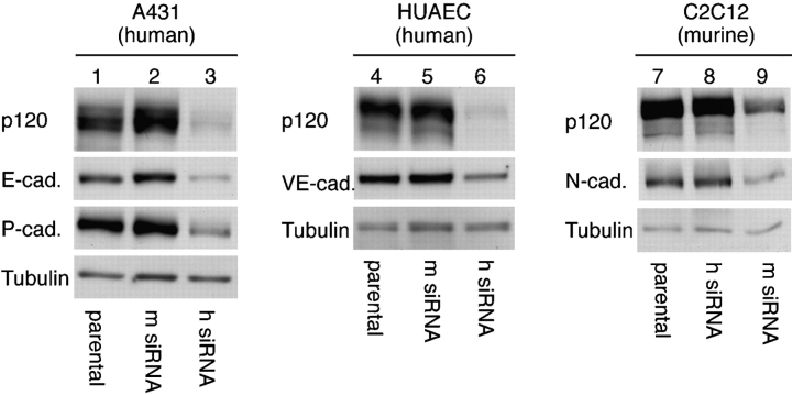
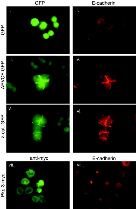
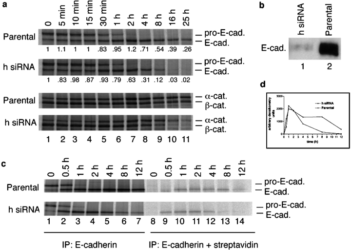
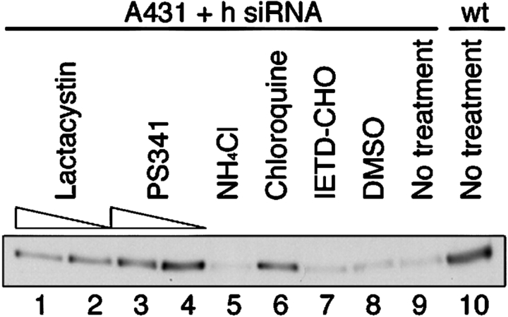
p120-catenin stabilizes epithelial cadherin (E-cadherin) in SW48 cells, but the mechanism has not been established. Here, we show that p120 acts at the cell surface to control cadherin turnover, thereby regulating cadherin levels. p120 knockdown by siRNA expression resulted in dose-dependent elimination of epithelial, placental, neuronal, and vascular endothelial cadherins, and complete loss of cell-cell adhesion. ARVCF and delta-catenin were functionally redundant, suggesting that proper cadherin-dependent adhesion requires the presence of at least one p120 family member. The data reveal a core function of p120 in cadherin complexes, and strongly predict a dose-dependent loss of E-cadherin in tumors that partially or completely down-regulate p120.


Figures

Figure 1.
Figure 1.
p120 knockdown eliminates the E-cadherin complex and abolishes adhesion. (a) Human and murine p120 siRNAs (h siRNA and m siRNA, respectively) were generated against homologous human and murine sequences that contain three mismatches at the nucleotide level (asterisks). (b) Schematic depicting a novel method for in vitro p120 knock-down and knock-up. Human p120 was knocked down using the retroviral vector pRS to express human-specific p120 siRNA, and stable cell lines were selected. p120 was then reexpressed (knock-up) by infecting the knock-down cell line with an LZRS retrovirus containing murine p120 cDNA. (c) Wild-type A431 cells (lane 1) were infected with virus carrying the control m siRNA (lane 2) or h siRNA (lane 3), and stable cell lines were isolated. p120 expression was restored (knock-up) by infecting h siRNA–expressing cells with retrovirus containing murine p120 (lane 4). The indicated cadherin complex proteins were analyzed by Western blotting whole cell lysates. E-cadherin, β-catenin, and α-catenin levels were substantially reduced in p120 knockdown cells, and restoring p120 reversed the effect. (d) p120 (i and vii), E-cadherin (ii and viii), β-catenin (iii and ix), α-catenin (iv and x), tubulin (v and xi), and vinculin (vi and xii) were localized by immunofluorescence in stable A431 cell lines expressing the control m siRNA (i–vi) or h siRNA (vii–xii). Cells were plated sparsely to allow colonies to emerge from single cells. Note that p120 knockdown cells lack cadherin complexes and have lost cell–cell adhesion. The cadherin complex is selectively targeted because the levels of tubulin and vinculin are unaffected.
Figure 2.
Figure 2.
The p120-associated destruction mechanism is common to multiple cadherins. A431 (human cervical carcinoma), HUAEC, and C2C12 (mouse myocyte) cells express E- and P-cadherin, VE-cadherin, and N-cadherin, respectively. Each cell line was infected with either human- or murine-specific p120 siRNA retrovirus to generate polyclonal knockdown cell lines, and levels of p120 or E-, P-, VE-, and N-cadherins were assayed by Western blotting of whole-cell lysates. Tubulin levels were used as a loading control. p120 knockdown reduced expression of all these cadherins, indicating that its function is common to most (if not all) p120-associated cadherins. Note that the effects of the h and m siRNAs used for knockdown and control in the human cell lines are reversed in the murine cell line C2C12.
Figure 3.
Figure 3.
p120 levels act as a set point mechanism for determining cadherin levels. (a) Assay of relationship between p120 and E-cadherin levels by immunofluorescent staining. A431 cells expressing p120 siRNA were infected with the murine p120 retrovirus and plated sparsely so that individual clones could emerge that expressed widely varying amounts of murine p120. Cells were costained by immunofluorescence to examine the p120–E-cadherin relationship and its affect on cell–cell adhesion. p120 loss (i) caused complete loss of E-cadherin (ii) and the cells were nonadhesive. Intermediate levels of p120 expression (panel iii) permitted intermediate levels of E-cadherin (panel iv), and cell–cell adhesion was partially restored. Higher than normal levels of p120 (panel v) strongly induced E-cadherin (vi) and cell–cell adhesion was robust. These experiments reveal a direct relationship between p120 and E-cadherin levels, and the extent of cell–cell adhesion is directly affected. (b) Quantitative assessment of relationship between p120 and E-cadherin levels. A polyclonal population of cells expressing p120 siRNA was generated by retroviral infection. Individual clones within the population express different levels of p120 depending on integration events that affect the efficiency of the siRNA expression. Using E-cadherin antibodies (HECD-1), the cells were separated by FACS® into pools expressing progressively lower levels of E-cadherin. Cell lysates from the samples were split and then Western blotted with anti-p120 (mAb pp120) or anti-E-cadherin (C-20820).
Figure 4.
Figure 4.
Redundant roles for p120 family members ARVCF and δ-catenin. A431 cells stably expressing human p120 siRNA were transiently transfected with ARVCF-GFP, δ-catenin–GFP, or myc-tagged plakophilin-3 (Pkp-3-myc). 24 h after transfection, cells were plated sparsely and individual colonies grew for 2 d. Levels of the transfected proteins and E-cadherin were then analyzed by immunofluorescence. GFP expression alone (i, eluminated cells) did not affect E-cadherin levels (ii). Both ARVCF-GFP (iii) and δ-catenin–GFP (v) substantially increased levels of E-cadherin (iv and vi) and rescued cell–cell adhesion. In contrast, plakophilin-3 (vii), a p120-related protein that does not bind classical cadherins, but had no effect on E-cadherin levels (viii) or cell–cell adhesion.
Figure 5.
Figure 5.
p120 regulates E-cadherin turnover at the cell membrane. (a) E-cadherin synthesis and processing in p120 knockdown cells. E-cadherin turnover was examined by pulse-chase analysis of parental and h p120 siRNA-expressing A431 cells. α- and β-Catenin processing from the same experiment are shown below. Chase times are indicated across the top. At chase time 0 (15 min after initiation of the pulse labeling), E-cadherin synthesis was identical in the presence and absence of p120 (a, compare E-cadherin bands). The processing of the pro- (pro-E-cad) and mature (E-cad.) forms were identical for at least 1 h. Soon thereafter, E-cadherin degradation was significantly accelerated in the absence of p120. (b) Analysis of total E-cadherin surface levels in parental and p120 knockdown A431 cells. The parental and p120 knockdown A431 cells were biotinylated for 20 min at 4°C to label surface cadherins. To specifically measure the surface levels, E-cadherin was first immunoprecipitated directly with E-cadherin mAb HECD-1. The sample was eluted with 0.5% SDS and then reprecipitated with streptavidin-coated beads to isolate the surface-labeled pool. E-cadherin levels at the surface in p120 knockdown cells (lane 1) are at least 100-fold diminished relative to the parental cells (lane 2). The result in lane 2 shows that the surface E-cadherin can be efficiently labeled (and detected) by this method. (c) Tracking the arrival of newly synthesized E-cadherin to the cell surface. The methods in a and b were combined to determine whether newly synthesized E-cadherin could transit to the cell surface in the absence of p120. The results in c were quantified by densitometry and represented graphically in d. Parental and p120 knockdown (h siRNA) cells were labeled with [35S]methionine for 15 min, chased at 37°C for the times indicated across top, and placed on ice (4°C) to suspend trafficking. Cell surface proteins were immediately biotinylated at 4°C for 20 min as in b. Surface E-cadherin was then isolated as in b, and the nascent [35S]methionine E-cadherin pool was visualized by SDS-PAGE and radiography. Nascent E-cadherin appeared at the surface at 30 min and peaked at 1 h. The absence of p120 had no effect on this result. Therefore, p120 is not required for E-cadherin synthesis or trafficking, but is essential to regulate E-cadherin turnover soon after its arrival at the cell surface.
Figure 6.
Figure 6.
Mechanism of E-cadherin degradation. The effects of various inhibitors known to influence cadherin stability were assayed in the stable p120 knockdown A431 cells. E-cadherin levels from samples treated for 24 h (lanes 1–9) were monitored by Western blotting whole-cell lysates and were compared with normal E-cadherin levels in the parental cell line (lane 10). Inhibitor concentrations were as follows: lactacystin 10 μM (lane 1), 3.3 μM (lane 2); PS341 100 nM (lane 3), 33 nM (lane 4), ammonium chloride 5 mM (lane 5), Chloroquine 33 μM (lane 6), and IETD-CHO 10 nM (lane 7). DMSO is the control condition (lane 8). The proteosome inhibitors lactacystin and PS341 increased E-cadherin levels (compare lanes 1–4 to lanes 8 and 9). The lower cadherin levels after 100 nM PS341 (lane 3) relative to the 33-nM treatment (lane 4) is due to toxicity at the higher concentration. Of the lysosomal inhibitors, chloroquine (lane 6), but not ammonium chloride (lane 5), increased E-cadherin levels. A caspase 8 inhibitor (lane 7) that has been shown to inhibit E-cadherin degradation in myeloma cells had no effect.
Figure 7.
Figure 7.
Model for p120 function in regulating cadherin turnover. The low affinity of p120 for cadherins, as judged by coimmunoprecipitation experiments, probably reflects the ability of p120 to rapidly alternate between cadherin-bound and -unbound states. (1) Our data suggest that the rate of cadherin turnover is controlled by cell surface events that transiently increase or decrease p120 affinity for cadherins. Thus, cadherin complexes exist in a dynamic equilibrium between p120-bound and -unbound states, which in turn may be regulated by p120 phosphorylation (not depicted). (2) Unbound cadherin is targeted for internalization, possibly via a Hakai-like ubiquitination mechanism (see Discussion). (3) We cannot yet rule out an alternative pathway where p120 binding is irrelevant for internalization, but mediates a sorting decision that recycles internalized cadherin back to the membrane. (4) Regardless of the exact decision point, unbound cadherin is targeted for degradation by the proteosome and/or lysosome. Considerable evidence indicates that signaling events at the cell surface modulate phosphorylation of the cadherin-bound pool of p120. The simplest interpretation of these observations is that p120 phosphorylation regulates its steady-state affinity for cadherins, which in turn regulates adhesion by controlling the rate of cadherin turnover. Note that α- and β-catenin are passive players in this model. They likely participate in clustering and certainly mediate the cytoskeletal interaction (not depicted), but their role may be secondary to regulating surface cadherin levels, which is almost completely determined by p120.

Comment in

Similar articles

Cited by

References

    1. Anastasiadis, P.Z., and A.B. Reynolds. 2000. The p120 catenin family: complex roles in adhesion, signaling and cancer. J. Cell Sci. 113:1319–1334. - PubMed
    1. Anastasiadis, P.Z., and A.B. Reynolds. 2001. Regulation of Rho GTPases by p120-catenin. Curr. Opin. Cell Biol. 13:604–610. - PubMed
    1. Anastasiadis, P.Z., S.Y. Moon, M.A. Thoreson, D.J. Mariner, H.C. Crawford, Y. Zheng, and A.B. Reynolds. 2000. Inhibition of RhoA by p120 catenin. Nat. Cell Biol. 2:637–644. - PubMed
    1. Baki, L., P. Marambaud, S. Efthimiopoulos, A. Georgakopoulos, P. Wen, W. Cui, J. Shioi, E. Koo, M. Ozawa, V.L. Friedrich, Jr., and N.K. Robakis. 2001. Presenilin-1 binds cytoplasmic epithelial cadherin, inhibits cadherin/p120 association, and regulates stability and function of the cadherin/catenin adhesion complex. Proc. Natl. Acad. Sci. USA. 98:2381–2386. - PMC - PubMed
    1. Berx, G., A.M. Cleton-Jansen, F. Nollet, W.J. de Leeuw, M. van de Vijver, C. Cornelisse, and F. van Roy. 1995. E-cadherin is a tumour/invasion suppressor gene mutated in human lobular breast cancers. EMBO J. 14:6107–6115. - PMC - PubMed

Publication types

MeSH terms