. 2003 Nov 10;163(3):535-45.
doi: 10.1083/jcb.200306001.

Cellular levels of p120 catenin function as a set point for cadherin expression levels in microvascular endothelial cells

Affiliations

Cellular levels of p120 catenin function as a set point for cadherin expression levels in microvascular endothelial cells

Kanyan Xiao et al. J Cell Biol. .

Abstract

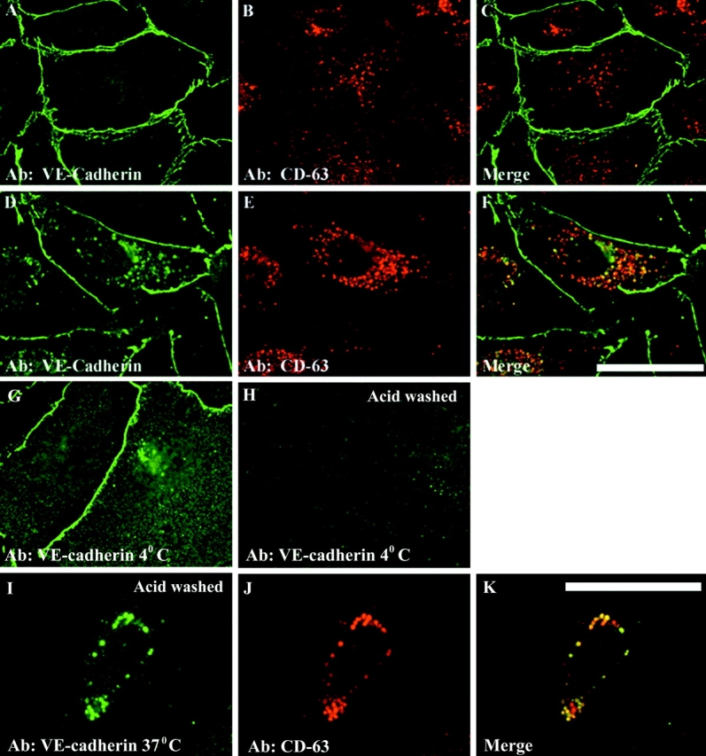
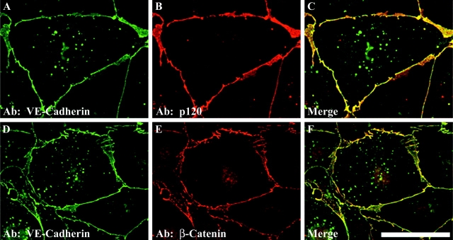
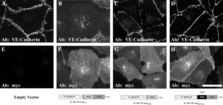
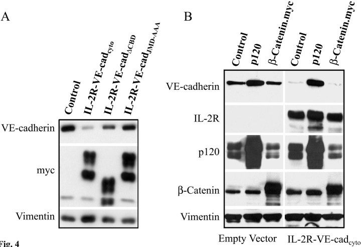
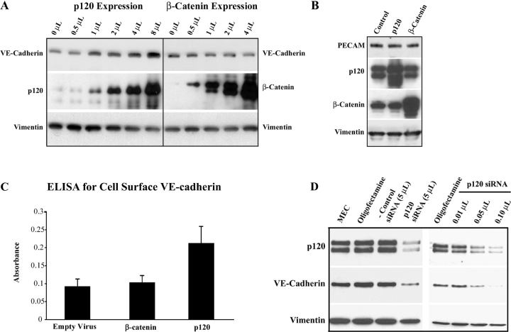
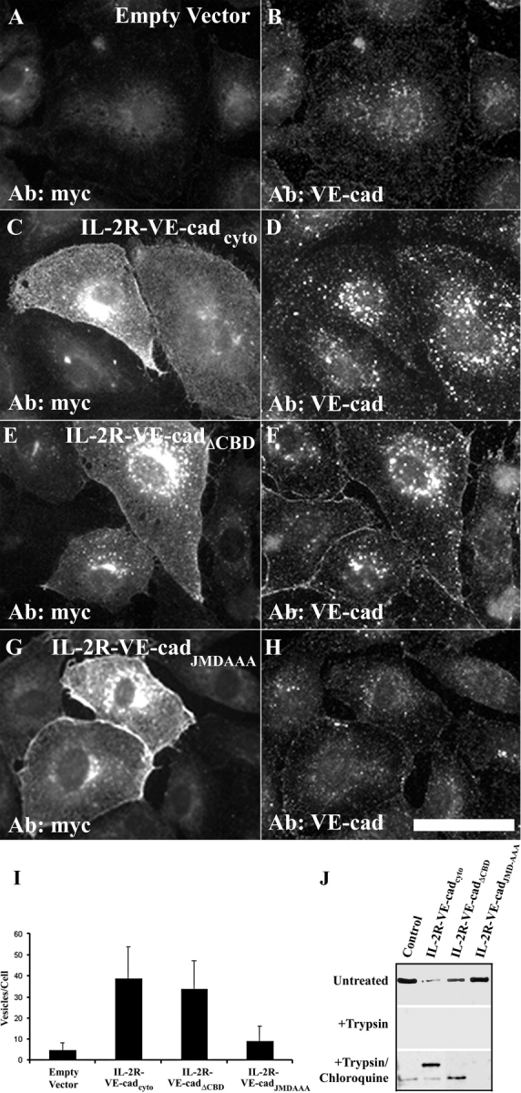
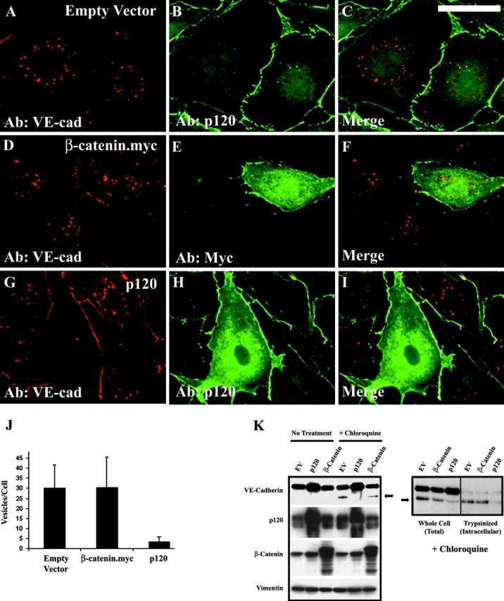
The mechanisms by which catenins regulate cadherin function are not fully understood, and the precise function of p120 catenin (p120ctn) has remained particularly elusive. In microvascular endothelial cells, p120ctn colocalized extensively with cell surface VE-cadherin, but failed to colocalize with VE-cadherin that had entered intracellular degradative compartments. To test the possibility that p120ctn binding to VE-cadherin regulates VE-cadherin internalization, a series of approaches were undertaken to manipulate p120ctn availability to endogenous VE-cadherin. Expression of VE-cadherin mutants that competed for p120ctn binding triggered the degradation of endogenous VE-cadherin. Similarly, reducing levels of p120ctn using siRNA caused a dramatic and dose-related reduction in cellular levels of VE-cadherin. In contrast, overexpression of p120ctn increased VE-cadherin cell surface levels and inhibited entry of cell surface VE-cadherin into degradative compartments. These results demonstrate that cellular levels of p120ctn function as a set point mechanism that regulates cadherin expression levels, and that a major function of p120ctn is to control cadherin internalization and degradation.


Figures

Figure 1.
Figure 1.
VE-Cadherin accumulates in an endosomal–lysosomal compartment in chloroquine-treated MEC. VE-Cadherin localization was examined by immuno–fluorescence microscopy and compared with CD63 localization in untreated MEC (A–C) and in MEC treated with the lysosomal inhibitor chloroquine for 4 h (D–F). Note the extensive accumulation of vesicular VE-cadherin in chloroquine-treated MEC (D) and the colocalization of this intracellular pool with CD63 (F), a marker for late endosomes and lysosomes. To determine if cell surface– derived VE-cadherin was internalized into CD63-positive vesicular compartments, cell surface VE-cadherin was labeled in living MEC at 4°C using an mAb (BV6) directed against the VE-cadherin extracellular domain. The cells were rinsed, fixed, and processed for immunofluorescence microscopy (G). Parallel cultures were labeled at 4°C and transferred to 37°C for 6 h in the presence of chloroquine to allow for cadherin internalization (I–K). After fixation, cells were incubated in an antibody directed against CD63, followed by processing for dual label immunofluorescence microscopy. A low pH wash (acid washed) was used to distinguish cell surface cadherin from internalized cadherin. Note that in H, cell surface VE-cadherin antibody was removed by the low pH wash, indicating that the vesicular VE-cadherin observed in I represents cadherin that was internalized from the cell surface. Bars, 50 μm.
Figure 2.
Figure 2.
The internalized pool of VE-cadherin does not colocalize with β-catenin or p120ctn. Dual label immunofluorescence analysis was performed on MEC treated with chloroquine for 4 h. The internalized pool of VE-cadherin exhibited limited colocalization with (A–C) p120ctn and (D–F) β-catenin. Bar, 50 μm.
Figure 3.
Figure 3.
Cadherin mutants that bind to p120ctn disrupt MEC intercellular junctions. MEC were infected by (A and E) empty adenoviral vector, (B and F) the IL-2R-VE-cadcyto, (C and G) IL-2R-VE-cadΔCBD, or (D and H) IL-2R-VE-cadJMD-AAA mutants, respectively, for 18 h. The IL-2R-VE-cadΔCBD mutant lacks the β-catenin binding site, whereas the IL-2R-VE-cadJMD-AAA mutant contains the β-catenin binding domain, but harbors a triple alanine substitution that abrogates p120ctn binding. The cells were fixed in methanol and processed for dual label immunofluorescence using antibodies directed against (A–D) endogenous VE-cadherin (cad-5 antibody) and (E–H) the myc epitope tag. Bar, 50 μM.
Figure 4.
Figure 4.
Cadherin mutants cause down-regulation of endogenous VE-cadherin by competing for p120ctn. (A) MEC were transduced with adenoviruses carrying the various IL-2R-VE-cad mutants and endogenous VE-cadherin levels were monitored by Western blot analysis. The mutant lacking the p120ctn binding site (IL-2R-VE-cadJMD-AAA) failed to cause down-regulation of endogenous VE-cadherin. (B) p120ctn and β-catenin were coexpressed with the IL-2R-VE-cadcyto mutant to determine which armadillo family proteins could prevent the down-regulation of endogenous VE-cadherin in response to the cadherin mutant. p120ctn was able to prevent the down-regulation of endogenous VE-cadherin by the IL-2R-VE-cadcyto mutant, but β-catenin expression did not rescue endogenous VE-cadherin.
Figure 5.
Figure 5.
Cellular levels of VE-cadherin are tightly coupled to p120ctn expression levels. (A) p120ctn or β-catenin were overexpressed in MEC using an adenoviral delivery system. Expression of exogenous p120ctn leads to a dose- dependent increase in VE-cadherin accumulation, whereas β-catenin has no affect on VE-cadherin levels. (B) Expression levels of PECAM-1 were unaltered in MEC overexpressing p120ctn or β-catenin. (C) Relative cell surface levels of VE-cadherin were monitored using an ELISA on unpermeabilized MEC. Exogenous expression of p120ctn increased cell surface levels of VE-cadherin. Error bars represent the SD. (D) siRNA was used to knock down p120ctn levels in MEC. Western blot analysis was performed to determine p120ctn, VE-cadherin, and vimentin levels in untreated and siRNA-treated MEC. The loss of p120ctn results in a corresponding and dose-related loss of VE-cadherin.
Figure 6.
Figure 6.
Sequestration of cytoplasmic p120ctn by exogenously expressed cadherin mutants causes internalization of cell surface VE-cadherin. The internalization of cell surface VE-cadherin was monitored in MEC expressing various IL-2R-VE-cadherin mutants. MEC were incubated with the BV6 antibody at 4°C and transferred to 37°C for 3 h in the presence of chloroquine. The cells were acid washed to remove surface bound VE-cadherin antibody, and fixed and processed for dual label immunofluorescence microscopy to detect both the internalized cadherin and the IL-2R-VE-cadherin mutants. Internalized VE-cadherin was detected in control MEC expressing empty adenoviral vector (A and B). VE-Cadherin internalization was dramatically increased in MEC expressing either the IL-2R-VE-cadcyto (C and D) or the IL-2R-VE-cadΔCBD mutant (E and F). However, the IL-2R-VE-cadJMDAAA mutant (G and H), which does not bind and compete for cytoplasmic p120ctn, failed to cause increased VE-cadherin internalization relative to cells expressing empty vector. (I) Quantitative representation of vesicular VE-cadherin detected in cells expressing the various cadherin mutants (results representative of greater than three independently conducted experiments. Error bars indicate the SD; n > 10 cells). (J) MEC were infected with empty virus, IL-2R-VE-cadcyto, IL-2R-VE-cadΔCBD, or the IL-2R-VE-cadJMD-AAA mutant and Western blot analysis was performed using an antibody against the extracellular domain of VE-cadherin to monitor endogenous VE-cadherin levels. Expression of the mutants was verified using the c-myc epitope tag (not depicted). Mutant cadherins that bind p120ctn cause down-regulation of endogenous VE-cadherin (top). In untreated cells, VE-cadherin was completely removed by trypsinization (middle). In MEC treated with chloroquine for 8 h, VE-cadherin is detected in trypsinized cells (bottom), indicating that this pool of VE-cadherin is intracellular. The results indicate that the IL-2R-VE-cadcyto and the IL-2R-VE-cadΔCBD mutant cause internalization of endogenous VE-cadherin, whereas the IL-2R-VE- cadJMD-AAA mutant does not trigger VE-cadherin internalization. Bar, 50 μm.
Figure 7.
Figure 7.
Expression of exogenous p120ctn decreases internalization of cell surface VE-cadherin. MEC were transduced with adenovirus carrying β-catenin or p120ctn overnight. The cells were surfaced labeled with the BV6 VE-cadherin mAB and transferred to 37°C for 6 h in the presences of chloroquine to allow for the accumulation of internalized VE-cadherin (Fig. 1). The cells were acid washed to remove cell surface cadherin and processed for immunofluorescence microscopy to detect both internalized VE-cadherin and the exogenously expressed catenins. Extensive vesicular accumulation of internalized VE-cadherin was apparent in chloroquine-treated MEC expressing (A–C) empty virus or (D–F) β-catenin. In contrast, expression of exogenous p120ctn dramatically inhibited (G–I) VE-cadherin internalization. (J) Quantitative representation of the results shown in A–I (results representative of three independent experiments. n > 10 cells. Error bars represent the SD. (K) MEC were transduced with adenovirus carrying p120ctn or β-catenin. The cells were chloroquine treated overnight and harvested for Western blot analysis. As reported previously (Xiao et al., 2003), chloroquine treatment results in the accumulation of an intracellular truncated form of VE-cadherin (arrows). To distinguish intracellular from cell surface cadherin, cells were trypsinized before harvesting the cells for Western blot analysis to remove cell surface pools of cadherin (K, right). p120ctn, but not β-catenin, inhibited the accumulation of this intracellular-processed form of VE-cadherin. Bar, 50 μm.

Comment in

References

    1. Aberle, H., S. Butz, J. Stappert, H. Weissig, R. Kemler, and H. Hoschuetzky. 1994. Assembly of the cadherin-catenin complex in vitro with recombinant proteins. J. Cell Sci. 107:3655–3663. - PubMed
    1. Aberle, H., A. Bauer, J. Stappert, A. Kispert, and R. Kemler. 1997. β-Catenin is a target for the ubiquitin-proteosome pathway. EMBO J. 16:3797–3804. - PMC - PubMed
    1. Akhtar, N., and N.A. Hotchin. 2001. RAC1 regulates adherens junctions through endocytosis of E-cadherin. Mol. Biol. Cell. 12:847–862. - PMC - PubMed
    1. Albelda, S.M., W.A. Muller, C.A. Buck, and P.J. Newman. 1991. Molecular and cellular properties of PECAM-1 (endoCAM/CD31): a novel vascular cell–cell adhesion molecule. J. Cell Biol. 114:1059–1068. - PMC - PubMed
    1. Anastasiadis, P.Z., and A.B. Reynolds. 2000. The p120 catenin family: complex roles in adhesion, signaling and cancer. J. Cell Sci. 113:1319–1334. - PubMed

Publication types

MeSH terms