. 2003 Nov 10;163(3):547-57.
doi: 10.1083/jcb.200305137.

p120 catenin associates with kinesin and facilitates the transport of cadherin-catenin complexes to intercellular junctions

Affiliations

p120 catenin associates with kinesin and facilitates the transport of cadherin-catenin complexes to intercellular junctions

Xinyu Chen et al. J Cell Biol. .

Abstract

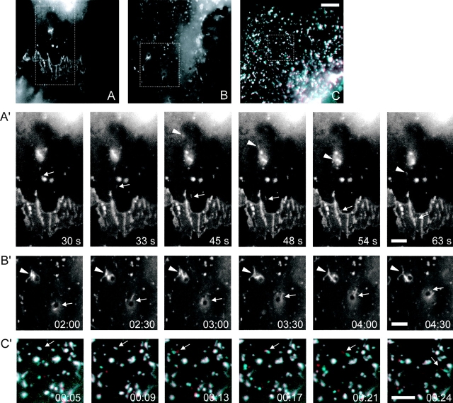
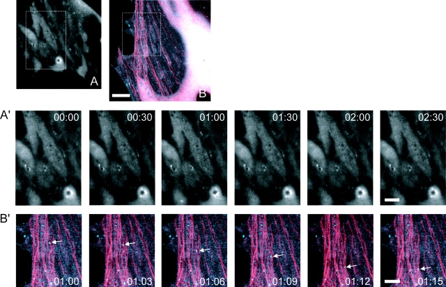
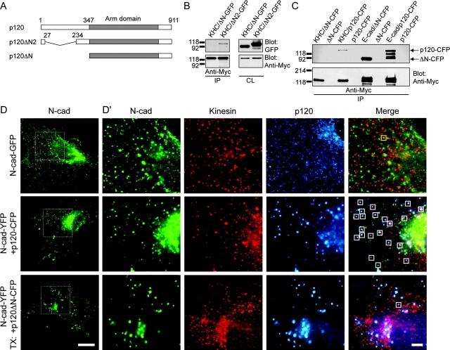
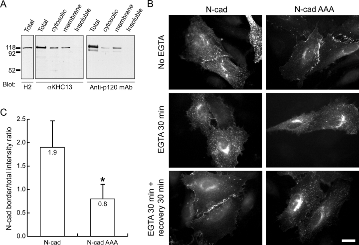
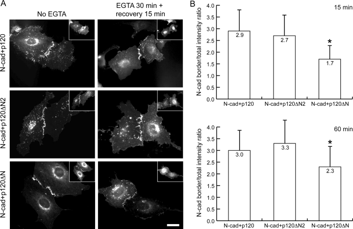
p120 catenin (p120) is a component of adherens junctions and has been implicated in regulating cadherin-based cell adhesion as well as the activity of Rho small GTPases, but its exact roles in cell-cell adhesion are unclear. Using time-lapse imaging, we show that p120-GFP associates with vesicles and exhibits unidirectional movements along microtubules. Furthermore, p120 forms a complex with kinesin heavy chain through the p120 NH2-terminal head domain. Overexpression of p120, but not an NH2-terminal deletion mutant deficient in kinesin binding, recruits endogenous kinesin to N-cadherin. Disruption of the interaction between N-cadherin and p120, or the interaction between p120 and kinesin, leads to a delayed accumulation of N-cadherin at cell-cell contacts during calcium-initiated junction reassembly. Our analyses identify a novel role of p120 in promoting cell surface trafficking of cadherins via association and recruitment of kinesin.


Figures

Figure 1.
Figure 1.
Dynamics of p120-GFP in REF52 cells. (A–C) The first frame of each movie in A′–C′ is shown. The boxed regions were enlarged and selected still frames of each movie are shown in A′–C′. (A′) p120-GFP particles move into existing cell–cell junctions (Video 1, available at http://www.jcb.org/cgi/content/full/jcb.200305137/DC1). A region of two contacting cells is shown. The arrow indicates a p120-GFP particle that moved toward and eventually incorporated into an existing junction. The arrowhead shows another p120-GFP dot traveling in an anterograde direction toward the cell–cell contact area. (B′) p120-GFP shows dynamic structural changes characteristic of vesicles (Video 2, available at http://www.jcb.org/cgi/content/full/jcb.200305137/DC1). Many p120-GFP particles appeared to associate with spherical structures of various sizes, and exhibited dynamic changes in their structure such as stretching and shortening. The arrow and arrowhead indicate two examples. (C′) p120-CFP and N-cad–YFP colocalize and move together (Video 3, available at http://www.jcb.org/cgi/content/full/jcb.200305137/DC1). White dots represent colocalization of p120-CFP (blue) and N-cad–YFP (red). Due to the 3-s gap between the images taken from the CFP and YFP channel, moving complexes of p120-CFP and N-cad–YFP appeared as two adjacent blue and red dots, whereas static complexes remained white. The arrow indicates a complex of p120-CFP and N-cad–YFP particles moving together. In all the above experiments, REF52 cells were subjected to time-lapse imaging 48–72 h after transfection. Images were taken at 3-s intervals for 5–10 min. The time stamp in the bottom right-hand corner of each still image is displayed in min:sec, except in A′, which is displayed in seconds. Bars: 10 μm (A–C), 5 μm (A′–C′).
Figure 2.
Figure 2.
Dependence of p120 dynamics on MTs. (A and B) The first frame of each movie in A′ and B′ is shown. The boxed regions were enlarged and selected still frames of each movie are shown in A′ and B′. (A′) Unidirectional movement and dynamic structural changes of p120-GFP are MT-dependent (Video 4, available at http://www.jcb.org/cgi/content/full/jcb.200305137/DC1). REF52 cells transiently express- ing p120-GFP were treated with nocodazole at 1 μg/ml for 30 min to disrupt MTs, followed by time-lapse imaging to observe p120-GFP dynamics. p120-GFP particles remained largely static with occasional random local jiggling. No unidirectional movements or structural changes of p120-GFP were observed. (B′) p120-CFP dots travel along MTs (Video 5, available at http://www.jcb.org/cgi/content/full/jcb.200305137/DC1). p120-CFP (blue) and YFP-α-tubulin (red) were cotransfected into REF52 cells. The arrow indicates one p120-CFP dot moving along MTs. In the above experiments, REF52 cells were subjected to time-lapse imaging 48–72 h after transfection. Images were taken at 3-s interval for 5–10 min. The time stamp in each still image is displayed in min:sec. Bars: 10 μm (A and B), 5 μm (A′, B′).
Figure 3.
Figure 3.
p120 associates with kinesin. (A) p120-GFP colocalizes with endogenous kinesin in the cytoplasm and at cell contacts. REF52 cells were transiently transfected with p120-GFP (green), followed by immunofluorescence using a monoclonal KHC antibody H1 (red). In some cells, cytoplasmic p120-GFP dots colocalized extensively with endogenous kinesin, which was also recruited to cell–cell contacts together with p120-GFP (arrow). Bottom panels are enlarged views of the boxed areas. (B) p120 associates with conventional KHC. Lysates of HEK293 cells transiently expressing p120 or in combination with HA-tagged KLC and Myc-tagged KHC as indicated were immunoprecipitated with monoclonal anti-HA antibody 3F10 or monoclonal anti-Myc antibody 9E10. The mouse IgG (mIgG) was used as a negative control. The band detected by the anti-HA antibody in the control immunoprecipitation is the mouse IgG heavy chain. (C) Detection of endogenous complexes of p120 and kinesin. An anti-KHC monoclonal antibody (anti-KHC) H2 was used to immunoprecipitate endogenous KHC and its associated proteins from a mouse brain high speed supernatant fraction. Beads alone (No Ab) or equal amount of nonspecific mouse IgG (mIgG) was used as a negative control. The immunocomplexes were analyzed by immunoblot using the rabbit anti-p120 antibody F1-SH and the anti-KHC peptide antibody αKHC13. TX, transfection; IP, immunoprecipitation; CL, cell lysate. Bars: 10 μm (A, top panels), 5 μm (A, bottom panels).
Figure 4.
Figure 4.
p120 recruits endogenous kinesin to the cadherin–catenin complexes. (A) Diagram of p120 and two of its NH2-terminal deletion mutants. (B) p120ΔN2 (but not p120ΔN) associates with kinesin. p120ΔN2-GFP or p120ΔN-GFP was transiently transfected together with Myc-tagged KHC into HEK293 cells, followed by coimmunoprecipitation using the monoclonal anti-Myc antibody 9E10. (C) p120ΔN-CFP was coimmunoprecipitated with E-cad, but not with KHC. p120ΔN-CFP (or p120-CFP) was transiently transfected together with Myc-tagged KHC or Myc-tagged E-cad into HEK293 cells, followed by coimmunoprecipitation using the monoclonal anti-Myc antibody 9E10. The multiple bands of p120-CFP coprecipitated with E-cad detected in the top anti-GFP blot are likely breakdown products. (D and D′) p120, but not p120ΔN, recruits kinesin to N-cadherin. REF52 cells transiently expressing N-cad–GFP alone (top panels) were stained with the anti-KHC antibody H1 (red) and the anti-p120 monoclonal antibody (blue). REF52 cells transiently expressing N-cad–YFP (pseudocolored green) together with p120-CFP or p120ΔN-CFP (pseudocolored blue) were stained with the anti-KHC antibody H1 (red). Boxed regions in D were enlarged and shown for N-cad, kinesin, p120, and merged view in D′. In the absence of ectopic p120, very few N-cad–GFP dots colocalized with endogenous kinesin (D′, small yellow box in merged view). Introduction of ectopic p120 (but not p120ΔN) led to the colocalization of endogenous kinesin with the cadherin–catenin complexes, shown in the merged pictures as indicated by the small white box in D′ (colocalization of all three components appears as white, colocalization of N-cadherin and kinesin appears as yellow). TX. transfection; IP, immunoprecipitation; CL, cell lysate. Bars: 20 μm (D), 5 μm (D′).
Figure 5.
Figure 5.
Disruption of p120 binding to N-cadherin causes delayed accumulation of N-cadherin at cell–cell contacts in the presence of ectopic p120. (A) Triple Ala mutation in the JMD domain of N-cadherin completely abolishes its ability to interact with p120. HEK293 cells transiently expressing N-cad–YFP (N-YFP) or N-cad AAA-YFP (N AAA-YFP) together with HA-tagged p120 were subjected to coimmunoprecipitation using a monoclonal anti-HA antibody or mouse IgG (mIgG) as control. (B) N-cad AAA-YFP is delayed in its accumulation at cell–cell contacts compared with N-cad–YFP during calcium switch. REF52 cells were transiently cotransfected with p120-CFP (insets) together with N-cad–YFP or N-cad AAA-YFP. 20 h after transfection, cells were incubated with growth medium containing 4 mM EGTA for 30 min, followed by incubation in complete growth medium (recovery). Only adjacent cells that were in close contact and expressed both N-cad and p120 were examined. (C) Quantification of N-cad–YFP or N-cad AAA-YFP accumulation at cell–cell contacts after 15 min of calcium recovery. Pairs of contacting cells expressing p120-CFP with either N-cad–YFP or N-cad AAA-YFP were randomly selected, and the level of cell border accumulation of N-cad–YFP or N-cad AAA-YFP is expressed as ratios of the average YFP fluorescent intensity at cell–cell contacts over the average total YFP fluorescent intensity within the two contacting cells. The number in each bar represents the mean value. The average accumulation of N-cad AAA-YFP (n = 23) at cell–cell contacts is 66% less than that of N-cad–YFP (n = 27) 15 min after calcium recovery. Asterisk denotes significant difference from cells coexpressing N-cad–YFP and p120-CFP (P = 3.8 × 10⁻¹² < 0.05) by t test. IP, immunoprecipitation; CL, cell lysate. Bar, 20 μm.
Figure 6.
Figure 6.
Disruption of p120 binding to N-cadherin causes delayed accumulation of N-cadherin at cell–cell contacts in the presence of only endogenous p120. (A) Sequential detergent extraction and immunoblot of endogenous kinesin and p120 in REF52 cells. A mouse anti-KHC antibody H2 and a rabbit anti-KHC antibody αKHC13 both detected a major band of KHC, which is mainly distributed in the saponin-soluble cytosolic fraction and the Triton X-100–soluble membrane fraction. Endogenous p120 is also primarily distributed within these two pools. (B) N-cad AAA-YFP is delayed in its accumulation at cell–cell contacts compared with N-cad–YFP during calcium switch. REF52 cells were transiently transfected with N-cad–YFP or N-cad AAA-YFP. 20 h after transfection, cells were incubated with growth medium containing 4 mM EGTA for 30 min, followed by incubation in complete growth medium (recovery). Only adjacent cells that were in close contact and that both expressed N-cad were examined. After 30 min of calcium recovery, N-cad–YFP localized prominently at cell–cell borders, whereas most N-cad AAA-YFP still remained in the cytoplasm. (C) Quantification of N-cad–YFP or N-cad AAA-YFP accumulation at cell–cell contacts after 30 min of calcium recovery. Pairs of contacting cells expressing N-cad–YFP or N-cad AAA-YFP were randomly selected, and the cell border accumulation level of N-cad–YFP or N-cad AAA-YFP is expressed as ratios of the average YFP fluorescent intensity at cell–cell contacts over the average total YFP fluorescent intensity within the two contacting cells. The number in each bar represents the mean value. The average accumulation of N-cad AAA-YFP (n = 25) at cell–cell contacts is 58% less than that of N-cad–YFP (n = 26) 30 min after calcium recovery. Asterisk denotes significant difference from cells expressing N-cad–YFP (P = 1.1 × 10⁻¹⁰ < 0.05) by t test. Bar, 20 μm.
Figure 7.
Figure 7.
Disruption of p120 binding to kinesin causes delayed accumulation of N-cadherin at cell–cell contacts during junction reassembly. (A) N-cad–YFP is delayed in its accumulation at cell– cell contacts when coexpressed with p120ΔN-CFP compared with N-cad–YFP coexpressed with p120-CFP or p120ΔN2-CFP during calcium switch. REF52 cells were transiently co-transfected with p120-CFP, p120ΔN2-CFP, or p120ΔN-CFP (insets) together with N-cad–YFP. 20 h after transfection, cells were incubated with growth medium containing 4 mM EGTA for 30 min, followed by incubation in complete growth medium (recovery). Only adjacent cells that were in close contact and expressed both N-cad and p120 were examined. (B) Quantification of N-cad–YFP accumulation at cell–cell contacts after 15 and 60 min of calcium recovery. Pairs of contacting cells expressing N-cad–YFP with either p120-CFP, p120ΔN2-CFP, or p120ΔN-CFP were randomly selected, and the level of cell border accumulation of N-cad–YFP was measured. The number in each bar represents the mean value. The accumulation of N-cad–YFP at cell–cell contacts when coexpressed with p120ΔN-CFP (n = 33 at 15 min; n = 27 at 60 min) is 42% less than that of N-cad–YFP coexpressed with p120-CFP (n = 32 at 15 min; n = 29 at 60 min) 15 min after calcium recovery, and is 24% less after 60 min of calcium recovery. There is no significant difference between the border accumulation of N-cad–YFP when coexpressed with p120-CFP or p120ΔN2-CFP (P = 0.32 > 0.05 at 15 min; P = 0.22 > 0.05 at 60 min). Asterisk denotes significant difference from cells coexpressing N-cad–YFP and p120-CFP (P = 1.1 × 10⁻⁷ < 0.05 at 15 min; P = 2.1 × 10⁻³ < 0.05 at 60 min) by t test. Bar, 20 μm.

Comment in

References

    1. Aberle, H., S. Butz, J. Stappert, H. Weissig, R. Kemler, and H. Hoschuetzky. 1994. Assembly of the cadherin-catenin complex in vitro with recombinant proteins. J. Cell Sci. 107:3655–3663. - PubMed
    1. Anastasiadis, P.Z., and A.B. Reynolds. 2000. The p120 catenin family: complex roles in adhesion, signaling and cancer. J. Cell Sci. 113:1319–1334. - PubMed
    1. Anastasiadis, P.Z., S.Y. Moon, M.A. Thoreson, D.J. Mariner, H.C. Crawford, Y. Zheng, and A.B. Reynolds. 2000. Inhibition of RhoA by p120 catenin. Nat. Cell Biol. 2:637–644. - PubMed
    1. Aono, S., S. Nakagawa, A.B. Reynolds, and M. Takeichi. 1999. p120(ctn) acts as an inhibitory regulator of cadherin function in colon carcinoma cells. J. Cell Biol. 145:551–562. - PMC - PubMed
    1. Brieher, W.M., and B.M. Gumbiner. 1994. Regulation of C-cadherin function during activin induced morphogenesis of Xenopus animal caps. J. Cell Biol. 126:519–527. - PMC - PubMed

Publication types

MeSH terms