. 2003 Nov 10;163(3):583-95.
doi: 10.1083/jcb.200305010.

ROCK-generated contractility regulates breast epithelial cell differentiation in response to the physical properties of a three-dimensional collagen matrix

Affiliations

ROCK-generated contractility regulates breast epithelial cell differentiation in response to the physical properties of a three-dimensional collagen matrix

Michele A Wozniak et al. J Cell Biol. .

Abstract

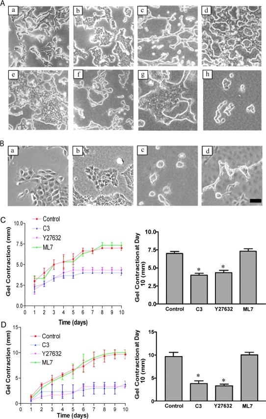
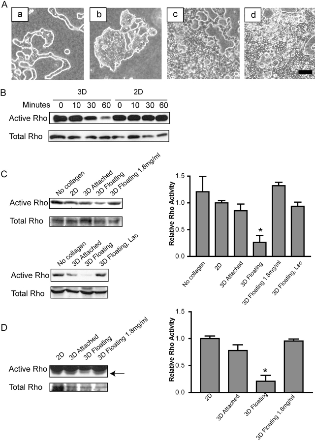
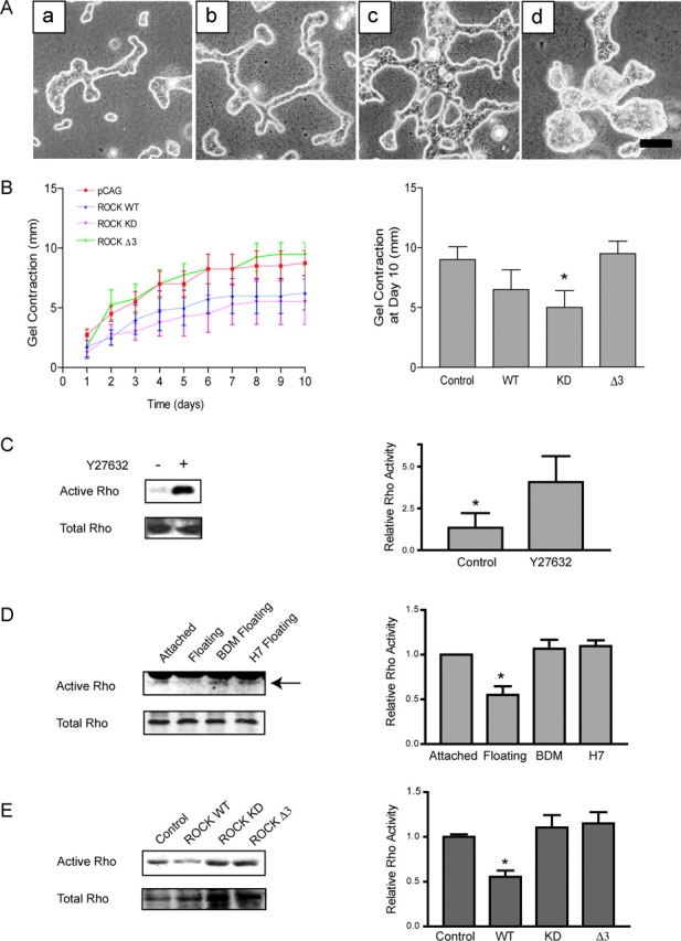
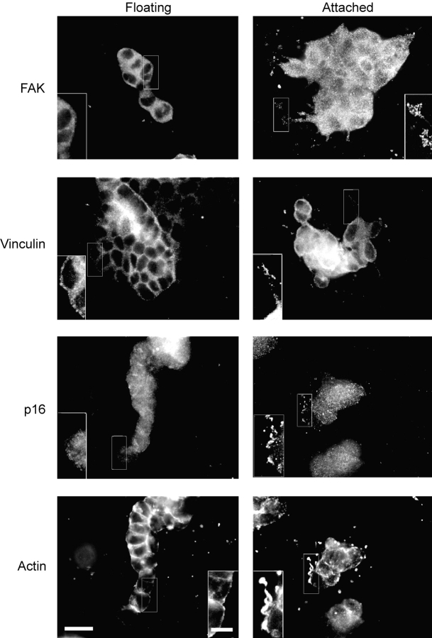
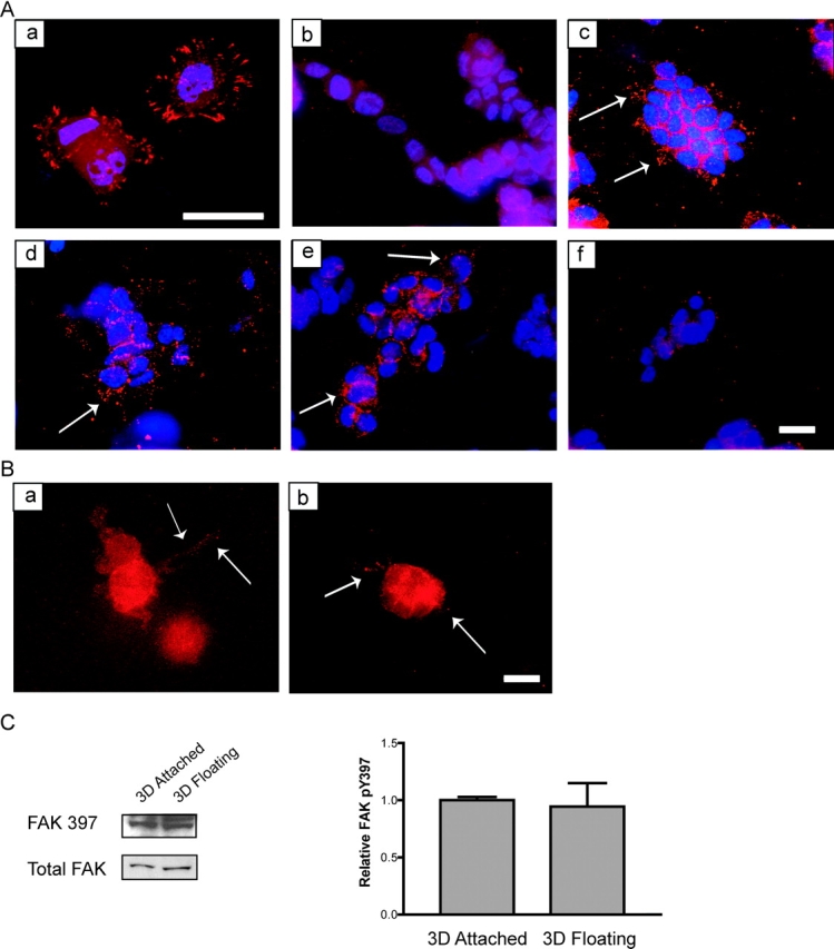
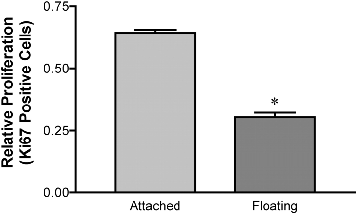
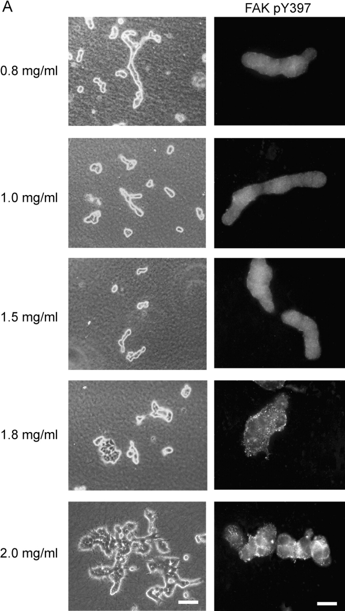
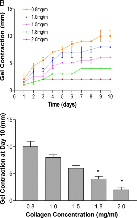
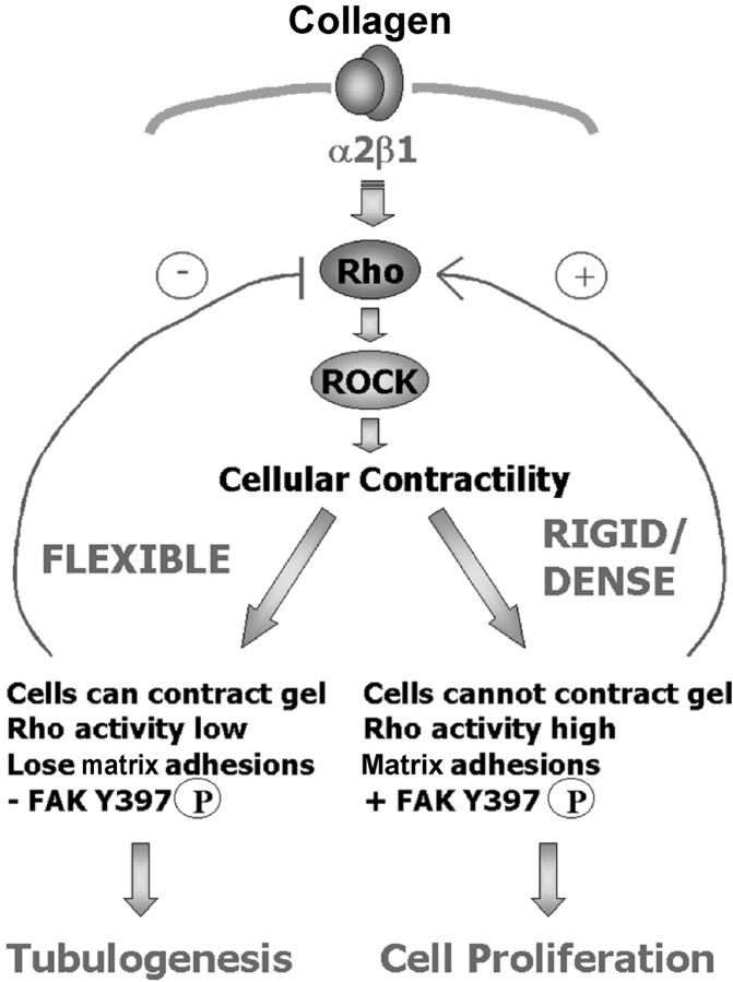
Breast epithelial cells differentiate into tubules when cultured in floating three-dimensional (3D) collagen gels, but not when the cells are cultured in the same collagen matrix that is attached to the culture dish. These observations suggest that the biophysical properties of collagenous matrices regulate epithelial differentiation, but the mechanism by which this occurs is unknown. Tubulogenesis required the contraction of floating collagen gels through Rho and ROCK-mediated contractility. ROCK-mediated contractility diminished Rho activity in a floating 3D collagen gel, and corresponded to a loss of FAK phosphorylated at Y397 localized to 3D matrix adhesions. Increasing the density of floating 3D collagen gels also disrupted tubulogenesis, promoted FAK phosphorylation, and sustained high Rho activity. These data demonstrate the novel finding that breast epithelial cells sense the rigidity or density of their environment via ROCK-mediated contractility and a subsequent down-regulation of Rho and FAK function, which is necessary for breast epithelial tubulogenesis to occur.


Figures

Figure 1.
Figure 1.
The rigidity of the collagen matrix regulates tubulogenesis through Rho- and ROCK-mediated contractility. (A) T47D breast epithelial cells were cultured on collagen-coated plastic (a), for 10 d in 3D collagen gels (1.3 mg/ml) that were left attached to the dish (b) or floated into the culture medium (c). Inhibition of Rho with C3 exoenzyme (10 μg/ml) (d) or ROCK with Y27632 (10 μM) (e) in floating 3D collagen gels blocked tubulogenesis, whereas inhibition of MLCK with ML7 (10 μM) did not alter tubulogenesis (f). Inhibition of actin–myosin contractility with BDM (20 mM) (g) or H-7 (300 μM) (h) also blocked tubulogenesis. (B) MCF10A cells do not differentiate when cultured on collagen-coated plastic (a) or in attached 3D gels (b), but do form acini in a floating collagen gel (c). Addition of HGF (50 ng/ml) promotes the formation of tubules in a floating gel (d). Bar: (A and B) 100 μm. (C) Inhibition of Rho with C3 (10 μg/ml) or ROCK with Y27632 (10 μM), but not MLCK with ML7 (10 μM), blocks contraction of floating collagen gels by T47D cells. The diameter of the gel was measured every day for 10 d, and graphed as total change in diameter in millimeters (left). The graph on the right shows the extent of gel contraction on the tenth day, expressed as total change in diameter. Data is expressed ±SEM for four separate experiments. Inhibition of Rho or ROCK significantly (*P < 0.05) decreased gel contraction. (D) Contraction of floating gels by MCF10A cells also shows significant (*P < 0.05) sensitivity to Rho and ROCK inhibition, but not MLCK inhibition.
Figure 2.
Figure 2.
Rho activity is regulated by ECM rigidity. (A) Compared with control cells (a), stable expression of constitutively activated Rho (63L) (b) or dominant–negative Rho (19N) (c) disrupted tubulogenesis. Expression of truncated, constitutively activated Lsc, an exchange factor specific for Rho, also disrupted tubule formation (d). Bar, 100 μm. (B) ECM rigidity regulates Rho activity. T47D cells were stimulated in suspension with collagen fibers (3D) or on a collagen coated plate (30 μg/ml) (2D). At the times indicated, cells were lysed, and 30 μg RBD–GST was incubated with the cell lysates to pull down active Rho. Active and total Rho was detected by Western blotting. (C) Rho activity is significantly down-regulated in a floating 3D collagen gel by 1 h. Quantitation was performed on five individual experiments, and is shown in the bar graph on the right (*P < 0.05 vs. 2D). (D) Rho activity significantly (*P < 0.05) remains down-regulated in a floating, but not attached, collagen gel after 24 h. Quantitation was performed on four individual experiments.
Figure 3.
Figure 3.
Proper ROCK regulation is required for tubulogenesis, collagen gel contraction, and Rho regulation. (A) Control cells (a) and cells expressing wild-type (WT) ROCK (b) form tubules in floating collagen gels. Misregulation of ROCK by the expression of constitutively activated ROCK (Δ3) (c) or kinase-dead ROCK (KD-IA) (d) alter tubulogenesis. Bar, 100 μm. (B) Expression of ROCK (KD-IA) decreases the contraction of floating 3D gels (*P = 0.066) compared with control cells. ROCK (WT) and (ROCK Δ3) do not significantly affect contraction (P = 0.253 and 0.750, respectively compared with control cells). (C) The presence of Y27632 (10 μM) for 1 h significantly (*P < 0.05) reduced the down-regulation of Rho activity, suggesting a negative feedback loop to Rho from its effector, ROCK. In the example shown, two noncontinuous lanes from the same gel have been moved next to one another to remove intervening lanes treated with other inhibitors. The quantitation shown (right) is a representation of four similar experiments. (D) Rho-GTP was determined in cells cultured in floating 3D collagen gels in the presence or absence of 20 mM BDM or 300 μM H7 for 60 min. Both inhibitors prevented the down-regulation of Rho activity. Quantitation was performed on eight individual experiments (bar graph; *P<0.05 vs. BDM and H7). (E) Expression of ROCK (WT) significantly decreases Rho activity in a floating 3D collagen gel, but misregulation of ROCK activity by expression of ROCK (Δ3) or (KD) increases Rho activity, suggesting proper regulation of ROCK is required to down-regulate Rho. The experiment was repeated five times and quantitation performed (bar graph; *P < 0.05 vs. control).
Figure 4.
Figure 4.
The rigidity of the matrix regulates 3D matrix adhesion formation in breast epithelial cells. Components of focal adhesions were analyzed by immunostaining T47D cells cultured in collagen gels (1.3 mg/ml). Breast cells in attached, but not floating, 3D gels show FAK, vinculin, and p16 localized to small punctate 3D matrix adhesions (insets). Actin fibers are also localized to these structures (insets). Bar: 50 μm; (insets) 10 μm.
Figure 5.
Figure 5.
The rigidity of the matrix and cellular contractility regulates the localization of FAK phosphorylated at Y397 at 3D matrix adhesions. (A) FAK phosphorylated at Y397 is localized to focal adhesions when T47D cells are plated on 2D collagen-coated coverslips (a). In a floating 3D gel, FAK phosphorylation at Y397 is minimal (b). Cells cultured in an attached 3D collagen gel localized FAK pY397 to punctate adhesions (c). Cells in floating 3D gels treated with C3 (10 μg/ml) (d) or Y27632 (10 μM) (e) localized FAK pY397 to small 3D matrix adhesions (see arrows), whereas cells treated with ML7 (10 μM) lost this localization (f). FAK pY397 is shown in red, and nuclei are shown in blue. Bar: (a) 25 μm; (b–f) 50 μm. (B) Cells treated with BDM (20 mM) (a) or H7 (300 μM) (b) also localized FAK pY397 to punctuate matrix adhesions. Bar, 50 μm. (C) The localization, and not phosphorylation, of FAK Y397 is regulated by ECM rigidity. Western blot analysis of FAK pY397 in attached vs. floating collagen gels (1.3 mg/ml) show statistically similar (P = 0.8014) levels of phosphorylation and FAK expression. The quantitation represents four individual experiments (right).
Figure 6.
Figure 6.
ECM rigidity regulates breast epithelial proliferation. T47D cells were cultured in floating and attached 3D collagen gels and allowed to grow for 7 d. The gels were then fixed and costained for Ki67, a marker of proliferation, and bisbenzimide, which will stain all nuclei regardless of cell cycle state. Cells in floating collagen gels have significantly (*P < 0.05) decreased proliferation compared with cells in attached gels. The graph is representative of three individual experiments.
Figure 7.
Figure 7.
Collagen density regulates tubulogenesis, the localization of FAK phosphorylation at Y397, and cellular contraction. (A) T47D cells were cultured in floating collagen gels composed of increasing concentrations of collagen, as indicated. Collagen concentrations equal to or above 1.8 mg/ml disrupt tubulogenesis (left panels). Bar, 100 μm. Increasing collagen concentration also increased FAK pY397 localized to 3D matrix adhesions (right panels). Bar, 50 μm. (B) Increasingly dense collagen gels significantly (*P < 0.05) decrease the contraction of floating 3D collagen gels by T47D breast epithelial cells. The data are an average of three experiments.
Figure 7.
Figure 7.
Collagen density regulates tubulogenesis, the localization of FAK phosphorylation at Y397, and cellular contraction. (A) T47D cells were cultured in floating collagen gels composed of increasing concentrations of collagen, as indicated. Collagen concentrations equal to or above 1.8 mg/ml disrupt tubulogenesis (left panels). Bar, 100 μm. Increasing collagen concentration also increased FAK pY397 localized to 3D matrix adhesions (right panels). Bar, 50 μm. (B) Increasingly dense collagen gels significantly (*P < 0.05) decrease the contraction of floating 3D collagen gels by T47D breast epithelial cells. The data are an average of three experiments.
Figure 8.
Figure 8.
Model for regulation of breast epithelial differentiation by the rigidity of 3D matrices. The α2β1 integrin binds to collagen, leading to Rho activation. Rho activates its effector, ROCK, to promote cellular contractility. If the cells are unable to contract their matrix; that is, if the matrix is rigid, attached, or dense, tension is generated within the cell. Rho activity remains high and promotes 3D matrix adhesion formation including FAK phosphorylated at Y397. This phosphorylation event links a rigid matrix to subsequent cell proliferation and the disruption of tubulogenesis. If the cells are able to contract their matrix (if the matrix is flexible, floating, or composed of low collagen density), ROCK down-regulates Rho activity. Matrix adhesions are changed and FAK phosphorylated at Y397 is not localized to adhesive structures, fundamentally altering subsequent signaling events and resulting in differentiation into tubules.

References

    1. Amano, M., K. Chihara, K. Kimura, Y. Fukata, N. Nakamura, Y. Matsuura, and K. Kaibuchi. 1997. Formation of actin stress fibers and focal adhesions enhanced by Rho-kinase. Science. 275:1308–1311. - PubMed
    1. Amano, M., M. Ito, K. Kimura, Y. Fukata, K. Chihara, T. Nakano, Y. Matsuura, and K. Kaibuchi. 1996. Phosphorylation and activation of myosin by Rho-associated kinase (Rho- kinase). J. Biol. Chem. 271:20246–20249. - PubMed
    1. Arthur, W.T., L.A. Petch, and K. Burridge. 2000. Integrin engagement suppresses RhoA activity via a c-Src-dependent mechanism. Curr. Biol. 10:719–722. - PubMed
    1. Balaban, N.Q., U.S. Schwarz, D. Riveline, P. Goichberg, G. Tzur, I. Sabanay, D. Mahalu, S. Safran, A. Bershadsky, L. Addadi, and B. Geiger. 2001. Force and focal adhesion assembly: a close relationship studied using elastic micropatterned substrates. Nat. Cell Biol. 3:466–472. - PubMed
    1. Blanchoin, L., K.J. Amann, H.N. Higgs, J.B. Marchand, D.A. Kaiser, and T.D. Pollard. 2000. Direct observation of dendritic actin filament networks nucleated by Arp2/3 complex and WASP/Scar proteins. Nature. 404:1007–1011. - PubMed

Publication types

MeSH terms