. 2003 Nov 10;163(3):609-23.
doi: 10.1083/jcb.200309042.

Defining BMP functions in the hair follicle by conditional ablation of BMP receptor IA

Affiliations

Defining BMP functions in the hair follicle by conditional ablation of BMP receptor IA

Krzysztof Kobielak et al. J Cell Biol. .

Abstract

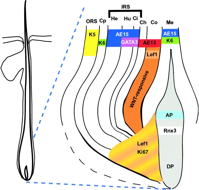
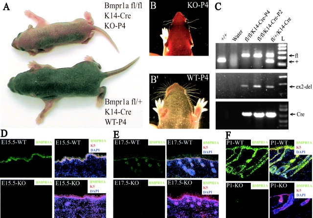
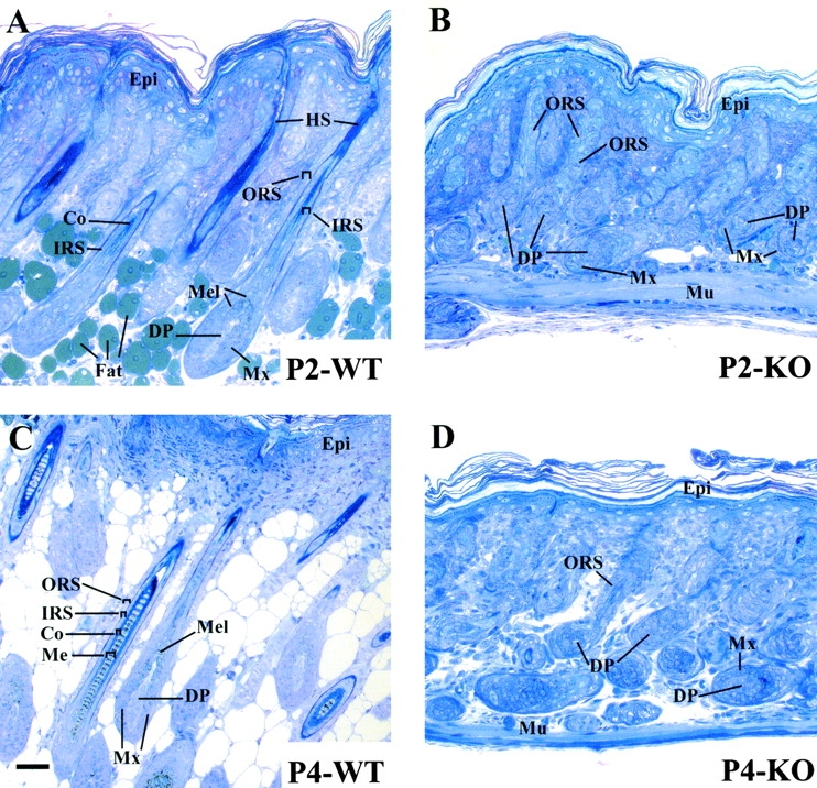
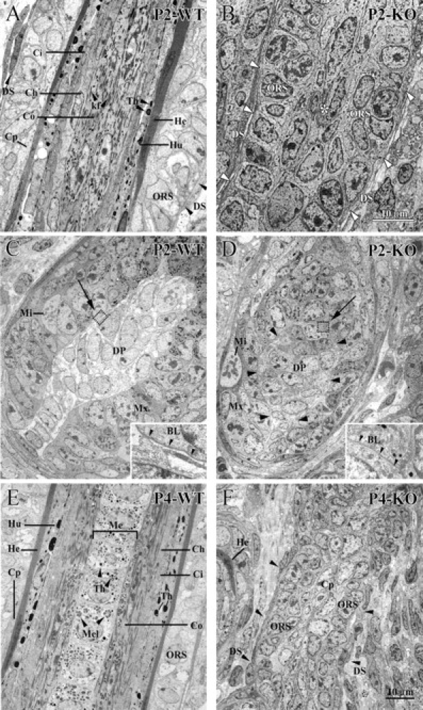
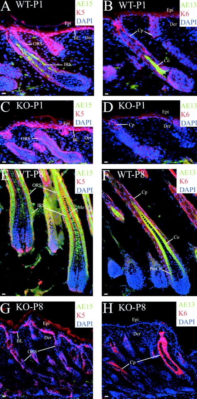
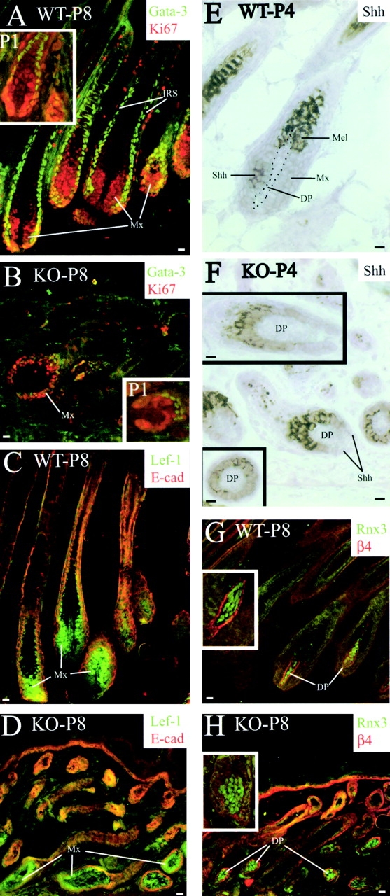
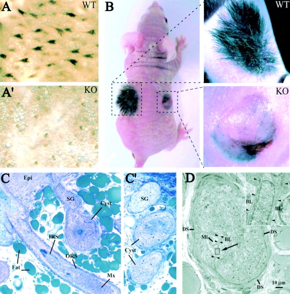
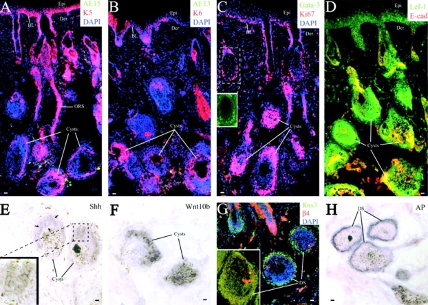
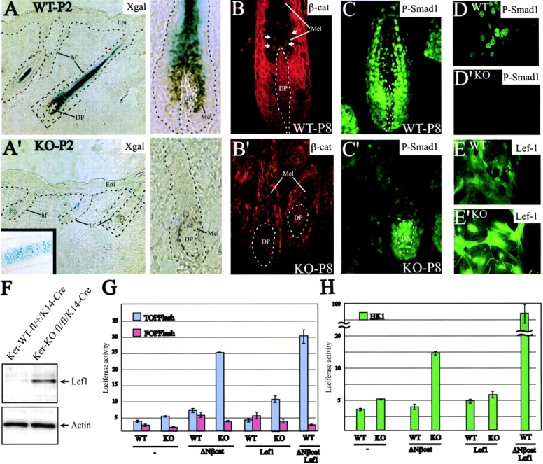
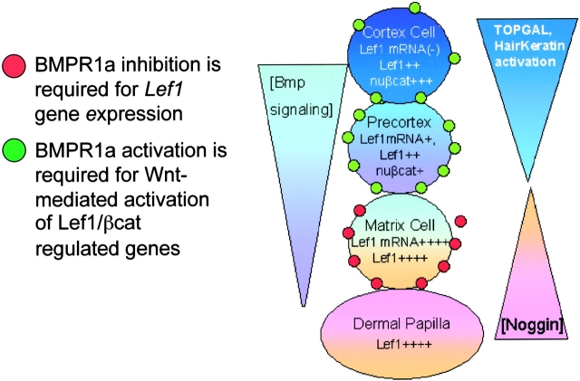
Using conditional gene targeting in mice, we show that BMP receptor IA is essential for the differentiation of progenitor cells of the inner root sheath and hair shaft. Without BMPRIA activation, GATA-3 is down-regulated and its regulated control of IRS differentiation is compromised. In contrast, Lef1 is up-regulated, but its regulated control of hair differentiation is still blocked, and BMPRIA-null follicles fail to activate Lef1/beta-catenin-regulated genes, including keratin genes. Wnt-mediated transcriptional activation can be restored by transfecting BMPRIA-null keratinocytes with a constitutively activated beta-catenin. This places the block downstream from Lef1 expression but upstream from beta-catenin stabilization. Because mice lacking the BMP inhibitor Noggin fail to express Lef1, our findings support a model, whereby a sequential inhibition and then activation of BMPRIA is necessary to define a band of hair progenitor cells, which possess enough Lef1 and stabilized beta-catenin to activate the hair specific keratin genes and generate the hair shaft.


Figures

Figure 1.
Figure 1.
Schematic of the hair follicle bulb. The diagram depicts the distinct cell layers of the hair follicle: ORS, outer root sheath; Cp, companion cell layer; IRS, inner root sheath; He, Henle's layer; Hu, Huxley's layer; Ci, cuticle of IRS; Ch, cuticle of hair shaft; Co, cortex of hair shaft; Me, medulla; and DP, dermal papilla. The illustration also summarizes patterns based on expression of antibodies against known markers that distinguish the complex programs of differentiation. Antibodies were against the proteins indicated except for the following: K6, Ab specific for the Cp K6; AE13, Ab against hair-specific keratins that are expressed in the Co; and AE15, Ab against trichohyalin, found in all three IRS layers and the Me.
Figure 2.
Figure 2.
Phenotype and genotype of K14-Cre, Bmpr1a fl/fl conditional knockout mice, which lack BMPRIA expression in skin epithelium. (A and B) Phenotypes of neonatal KO and WT pups. Mice lacked whiskers if their skin epithelium was null for Bmpr1a; those with a few aberrant whiskers (shown) were mosaic. (C) PCR analysis of DNAs isolated from dispase-separated epidermises of mice harboring the genotypes indicated. Primer sets used were diagnostic for: (top) both the floxed and WT alleles; (middle) the Cre-mediated deletion of exon 2; (bottom) the presence of the K14-Cre transgene. (D–F) 8-μm frozen skin sections of WT or KO animals at the ages indicated were subjected to indirect immunofluorescence with anti-BMPRIA and anti-K5. Color codings are according to the fluor tag of the secondary antibodies or DAPI (blue).
Figure 3.
Figure 3.
Histological abnormalities in Bmpr1a -null follicles. Matched P2 and P4 WT and Bmpr1a-null (KO) skin grafts were fixed, embedded in Epon, and sectioned (1 μm). Sections were stained with toluidine blue and subjected to light microscopic analysis. Mx, matrix; HS, hair shaft; Me, medulla; IRS, inner root sheath; ORS, outer root sheath; DP, dermal papilla; Co, cortex; epi, epidermis; Mu, muscle; and Mel, melanin granules (although skin epithelium does not make melanin granules, the Mx cells of the follicle and basal cells of the epidermis take up melanosomes from the cellular processes of the melanocytes). Note the lack of Me and paucity of IRS structures in the KO follicles. The paucity of fat in the dermis could be a reflection of malnutrition arising from oral complications. Bar, 20 μm, applies to all panels.
Figure 4.
Figure 4.
Ultrastructural abnormalities in Bmpr1 a-null follicles. Backskins of WT and KO animals were processed for transmission EM. All follicles are oriented with the skin surface at the top. (A) Sagittal section of the mid-segment of a P2 WT follicle, depicting the thin Henle's layer (He) with uniform density due to keratinization. This layer is flanked externally by the companion layer (Cp) and ORS and finally the dermal sheath (DS; arrowheads). Internally, the Henle's layer (He) is flanked by the trichohyalin granule (Th)–rich Huxley's layer (Hu) of the IRS, and the thin IRS cuticle (Ci). The hair shaft is internal to the IRS, and at this stage most shafts are composed only of two major layers: the cortex (Co) rich with keratin filaments (kf), and the cuticle of the hair shaft (Ch). (B) Sagittal sections from Bmpr1a-null P2 follicles. View from the mid-region above the follicle bulb. Note mostly ORS cells. Note the lack of hair shaft or IRS structures. Arrowheads show DS and asterisk denotes Cp. (C and D) Sagittal sections of bulb from a WT and KO P2 follicle. Note mitosis (Mi) in Mx cell. Note also the central strand of DP, whose cytoplasmic density is less than the surrounding matrix (Mx) of WT follicles. Note that distinctions between DP and Mx are less obvious in KO follicle, despite their clear separation by an intact basal lamina (BL). Boxed areas are magnified in the insets. Arrowheads point to BL. (E and F) P4, sagittal sections of mid-segments of a P4 WT (E) and KO (F) follicle. Note that at this age, most WT follicles display a well-developed hair shaft composed of an inner core of medulla (Me) cells with trichohyalin granules (Th) and melanin granules (Mel; arrowheads); otherwise the morphology is largely similar to P2. In contrast, note that the P4 KO follicle is still undeveloped, and appears more analogous to its P2 state. Bar in B is valid for A and B; bar in F is valid for C–F.
Figure 5.
Figure 5.
Defects in IRS and hair shaft differentiation in Bmpr1a -null skin epithelium. 8-μm frozen sections of P8 and P1 skins from Bmpr1a-null mice (KO) or their control littermates (WT) were subjected to indirect immunofluorescence using the antibodies indicated. Color coding is according to the secondary antibodies used: AE15, specific for all three layers of the IRS and medulla (Me); AE13, specific for the hair keratins of the cortex (Co) and precortex (Pre-Co); K6, specific for the K6 expressed in the companion layer (Cp); and DAPI, a fluorescent dye, which intercalates into DNA. Der, dermis; Epi, epidermis; BL, basal layer; ORS, outer root sheath; and K5, keratin 5. Bars, 10 μm.
Figure 6.
Figure 6.
Defects in transcriptional regulators of the IRS and hair shaft and epithelial–mesenchymal alterations in Bmpr1a -null skin epithelium. 8-μm frozen sections of P8 or P4 skins from Bmpr1a-null mice (KO) or control littermates (WT) were subjected to either indirect immunofluorescence using the antibodies indicated (color coding according to the secondary antibodies) or in situ hybridization. Antibodies/probes were: GATA-3, a differentiation regulator of Hu and cuticle layers of the IRS; Ki67, a marker of cycling (proliferating) cells; Lef1, specific for Mx, pre-Co, Co, and DP; E-cad, specific for E-cadherin, present throughout skin epithelium, but reduced in Mx and its progeny; a digoxygenin-labeled cRNA probe for Sonic hedgehog (Shh), thought to be a proliferation regulator in skin; Rnx3, Runx3, a transcriptional regulator found in DP; β4 integrin, specific for hemidesmosomes attached to an underlying basement membrane; and DAPI, a fluorescent dye, which intercalates into DNA. DP, dermal papilla; Mx, matrix; IRS, inner root sheath; and Mel, melanin granules. Bars: (A–D, G, and H) 10 μm; (E, F, and inset) 20 μm.
Figure 7.
Figure 7.
Bmpr1a-null skin grafts develop strikingly abnormal hairs. Full-thickness skins from WT and Bmpr1a-null (KO) P2 C57/Bl6 mice were grafted onto the left and right sides, respectively, of nu/nu mice. (A and A′) Skin of WT animals displayed black hairs, which protruded from the skin surface. In contrast, black melanin granule spots, indicative of underlying, developing hair placodes (pigmented) were visible, but no hairs were detected. Sectioning and staining of adjacent regions confirmed the paucity of BMPRIA in KO skin. (B) After engraftment, hairs (black) appeared in WT grafts but appeared absent in Bmpr1a-null grafts. By 24 d (shown), pelage hairs were only seen on WT grafts; KO grafted skin displayed a taut, shiny surface; the grayish tone was due to melanin granules from melanocytes present in the full thickness grafts. (C) Skins from 24-d grafts were fixed in glutaraldehyde, embedded in epon, and sectioned (1 μm). Sections were stained with toluidine blue. Note that normal follicles, replete with IRS and medulla, were seen in WT grafted skin (not depicted). Here, we focus on the cystlike follicular structures, prevalent in KO grafted skin. Some cysts emanated from the epidermis, indicating that these structures reflected aberrant folliculogenesis, rather than transverse sections of follicles. Note the presence of larger follicle, rare in KO skin. Sg, sebaceous gland; Epi, epidermis; ORS, outer root sheath; IRS, inner root sheath; and Mx, matrix. (D) Ultrastructural analyses of KO cyst. Note mitoses (Mi) in cysts, and also invaginations of DP-like cells (arrowheads). These pockets of dermal cells are surrounded by a basal lamina (BL; inset), which extends to and encases the cyst surface. DS, dermal sheath. The arrow indicates the area, which is magnified in the inset.
Figure 8.
Figure 8.
Biochemical analyses of cysts generated in grafted Bmpr1 a-null skin epithelium. 8-μm frozen sections of grafted P2 skins (24-d grafts) of Bmpr1a-null mice (KO) or their control littermates (WT) were subjected to indirect immunofluorescence or in situ hybridizations as indicated. Color coding is according to the secondary antibodies used: AE15, specific for IRS and Me; AE13, specific for hair keratins of the cortex; GATA-3 specific for the Huxley's and cuticle layers of the IRS (boxed area shows only the green channel to reveal a low level of GATA-3 expression in some cyst cells); Ki67, a marker of cycling (proliferating) cells; K6, specific for the K6 expressed in the Cp; Lef1, specific for the matrix, precortex, cortex, and DP (note its aberrant up-regulation in epidermis lacking BMPRIA); E-cad, specific for E-cadherin, present throughout skin epithelium, but reduced in the matrix; Runx3 and alkaline phosphatase (AP), present in DP cells; β4 integrin, specific for hemidesmosomes attached to an underlying basement membrane; DAPI, a fluorescent dye, which intercalates into DNA; Shh, a cRNA probe against sonic hedgehog (arrows denote Shh expression; boxed region is magnified to see expression); and Wnt10b, a cRNA probe against this Wnt, expressed in the follicle bulb of WT skin (Reddy et al., 2001). Note that larger brown grains are melanin granules. The DAPI channel was removed for the inset shown in G, to better visualize the internal β4-positive cells, which mark dermal invaginations. Epi, epidermis; Der, dermis; BL, basal layer; DS, dermal sheath; ORS, outer root sheath; and K5, keratin 5. Bars, 10 μm.
Figure 9.
Figure 9.
Failure to activate Wnt-responsive genes and accumulate nuclear β-catenin when BMPRIA signaling is blocked. (A and A') TOPGAL activity in postnatal follicles. Note activity in WT follicles, absent in Bmpr1a-null follicles. Inset shows control TOPGAL activity in cartilage, verifying TOPGAL activation in non-K14Cre expressing cells. Mel, melanin granules. (B and B') Nuclear β-catenin in postnatal (P8) WT follicles (double arrows), absent in KO follicles. (C and C') Immunoreactivity against nuclear phospho-Smad1 (induced upon BMPRIA activation) in developing IRS/hair in WT but not KO. (D–E′) Immunoreactivity of primary cultured keratinocytes to either antiphospho-Smad1 or anti-Lef1, as indicated. (F) Anti-Lef1 and anti–β-actin (control) immunoblot of lysates from WT and KO cells. (G and H) Effect of Bmpr1a ablation on activity of Wnt-regulated promoters. Two Wnt-responsive luciferase reporter genes were used for the work: TOPFlash (Korinek et al., 1997) and the murine hair keratin promoter equivalent, HK1Flash (Zhou et al., 1995; Merrill et al., 2001). The TOP promoter with a mutation in the TCF/Lef1 binding site (FOPFlash) was used as a control for binding specificity. Constructs were transiently transfected ± K14-ΔNβ-catenin (Gat et al., 1998) ± K14-Lef1 into keratinocytes, and 48 h after transfection, cells were lysed and processed for luciferase activity. CMV-renilla was used to control for transfection efficiency, and the results are the average of three independent experiments performed in triplicate, with the standard mean of deviation provided. Luciferase activities are given in arbitrary units relative to control. ≈, represents a gap in the scale of the histogram.
Figure 10.
Figure 10.
Model for how opposing signaling through BMPR1A controls Lef1/βcat signaling in stem cell lineage differentiation of the hair shaft. According to the model, in the postnatal follicle, epithelial stem cells are activated through interactions with DP to generate matrix cells (Cotsarelis et al., 1990). DP cells produce a noggin signal, which inhibits BMP signaling, leading to elevated Lef1 mRNA expression and proliferation, maintaining the cells in an undifferentiated state (Zhou et al., 1995; Botchkarev et al., 1999; Jamora et al., 2003). Despite the expression of Lef1 and some Wnts, hair cell progenitors do not activate Lef1/β-catenin when they are inhibited from BMPRIa activation (this paper). Whether the inhibition of BMP signaling prevents Wnt receptor expression (Soshnikova et al., 2003), prevents nuclear β-catenin from being stabilized, or blocks some earlier step in differentiation is unknown. However, matrix cells exposed to Noggin begin to express BMP4 (Kulessa et al., 2000), and in combination with their upward movement, the cells are likely to activate their BMPRIa receptor. The predicted outcome is attenuation of Lef1 mRNA expression, but a window of differentiation (the precortex) may exist where cells have stabilized nuclear β-catenin and still have sufficient Lef1 protein to activate Wnt-mediated genes. Natural Wnt target genes include the hair-specific keratin genes and Foxn1, another transcriptional regulator of hair keratin gene transcription (Zhou et al., 1995; DasGupta and Fuchs, 1999; Merrill et al., 2001; Andl et al., 2002; Balciunaite et al., 2002). Wnt/Lef1-mediated active chromatin may further act to stabilize Lef1 to enable target genes to remain active in the cortex after Lef1 mRNA is down-regulated.

Similar articles

Cited by

References

    1. Ahn, K., Y. Mishina, M.C. Hanks, R.R. Behringer, and E.B. Crenshaw, III. 2001. BMPR-IA signaling is required for the formation of the apical ectodermal ridge and dorsal-ventral patterning of the limb. Development. 128:4449–4461. - PubMed
    1. Alonso, L., and E. Fuchs. 2003. Stem cells in the skin: waste not, Wnt not. Genes Dev. 17:1189–1200. - PubMed
    1. Andl, T., S.T. Reddy, T. Gaddapara, and S.E. Millar. 2002. WNT signals are required for the initiation of hair follicle development. Dev. Cell. 2:643–653. - PubMed
    1. Balciunaite, G., M.P. Keller, E. Balciunaite, L. Piali, S. Zuklys, Y.D. Mathieu, J. Gill, R. Boyd, D.J. Sussman, and G.A. Hollander. 2002. Wnt glycoproteins regulate the expression of Foxn1, the gene defective in nude mice. Nat. Immunol. 3:1102–1108. - PubMed
    1. Barrow, J.R., K.R. Thomas, O. Boussadia-Zahui, R. Moore, R. Kemler, M.R. Capecchi, and A.P. McMahon. 2003. Ectodermal Wnt3/beta-catenin signaling is required for the establishment and maintenance of the apical ectodermal ridge. Genes Dev. 17:394–409. - PMC - PubMed

Publication types

MeSH terms

Substances