. 2003 Nov 10;163(3):625-35.
doi: 10.1083/jcb.200302152.

Processing by proprotein convertases is required for glypican-3 modulation of cell survival, Wnt signaling, and gastrulation movements

Affiliations

Processing by proprotein convertases is required for glypican-3 modulation of cell survival, Wnt signaling, and gastrulation movements

Bart De Cat et al. J Cell Biol. .

Abstract

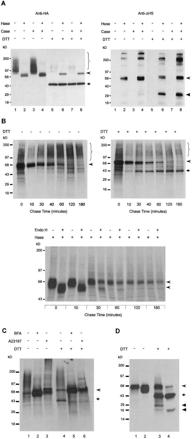
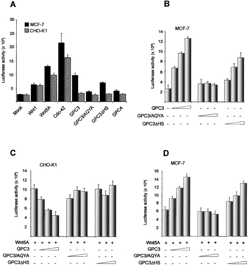
Glypican (GPC)-3 inhibits cell proliferation and regulates cell survival during development. This action is demonstrated by GPC3 loss-of-function mutations in humans and mice. Here, we show that the GPC3 core protein is processed by a furinlike convertase. This processing is essential for GPC3 modulating Wnt signaling and cell survival in vitro and for supporting embryonic cell movements in zebrafish. The processed GPC3 core protein is necessary and sufficient for the cell-specific induction of apoptosis, but in vitro effects on canonical and noncanonical Wnt signaling additionally require substitution of the core protein with heparan sulfate. Wnt 5A physically associates only with processed GPC3, and only a form of GPC3 that can be processed by a convertase is able to rescue epiboly and convergence/extension movements in GPC3 morphant embryos. Our data imply that the Simpson-Golabi-Behmel syndrome may in part result from a loss of GPC3 controls on Wnt signaling, and suggest that this function requires the cooperation of both the protein and the heparan sulfate moieties of the proteoglycan.


Figures

Figure 1.
Figure 1.
Posttranslational modification of GPC3 in MDCK cells. Total proteoglycan or GPC3 isolated from stable transfectant cells was treated with heparitinase (Hase), chondroitinase ABC (Case), or endoglycosidase H (Endo H) as indicated, and fractionated by SDS-PAGE under reducing (DTT) or nonreducing conditions. (A) Two-subunit structure of GPC3. Western blots of total proteoglycan extract, using rat anti-HA mAb 3F10 to detect GPC3, and anti-ΔHS mAb 3G10 to detect the desaturated uronates that are generated by heparitinase and that remain in association with the core proteins. (B) Time course of the GPC3 maturation. Cells were pulse labeled with [35S]cysteine-methionine for 10 min and chased for the indicated time periods. GPC3 from cell lysates, isolated using anti-HA antibody, was detected by autoradiography. Unreduced (left) and reduced (right) non enzyme-treated samples (top), and reduced glycosidase-treated samples (bottom) reveal that the HS substitution, proteolytic processing, and Endo H–resistant N-glycosylation of GPC3 follow similar time courses. (C) Inhibition of GPC3 processing by blocking ER export or calcium depletion. Cells were incubated for 6 h with (+) or without (−) 30 μM BFA or 2 μM A23187, pulse-labeled for 10 min, and chased for 60 min. GPC3 immunopurified from cell lysate was detected by autoradiography. (D) Endoproteolytic processing of GPC3ΔHS. Labeled HA-GPC3ΔHS was immunoprecipitated from the cell lysate (lanes 1 and 3) and the conditioned medium (lanes 2 and 4). Braces show glycanated GPC3, curved arrowheads indicate the GPC3 core protein, arrows indicates the ∼40-kD NH2-terminal (HA-tagged) α-subunit, and arrowheads indicate the COOH-terminal β subunit that is separated from the α-subunit by reduction. Numbers on the left represent molecular mass markers.
Figure 2.
Figure 2.
Identification of the cleavage site in GPC3. (A) Schematic representation of GPC3. The GPC3 domains are depicted as shaded boxes. Lines within the boxes denote cysteine residues of the CRD, as conserved in all glypicans (GPCs). Open arrowheads denote potential N-glycosylation sites; closed arrowheads indicate the positions of the HS attachment sites. The curved arrowhead indicates the position of the proteolytic cleavage site. This latter site occurs in a region of the CRD that shows low sequence similarity to corresponding regions in other GPCs. Amino acid substitutions, as indicated in bold, were introduced into this unconserved region (UR) and in the HS substitution domain (GAG), either alone or in combination. SP, signal peptide for membrane translocation; HA, hemagglutinin tag; CRD, cysteine-rich domain; GPI, signal peptide for glypiation. (B) Endoproteolytic processing of the GPC3 mutants. Whole extracts of CHO-K1 cells transiently transfected with a control vector, wild-type GPC3 or mutant forms of GPC3, were fractionated by SDS-PAGE under reducing conditions, and analyzed by Western blotting using rat anti-HA mAb 3F10. (C–F) Subcellular localization of GPC3 and GPC3/AQYA. Horizontal confocal sections of stable MDCK clones expressing GPC3 (top) or GPC3/AQYA (bottom), fixed and stained with rat anti-HA mAb 3F10, without (left) or after permeabilization (right). Bar, 10 μm. (G) Glycanation and endoproteolytic processing of GPC3/AQYA. Proteoglycan isolated from stably transfected MDCK cells was treated with the indicated enzymes, fractionated by SDS-PAGE under reducing and nonreducing conditions, and analyzed by Western blotting using rat anti-HA mAb 3F10. (H) Maturation of GPC3/AQYA. Stable transfectant MDCK cells were pulse labeled with [35S]cysteine-methionine for 10 min and chased for the indicated time periods. Mutant GPC3, immunopurified from cell lysate, was treated with the indicated enzymes, fractionated by SDS-PAGE under reducing conditions, and detected by auroradiography. Braces show glycanated GPC3/AQYA; curved arrowheads indicate the GPC3/AQYA core protein. Numbers on the left represent molecular mass markers.
Figure 2.
Figure 2.
Identification of the cleavage site in GPC3. (A) Schematic representation of GPC3. The GPC3 domains are depicted as shaded boxes. Lines within the boxes denote cysteine residues of the CRD, as conserved in all glypicans (GPCs). Open arrowheads denote potential N-glycosylation sites; closed arrowheads indicate the positions of the HS attachment sites. The curved arrowhead indicates the position of the proteolytic cleavage site. This latter site occurs in a region of the CRD that shows low sequence similarity to corresponding regions in other GPCs. Amino acid substitutions, as indicated in bold, were introduced into this unconserved region (UR) and in the HS substitution domain (GAG), either alone or in combination. SP, signal peptide for membrane translocation; HA, hemagglutinin tag; CRD, cysteine-rich domain; GPI, signal peptide for glypiation. (B) Endoproteolytic processing of the GPC3 mutants. Whole extracts of CHO-K1 cells transiently transfected with a control vector, wild-type GPC3 or mutant forms of GPC3, were fractionated by SDS-PAGE under reducing conditions, and analyzed by Western blotting using rat anti-HA mAb 3F10. (C–F) Subcellular localization of GPC3 and GPC3/AQYA. Horizontal confocal sections of stable MDCK clones expressing GPC3 (top) or GPC3/AQYA (bottom), fixed and stained with rat anti-HA mAb 3F10, without (left) or after permeabilization (right). Bar, 10 μm. (G) Glycanation and endoproteolytic processing of GPC3/AQYA. Proteoglycan isolated from stably transfected MDCK cells was treated with the indicated enzymes, fractionated by SDS-PAGE under reducing and nonreducing conditions, and analyzed by Western blotting using rat anti-HA mAb 3F10. (H) Maturation of GPC3/AQYA. Stable transfectant MDCK cells were pulse labeled with [35S]cysteine-methionine for 10 min and chased for the indicated time periods. Mutant GPC3, immunopurified from cell lysate, was treated with the indicated enzymes, fractionated by SDS-PAGE under reducing conditions, and detected by auroradiography. Braces show glycanated GPC3/AQYA; curved arrowheads indicate the GPC3/AQYA core protein. Numbers on the left represent molecular mass markers.
Figure 3.
Figure 3.
Identification of PCs as GPC3-processing enzymes. (A) PC-dependent processing in CHO-K1 cells. CHO-K1 cells were transfected with empty expression vector alone (lane 1), with GPC3 or GPC3ΔHS and empty vector, or with GPC3 or GPC3ΔHS, and with an α1-PDX expression vector. Total cell extracts were fractionated by SDS-PAGE under reducing conditions and analyzed by Western blotting using anti-HA mAb. (B) Furin-mediated processing in RPE.40 cells. RPE.40 cells were transfected with empty vector, GPC3 or GPC3/AQYA, alone or in combination with a furin expression vector. Western blot analysis of reduced samples using anti-HA antibody. (C) PC-mediated processing in RPE.40 cells. RPE.40 cells were transfected with empty vector only (lane 1) or with GPC3 together with LPC, PC6A, PC6B, and PACE4. Western blot analysis of reduced samples using anti-HA antibody. (D) In vitro digestion of GPC3 with recombinant furin. GPC3 was immunoprecipitated from BFA-treated metabolically labeled transfectant MDCK cells. The immunoprecipitates were mock-digested or digested with recombinant furin, fractionated by SDS-PAGE under reducing conditions, and detected by autoradiography. The curved arrowhead indicates the core protein, the arrow indicates the α-subunit, and the arrowhead indicates the COOH-terminal β subunit. Numbers on the left represent molecular mass markers.
Figure 4.
Figure 4.
GPC3-induced apoptosis. (A and B) Apoptosis depends on GPC3 processing. MCF-7 cells were transiently transfected with a β-galactosidase expression vector and a fivefold excess of control vector, or vectors encoding GPC3, GPC3/AQYA, or GPC3ΔHS. (A) Apoptosis scored by nuclear morphology. The results (mean ± SEM) are shown as a percentage of apoptotic cells (the total number of scored cells taken as 100%). (B) Apoptosis scored by cell death ELISA assay. The results (mean ± SEM) are shown as fold increase in apoptosis, compared with cells transfected with control vector. (C) Processed GPC3 activates JNK. MCF-7 cells were transfected as in A and B. Normalized cell extracts were analyzed by Western blotting, using either anti–phospho-MAPK antibodies or the respective anti-MAPK antibodies. Total cell extracts from UV-treated NIH/3T3 cells were taken as positive control. (D) Apoptosis depends on JNK activation. MCF-7 cells were triple transfected with β-galactosidase expression vector, pcDNA3.1 containing either a dominant-negative MKK4 construct or no insert, and a control vector or a vector encoding GPC3. Apoptosis was measured 48 h after transfection by the cell death ELISA assay, as in B.
Figure 5.
Figure 5.
Processed GPC3 modulates Wnt-initiated canonical signaling. (A) Activation of β-catenin/TCF-dependent transcription by Wnts and Wnt signal transduction components. CHO-K1 cells were transfected with 0.2 μg of the TOPFLASH or FOPFLASH reporter plasmid, 0.2 μg of β-galactosidase expression vector, and 0.4 μg of the indicated expression plasmids. (B and C) GPC3 overexpression inhibits Wnt signaling, upstream of Dishevelled. CHO-K1 cells were transfected with 0.2 μg of the TOPFLASH reporter plasmid, 0.2 μg of β-galactosidase vector, and 0.4 μg Wnt1, β-cat, or DVL expression vector, along with (from left to right) either 0.2, 0.4, or 0.8 μg of GPC3 or mutant forms of GPC3, as indicated. Empty pDisplay plasmid was added to equalize the total amount of DNA used for transfection. (D) Inhibition of Wnt signaling depends on PC processing. RPE.40 cells were transfected with 0.2 μg of the TOPFLASH or FOPFLASH reporter plasmid, 0.2 μg of β-galactosidase expression vector, and 0.4 μg of furin expression vector, along with varying amounts of GPC3 and empty pDisplay plasmid, as in B and C. Data represent mean ± standard error.
Figure 6.
Figure 6.
Processed GPC3 modulates noncanonical Wnt signaling. (A) Activation of AP-1–dependent transcription by Wnts and GPC3. MCF-7 or CHO-K1 cells were transfected with 0.2 μg of the AP-1–responsive reporter and 0.2 μg of β-galactosidase expression vector, along with 0.4 μg of the indicated expression plasmids. (B) GPC3 dose-dependent activation of AP-1 in MCF-7 cells depends on the processed core protein. MCF-7 cells were transfected with AP-1 reporter plasmid and β-galactosidase (as in A), and (from left to right) 0.2, 0.4, or 0.8 μg of GPC3 or its mutants, as indicated. Empty pDisplay plasmid was added to equalize the total amount of DNA used for transfection. (C and D) GPC3 effects on noncanonical Wnt signaling. CHO-K1 cells or MCF-7 cells were transfected with the AP-1–responsive reporter and β-galactosidase (as in A) and 0.4 μg of Wnt5A expression vector (only 0.2 μg for MCF-7 cells), along with varying amounts of GPC3 or its mutants, and empty pDisplay plasmid (as in B). Wnt inhibition in CHO cells depends on processing and on HS substitution. In MCF-7 cells, processed proteoglycan or core protein further enhances AP-1–dependent transcription. (E) Inhibition of noncanonical Wnt signaling depends on PC processing. RPE.40 cells were transfected with 0.2 μg of the AP-1–responsive reporter, 0.2 μg of β-galactosidase expression vector, 0.4 μg of furin expression plasmid along with varying amounts of GPC3 and empty pDisplay plasmid (as in B). (A–E) Data represent mean ± standard error. (F) Wnt–GPC3 association depends on processing and on HS substitution. MCF-7 cells were cotransfected with HA-tagged Wnt5A or Wnt7A in combination with HA-tagged wild-type GPC3, GPC3/AQYA, GPC3ΔHS, or GPC4. Immunoprecipitates obtained with anti-GPC3/4 polyclonal antibodies were fractionated by SDS-PAGE under reducing conditions, and analyzed by Western blotting, using monoclonal anti-HA antibody. The positive controls are total extracts from Wnt5A/7A-transfected cells. Note that like GPC3, GPC4 is also composed of two disulfide-linked subunits. The upper smear (brace) represents the glycanated form of GPC3. The curved arrowhead indicates the unprocessed core protein, and the arrow indicates the α-subunit. The open arrowhead points to the HA-tagged form of Wnt5A, and the asterisk denotes the IgG heavy chain. Numbers represent molecular mass markers.
Figure 6.
Figure 6.
Processed GPC3 modulates noncanonical Wnt signaling. (A) Activation of AP-1–dependent transcription by Wnts and GPC3. MCF-7 or CHO-K1 cells were transfected with 0.2 μg of the AP-1–responsive reporter and 0.2 μg of β-galactosidase expression vector, along with 0.4 μg of the indicated expression plasmids. (B) GPC3 dose-dependent activation of AP-1 in MCF-7 cells depends on the processed core protein. MCF-7 cells were transfected with AP-1 reporter plasmid and β-galactosidase (as in A), and (from left to right) 0.2, 0.4, or 0.8 μg of GPC3 or its mutants, as indicated. Empty pDisplay plasmid was added to equalize the total amount of DNA used for transfection. (C and D) GPC3 effects on noncanonical Wnt signaling. CHO-K1 cells or MCF-7 cells were transfected with the AP-1–responsive reporter and β-galactosidase (as in A) and 0.4 μg of Wnt5A expression vector (only 0.2 μg for MCF-7 cells), along with varying amounts of GPC3 or its mutants, and empty pDisplay plasmid (as in B). Wnt inhibition in CHO cells depends on processing and on HS substitution. In MCF-7 cells, processed proteoglycan or core protein further enhances AP-1–dependent transcription. (E) Inhibition of noncanonical Wnt signaling depends on PC processing. RPE.40 cells were transfected with 0.2 μg of the AP-1–responsive reporter, 0.2 μg of β-galactosidase expression vector, 0.4 μg of furin expression plasmid along with varying amounts of GPC3 and empty pDisplay plasmid (as in B). (A–E) Data represent mean ± standard error. (F) Wnt–GPC3 association depends on processing and on HS substitution. MCF-7 cells were cotransfected with HA-tagged Wnt5A or Wnt7A in combination with HA-tagged wild-type GPC3, GPC3/AQYA, GPC3ΔHS, or GPC4. Immunoprecipitates obtained with anti-GPC3/4 polyclonal antibodies were fractionated by SDS-PAGE under reducing conditions, and analyzed by Western blotting, using monoclonal anti-HA antibody. The positive controls are total extracts from Wnt5A/7A-transfected cells. Note that like GPC3, GPC4 is also composed of two disulfide-linked subunits. The upper smear (brace) represents the glycanated form of GPC3. The curved arrowhead indicates the unprocessed core protein, and the arrow indicates the α-subunit. The open arrowhead points to the HA-tagged form of Wnt5A, and the asterisk denotes the IgG heavy chain. Numbers represent molecular mass markers.
Figure 7.
Figure 7.
Interference with GPC3 expression disrupts gastrulation movements in zebrafish. (A) Predicted domain structure of GPC3 and similarities to GPC3 sequences from other vertebrates. (B) Endoproteolytic processing of the zebrafish GPC3. Whole extracts of CHO-K1 cells transiently transfected with wild-type zebrafish GPC3 or mutant forms, designed not to interact with the morpholinos (GPC3*) and not to be proteolytically processed (GPC3*/AVSA), were analyzed as described in Fig. 2 B. The upper smear (brace) represents the glycanated form of GPC3. The curved arrowhead indicates the unprocessed core protein, and the arrow indicates the α-subunit. Numbers represent molecular mass markers. (C) Expression of GPC3 at 50% epiboly spreads throughout the animal pole. Detection by in situ hybridization, lateral view with animal pole up. (D–M) Analysis of the GPC3 morphant phenotype at 10 hpf. (D–G) Lateral views of living embryos injected with control mispair (5-mis MO) and antisense morpholinos (MO). Animal pole is to the left and dorsal is up. Note the arrest of blastoderm movement toward the vegetal pole (brace). (H–M) Dorsal views of control (H, J, and L) and morphant (I, K, and M) embryos. The expressions of three marker genes, indicated at top right, are analyzed by in situ hybridization. Animal pole is to the left. No tail (ntl) staining reveals marked shortening (arrows), thickening (curved arrowheads), and undulations of the notochord in the morphants. The shape of the neuroectoderm is outlined by the expression of distal-less3 (dlx3). Note the broadened (curved arrowheads) and shortened neural plate. Widening of paraxial protocadherin (papc) expression reflects a reduced convergence of presomitic mesoderm. (N–P) Correction of the effects of antisense morpholino MO1 by coinjection of GPC3 mRNA lacking MO-target sequences. Injection of MO1 causes gastrulation defects (less than 80% epiboly at 10 hpf), and the majority of the embryos are dead by 12 hpf. GPC3* mRNA rescues gastrulation and embryo survival (N), and corrects the expression pattern of the ntl marker gene (O). GPC3*/AVSA mRNA, encoding a GPC3 protein that cannot be processed, fails to do so (N and P). Results (means and standard errors) of three separate experiments (n, total number of embryos analyzed). Bars, 250 μm.

Similar articles

Cited by

References

    1. Baeg, G.H., X. Lin, N. Khare, S. Baumgartner, and N. Perrimon. 2001. Heparan sulfate proteoglycans are critical for the organization of the extracellular distribution of Wingless. Development. 128:87–94. - PubMed
    1. Cano-Gauci, D.F., H.H. Song, H. Yang, C. McKerlie, B. Choo, W. Shi, R. Pullano, T.D. Piscione, S. Grisaru, S. Soon, et al. 1999. Glypican-3–deficient mice exhibit developmental overgrowth and some of the abnormalities typical of Simpson–Golabi–Behmel syndrome. J. Cell Biol. 146:255–264. - PMC - PubMed
    1. Chen, R.L., and A.D. Lander. 2001. Mechanisms underlying preferential assembly of heparan sulfate on glypican-1. J. Biol. Chem. 276:7507–7517. - PubMed
    1. Chiao, E., P. Fisher, L. Crisponi, M. Deiana, I. Dragatsis, D. Schlessinger, G. Pilia, and A. Efstratiadis. 2002. Overgrowth of a mouse model of the Simpson–Golabi–Behmel syndrome is independent of IGF signaling. Dev. Biol. 243:185–206. - PubMed
    1. Creemers, J.W., D. Ines Dominguez, E. Plets, L. Serneels, N.A. Taylor, G. Multhaup, K. Craessaerts, W. Annaert, and B. De Strooper. 2001. Processing of beta-secretase by furin and other members of the proprotein convertase family. J. Biol. Chem. 276:4211–4217. - PubMed

Publication types

MeSH terms