. 2003 Dec;77(23):12466-78.
doi: 10.1128/jvi.77.23.12466-12478.2003.

Modulation of minute virus of mice cytotoxic activities through site-directed mutagenesis within the NS coding region

Affiliations

Modulation of minute virus of mice cytotoxic activities through site-directed mutagenesis within the NS coding region

Laurent Daeffler et al. J Virol. 2003 Dec.

Abstract

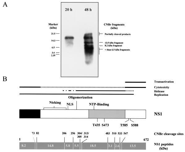
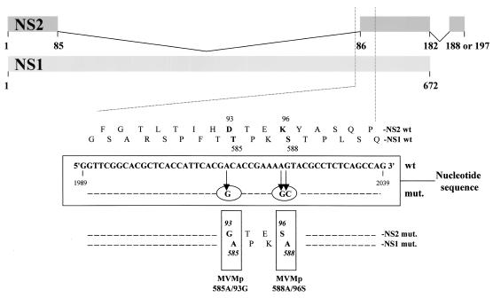
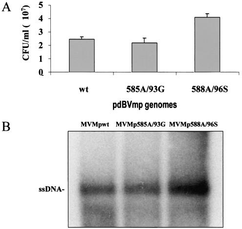
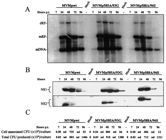
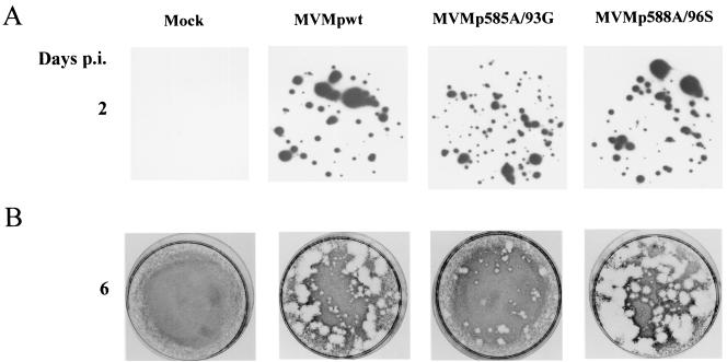
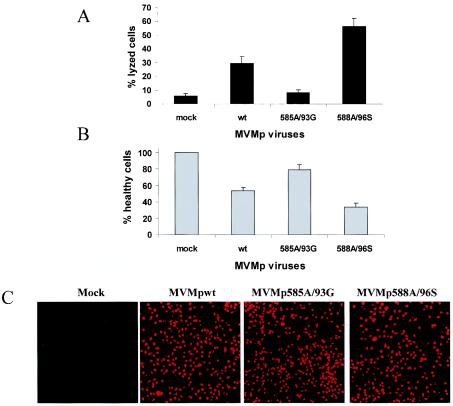
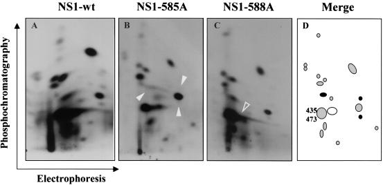
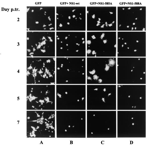
Late in infection, parvovirus minute virus of mice (MVMp) induces the lysis of mouse A9 fibroblasts. This effect depends on the large nonstructural phosphoprotein NS1, which plays in addition a major role in viral DNA replication and progeny particle production. Since the NS1 C-terminal region is subjected to late phosphorylation events and protein kinase C (PKC) family members regulate NS1 replicative activities, the present study was conducted to determine the impact of PKCs on NS1 cytotoxic functions. To this end, we performed site-directed mutagenesis, substituting alanine residues for two consensus PKC-phosphorylation sites located within the NS1 C-terminal region, T585 and S588. Although these substitutions had no detectable effect on virus multiplication in a single-round infection, the NS1-585A mutant virus was significantly less toxic to A9 cells than wild-type MVMp, whereas the NS1-588A mutant virus was endowed with a higher killing potential. These alterations correlated with specific changes in the late phosphorylation pattern of the mutant NS1 proteins compared to the wild-type polypeptide. Since the mutations introduced in this region of the viral genome also made changes in the minor nonstructural protein NS2, a contribution of this polypeptide to the above-mentioned phenotypes of mutant viruses cannot be excluded at present. However, the involvement of NS1 in these phenotypes was directly supported by the respective reduced and enhanced capacity of NS1-585A and NS1-588A recombinant proteins for inducing morphological alterations and cell detachment in transfected A9 cultures. Altogether, these data suggest that late-occurring phosphorylation of NS1 specifically regulates the cytotoxic functions of the viral product and that residues T585 and S588 contribute to this control in an antagonistic way.


Figures

FIG. 1.
FIG. 1.
Determination of NS1 regions phosphorylated late in infection. (A) CNBr cleavage pattern of wild-type NS1 polypeptides that were metabolically 32P labeled 20 h (productive phase) or 48 h (cytopathic phase) after the release of serum-starved infected A9 cells into S phase. (B) Alignment of the functional map of NS1 with the predicted CNBr cleavage pattern of the polypeptide. Functional map: NLS, nuclear localization signal; NTP, nucleotide binding site; T435 and S473, previously determined PKCλ phosphorylation sites; T585 and S588, consensus PKC phosphorylation sites chosen as targets for mutagenesis in the present study. CNBr map: predicted cleavage sites (amino acid numbers) and sizes of generated peptides (in kilodaltons).
FIG. 2.
FIG. 2.
Generation of MVMp585A/93G and MVMp588A/96S mutant viruses by site-directed mutagenesis. The upper diagram represents the NS1 and NS2 proteins, according to the splicing pattern of the respective transcripts and gives the amino acid sequence of both proteins in the region serving as a target for mutagenesis. The corresponding nucleic acid sequence is given in the central frame, together with the mutations (circled) introduced through chimeric PCRs to produce MVMp585A/93G and MVMp588A/96S, respectively. The resulting amino acid substitutions in NS1 and NS2 are shown at the bottom (boldface residues in the mutant versus wild-type sequences).
FIG. 3.
FIG. 3.
Production of wild-type and mutant MVMp virus stocks. Viruses were collected from 293T cells 3 days after transfection with either wild-type (wt; pdBMVp) or mutant (pMVMp585A/93G [585A/93G] and pMVMp588A/96S [588A/96S]) MVMp DNA clones. (A) Virus yields were determined by DNA hybridization assays after infection of A9 cell monolayers and are expressed in replicative CFU/ml of stock (average values and standard deviation bars from five independent transfection experiments). (B) Aliquots of the primary virus stocks (107 CFU) were compared for their contents in full virions as determined by Southern blotting detection of single-stranded genomic DNA (ssDNA).
FIG. 4.
FIG. 4.
Wild-type and mutant MVMp virus replication in a single-round infection. A9 cultures were infected with 5 CFU of primary stocks of either wild-type (wt) or mutant (585A/93G or 588A/96S) MVMp virus/cell and further incubated in culture medium supplemented with neuraminidase (from 6 h p.i. on) to prevent new rounds of infection. Cells were harvested at the indicated times p.i. and processed for the analysis of virus replication. (A) Southern blotting analysis of viral DNA intermediates. mRF, monomer RF; dRF, dimer RF; ssDNA, single-stranded genomic DNA. (B) Western blotting analysis of viral nonstructural protein production. The brackets encompass the phosphorylated and un(der)phosphorylated forms of the respective polypeptides. (C) Titration of progeny viruses by replication center assays on A9 indicator cell monolayers. Lane 1, amount of progeny viruses in adherent A9 monolayers; lane 2, total amount of viral particles present in adherent and detached cells, as well as in the culture medium; nd, not determined.
FIG. 5.
FIG. 5.
Impact of 585A/93G and 588A/96S mutations on the ability of MVMp viruses to form lysis plaques in A9 cell monolyaers. A9 indicator cells were infected with primary stocks of either wild-type or mutant MVMp at a multiplicity of 10-3 CFU/cell and further processed for hybridization (replicative centers [A]) and plaque (B) assays. The figure is representative of five experiments performed with five different stocks of each virus.
FIG. 6.
FIG. 6.
Varying cytotoxicity of wild-type and mutant MVMp viruses. The indicated virus stocks were tested for their ability to jeopardize the survival of A9 fibroblasts after infection at a multiplicity of 10 CFU/cell and further incubation for 3 days. (A) The virus lytic activity was measured through quantification of the cytoplasmic LDH released into the medium, expressed as a percentage of total LDH (determined after lysis of the whole culture with detergent). (B) The cell-killing activity of the different viruses was assessed by determining the reduction in the number of living cells (still able to reduce MTT) in the infected population, a value expressed as the percentage of the value for mock-treated cultures. The same cultures were used for panels A and B. The data shown are means with standard deviation bars from 10 independent experiments carried out each in triplicate. (C) NS1 produced by wild-type and mutant MVMp viruses was detected by indirect immunofluorescence in parallel cultures. Images were obtained by using a ×16 magnification lens.
FIG. 7.
FIG. 7.
Alteration of the late phosphorylation pattern of T585A and S588A mutant forms of NS1. A9 cultures arrested in G0/G1 were infected with indicated viruses, released into the cell cycle for 48 h and metabolically 32P labeled. NS1 proteins were isolated, purified, and processed for the analysis of tryptic phosphopeptides by two-dimensional electrophoresis and/or chromatography. Compared to wild-type NS1 (A), the NS1-585A (B) and NS1-588A (C) mutants lacked specific phosphorylation event(s), as evidenced by the disappearance of distinct phosphopeptide(s) (arrowheads). (D) Schematic representation of the “late” NS1 tryptic phosphopeptide pattern, in which the spots absent from the NS1-585A and NS1-588A maps are indicated with filled black and open circles, respectively, and the previously assigned PKCλ phosphorylation targets T435 and S473 are positioned.
FIG. 8.
FIG. 8.
Induction of morphological alterations by wild-type and mutant NS1 proteins. A9 cells competent for transfection with plasmid pP4-NS1x-P4-GFP were identified on the basis of GFP fluorescence and examined over a period of 7 days to detect phenotypic alterations induced by wild-type (B), 585A (C), or 588A (D) NS1. Parallel cultures transfected with plasmid pP4-GFP served as negative control (A). Images of GFP-expressing cells were obtained by using a ×16 magnification lens.

References

    1. Anouja, F., R. Wattiez, S. Mousset, and P. Caillet-Fauquet. 1997. The cytotoxicity of the parvovirus minute virus of mice nonstructural protein NS1 is related to changes in the synthesis and phosphorylation of cell proteins. J. Virol. 71:4671-4678. - PMC - PubMed
    1. Bodendorf, U., C. Cziepluch, J. C. Jauniaux, J. Rommelaere, and N. Salomé. 1999. Nuclear export factor CRM1 interacts with nonstructural proteins NS2 from parvovirus minute virus of mice. J. Virol. 73:7769-7779. - PMC - PubMed
    1. Brandenburger, A., D. Legendre, B. Avalosse, and J. Rommelaere. 1990. NS-1 and NS-2 proteins may act synergistically in the cytopathogenicity of parvovirus MVMp. Virology 174:576-584. - PubMed
    1. Brockhaus, K., S. Plaza, D. J. Pintel, J. Rommelaere, and N. Salomé. 1996. Nonstructural proteins NS2 of minute virus of mice associate in vivo with 14-3-3 protein family members. J. Virol. 70:7527-7534. - PMC - PubMed
    1. Caillet-Fauquet, P., M. Perros, A. Brandenburger, P. Spegelaere, and J. Rommelaere. 1990. Programmed killing of human cells by means of an inducible clone of parvoviral genes encoding nonstructural proteins. EMBO J. 9:2989-2995. - PMC - PubMed

Publication types

LinkOut - more resources