. 2003 Dec;77(23):12562-71.
doi: 10.1128/jvi.77.23.12562-12571.2003.

Mutations within the P2 domain of norovirus capsid affect binding to human histo-blood group antigens: evidence for a binding pocket

Affiliations

Mutations within the P2 domain of norovirus capsid affect binding to human histo-blood group antigens: evidence for a binding pocket

Ming Tan et al. J Virol. 2003 Dec.

Erratum in

  • J Virol. 2004 Mar;78(6):3200

Abstract

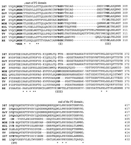
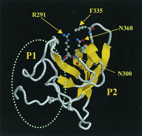
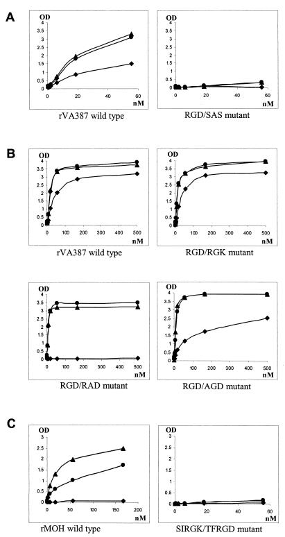
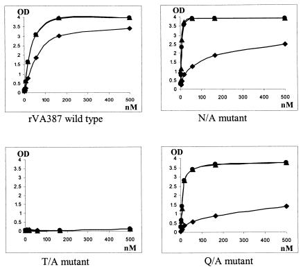
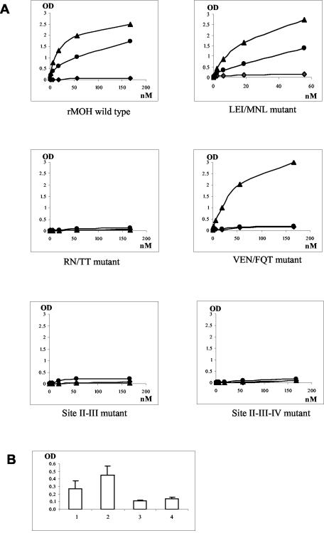
Noroviruses (NORs) are an important cause of acute gastroenteritis. Recent studies of NOR receptors showed that different NORs bind to different histo-blood group antigens (HBGAs), and at least four distinct binding patterns were observed. To determine the structure-function relationship for NORs and their receptors, two strains representing two of the four binding patterns were studied. Strain VA387 binds to HBGAs of A, B, and O secretors, whereas strain MOH binds to HBGAs of A and B secretors only. Using multiple sequence alignments, homology modeling, and structural analysis of NOR capsids, we identified a plausible "pocket" in the P2 domain that may be responsible for binding to HBGA receptors. This pocket consists of a conserved RGD/K motif surrounded by three strain-specific hot spots (N(302), T(337), and Q(375) for VA387 and N(302), N(338), and E(378) for MOH). Subsequent mutagenesis experiments demonstrated that all four sites played important roles in binding. A single amino acid mutation at T(337) (to A) in VA387 or a double amino acid mutation at RN(338) (to TT) in MOH abolished binding completely. Change of the entire RGD motif to SAS abolished binding in case of VA387, whereas single amino acid mutations in that motif did not have an apparent effect on binding to A and B antigens but decreased binding to H antigen. Multiple mutations at the RGK motif of MOH (SIRGK to TFRGD) completely knocked out the binding. Mutation of N(302) or Q(375) in VA387 affected binding to type O HBGA only, while switch mutants with three amino acid changes at either site from MOH to VA387 resulted in a weak binding to type O HBGAs. A further switch mutant with three amino acid changes at E(378) from MOH to VA387 diminished the binding to type A HBGA only. Taken together, our data indicate that the binding pocket likely exists on NOR capsids. Direct evidence of this hypothesis requires crystallography studies.


Figures

FIG. 1.
FIG. 1.
Sequence comparison of the P2 domain of NOR capsids. The strains representing the four binding patterns to human HBGAs are VA387, NV, MOH, and VA207. Four sites (I to IV) that are potentially responsible for building up a putative binding pocket are in bold. The NGR motif upstream of the P2 domain is also in bold. Strains VA387 (387) and Grimsby virus (GrV) bind to A-, B-, and O-type saliva. The binding patterns of the Bristol (BV) and Lordsdale viruses (LV) are unknown but they share over 95% amino acid identity with VA387 and Grimsby virus. The prototype NV is the only strain known to bind to A and O saliva. MOH and Mexico virus (MxV) bind to type A and B saliva. The binding patterns of their homologous strains Hillingdon virus (HIL) and Toronto virus (TV) remain to be determined. VA207 (207) binding to the Lewis epitope of secretors and nonsecretors. According to our preliminary results, a new cluster representative strain within genogroup I, Boxer (BX), also binds to the Lewis epitope (unpublished data), but additional characterization of this strain is necessary. The stars indicate conserved residues for all strains. Numbers on the right indicate the sequence position of capsid proteins.
FIG. 2.
FIG. 2.
Computational prediction of a plausible binding pocket on the surface of the NV capsid protein. The predicted pocket is located on top of the P2 domain and is composed of a conserved RGD-like motif (R291) and three strain-specific hot spots, N300, F335, and N368, that are located in close spatial proximity (see the text for details). Ball-and-stick models of the side chains indicate the critical residues surrounding the putative binding pocket. The oval represents the P1 domain of the capsid protein. The S domain is not shown. The Rasmol visualization program was used to prepare the figure.
FIG. 3.
FIG. 3.
Schematic representation of mutation constructs of NOR capsids used in this study. (A) Graphic representation of the P domains with emphasis on P2 domain. Arrows indicate the positions of the four sites that are predicted to build up the binding pocket. The conserved NGR motif is also indicated. (B) Mutants with mutations in the NGR motif and the four sites of the VA387 and MOH capsid proteins.
FIG. 4.
FIG. 4.
Western blot analysis of mutant capsid proteins expressed in Sf9 culture. (A) Mutants from VA387; (B) mutants from MOH. Each sample contained partially purified VLPs corresponding to an equal amount of original insect culture. The proteins were detected by hyperimmune guinea pig antibodies against recombinant wild-type VA387 (rVA387) (A) and MOH (rMOH) (B) capsids. In most cases, two major bands at ∼58 and ∼50 kDa were observed for each recombinant capsid. Arrows show the background bands from baculovirus. (C) VLPs from mutated capsids. A, VA387 wild type; B, VA387 RGD/AGD mutant; C, VA387 RGD/RAD mutant; D, VA387 N/A mutant; E, VA387 T/A mutant; F, VA387 Q/A mutant; G, MOH LEI/MNL mutant; H, MOH TN/TT mutant; I, MOH VEN/FQT mutant, J, MOH wild type. Magnification, ×31,500.
FIG. 5.
FIG. 5.
Binding curves of mutants with mutations related to the RGD-like motif. The x axes indicate the concentration of the capsid proteins, and the y axes indicate the ODs obtained from the saliva binding assay. (A) Mutants with amino acid changes of the entire RGD-like motif; (B) mutants with single amino acid change in the RGD-like motif; (C) mutant with longer sequences shifting from MOH to VA387. Data were averaged from at least two independent experiments. •, A antigen; ▴, B antigen; ⧫, H antigen (type O saliva).
FIG. 6.
FIG. 6.
Binding curves of mutants with single amino acid modification at sites II, III, and IV. The axes are the same as in Fig. 5. Data were averaged from at least two independent experiments. •, A antigen; ▴, B antigen; ⧫, H antigen (type O saliva).
FIG. 7.
FIG. 7.
Binding curves of shift mutants with sequence modifications from MOH to VA387 at sites II, III, and IV (A) and comparison of binding to H antigen with that of the wild type (B). (A) The axes are the same as in Fig. 5. Data were averaged from at least two independent experiments. •, A antigen; ▴, B antigen; ⧫, H antigen (type O saliva). (B) 1, LEI/MNL mutant; 2, VEN/FQT mutant; 3, RN/TT mutant; 4, MOH wild-type capsid. The y axis shows the OD of the saliva-binding assay.
FIG. 8.
FIG. 8.
Binding curves of mutants from VA387 (A) and MOH (B) with mutations related to NGR motif. The axes are the same as in Fig. 5. Data were averaged from at least two independent experiments. •, A antigen; ▴, B antigen; ⧫, H antigen (type O saliva).

References

    1. Bertolotti-Ciarlet, A., L. J. White, R. Chen, B. V. Prasad, and M. K. Estes. 2002. Structural requirements for the assembly of Norwalk virus-like particles. J. Virol. 76:4044-4055. - PMC - PubMed
    1. Blackwell, C. C., F. Z. Aly, V. S. James, D. M. Weir, A. Collier, A. W. Patrick, C. G. Cumming, D. Wray, and B. F. Clarke. 1989. Blood group, secretor status and oral carriage of yeasts among patients with diabetes mellitus. Diabetes Res. 12:101-104. - PubMed
    1. Blackwell, C. C., S. J. May, R. P. Brettle, C. J. MacCallum, and D. M. Weir. 1987. Secretor state and immunoglobulin levels among women with recurrent urinary tract infections. J. Clin. Lab. Immunol. 22:133-137. - PubMed
    1. Boren, T., P. Falk, K. A. Roth, G. Larson, and S. Normark. 1993. Attachment of Helicobacter pylori to human gastric epithelium mediated by blood group antigens. Science 262:1892-1895. - PubMed
    1. Boren, T., S. Normark, and P. Falk. 1994. Helicobacter pylori: molecular basis for host recognition and bacterial adherence. Trends Microbiol. 2:221-228. - PubMed

Publication types