. 2003 Dec;77(23):12852-64.
doi: 10.1128/jvi.77.23.12852-12864.2003.

Varicella-zoster virus-infected human sensory neurons are resistant to apoptosis, yet human foreskin fibroblasts are susceptible: evidence for a cell-type-specific apoptotic response

Affiliations

Varicella-zoster virus-infected human sensory neurons are resistant to apoptosis, yet human foreskin fibroblasts are susceptible: evidence for a cell-type-specific apoptotic response

C Hood et al. J Virol. 2003 Dec.

Abstract

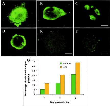
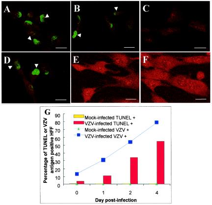
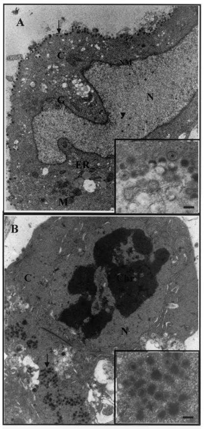
The induction of apoptosis or programmed cell death in virus-infected cells is an important antiviral defense mechanism of the host, and some herpesviruses have evolved strategies to modulate apoptosis in order to enhance their survival and spread. In this study, we examined the ability of varicella-zoster virus (VZV) to induce apoptosis in primary human dorsal root ganglion neurons and primary human foreskin fibroblasts (HFFs). Three independent methods (annexin V, TUNEL [terminal deoxynucleotidyltransferase-mediated dUTP-biotin nick end labeling] staining, and electron microscopy) were used to assess apoptosis in these cells on days 1, 2, and 4 postinoculation. By all three methods, apoptosis was readily detected in VZV-infected HFFs. In stark contrast, apoptosis was not detected during productive VZV infection of neurons. The low-passage clinical isolate Schenke and the tissue culture-adapted ROka strain both induced apoptosis in HFFs but not in neurons, suggesting that this cell-type-specific apoptotic phenotype was not VZV strain specific. These data show that the regulation of apoptosis differs markedly between HFFs and neurons during productive VZV infection. Inhibition of apoptosis during infection of neurons may play a significant role in the establishment, maintenance, and reactivation of latent infection by promoting survival of these postmitotic cells.


Figures

FIG. 1.
FIG. 1.
Light microscopy of a typical dissociated human fetal DRG neuronal culture. (A) Neuronal culture immediately after dissociation containing spherical or tear-shaped neurons ranging from 10 to 50 μm (arrow). (B) Neuronal culture 3 days later containing neurons with extensive axonal networks (boxed area) and neurons congregating (arrows). Schwann cells and fibroblasts are planar and spindle-shaped (arrowheads).
FIG. 2.
FIG. 2.
Immunofluorescence analysis of viral antigen expression in VZV-infected neurons. At 2 days p.i., neurons infected with VZV strain Schenke (A to D and F) and mock infected (E) were incubated with rabbit polyclonal antibodies to ORF62 (A and F), ORF4 (B), ORF29 (C), and a hyperimmune serum that predominantly detects glycoproteins (D). Rabbit and human anti-VZV antibodies were detected by using FITC-conjugated secondary antibodies (green fluorescence). Negative controls were mock-infected neurons (E) or VZV-infected neurons incubated with an isotype control antibody (F). Negative control images were obtained by increasing the laser voltage to enable the visualization of cells. Bar, 10 μm. (G) Percentages of cells stained with the VZV hyperimmune polyclonal serum (VZV antigen-positive cells) for VZV-infected neuronal and HFF cultures over the 4-day time period.
FIG. 3.
FIG. 3.
Visualization and quantitation of apoptotic cells by the TUNEL assay in VZV-infected neuronal cultures. Neurons inoculated with VZV-infected HFFs (A to C) or uninfected HFFs (D to F) were harvested on day 2 postinoculation and processed for the TUNEL assay. No TUNEL-specific staining was detected in VZV-infected (B) or mock-infected (E) neuronal cultures. Positive controls were treated with DNase prior to labeling (A and D), and negative controls were treated with TdT reaction mix containing no TdT (C and F). Bars, 10 μm. Each arrow indicates the neuronal axons, and each arrowhead indicates the apoptotic nuclei (green fluorescence). (G) Percentages of neurons labeled for apoptotic nuclei (TUNEL+) or VZV antigens (VZV+) over the 4-day time course in mock- and VZV-infected neuronal cultures.
FIG. 4.
FIG. 4.
Visualization and quantitation of apoptotic cells by the TUNEL assay in VZV-infected HFFs. VZV-infected HFFs (A to C) or uninfected HFFs (D to F) were harvested on day 2 postinoculation and processed for the TUNEL assay. (B) TUNEL-specific staining was detected in VZV-infected HFFs. (E) No TUNEL staining was detected in mock-infected HFFs. Positive controls were treated with DNase prior to labeling (A and D), and negative controls were treated with TdT reaction mix containing no TdT (C and F). Bars, 10 μm. Each arrowhead indicates the apoptotic nuclei (green fluorescence). (G) Percentages of HFFs labeled for apoptotic nuclei (TUNEL+) or VZV antigens (VZV+) over the 4-day time course in mock- and VZV-infected HFF cultures.
FIG. 5.
FIG. 5.
Immunofluorescence analysis of membrane PS exposure associated with apoptosis by annexin V staining in VZV-infected neuronal cultures. At 2 days p.i., neurons inoculated with VZV-infected HFFs (A and B) or mock-infected HFFs (C and D) were incubated with annexin V-Alexa Fluor 594 conjugate (red fluorescence) and a nuclear counterstain DAPI (blue fluorescence) and then analyzed by laser confocal microscopy. Positive controls were VZV-infected (A) and mock-infected (C) cells treated with 5% ethanol prior to staining. No specific annexin V staining was detected in VZV-infected (B) or mock-infected (D) neuronal cultures. Bar, 10 μm. Neuronal axon hillocks are indicated by arrows, and arrowheads indicate annexin V staining. (E) Percentages of neurons staining for annexin V (annexin V+) or VZV antigens (VZV+) over the 4-day time course in mock- and VZV-infected neuronal cultures.
FIG. 6.
FIG. 6.
Immunofluorescence analysis of membrane PS exposure associated with apoptosis by annexin V staining in VZV-infected HFFs. Two days p.i. VZV-infected HFFs (A and B) or mock-infected HFFs (C and D) were incubated with annexin V-Alexa Fluor 594-conjugate (red fluorescence) and a nuclear counterstain DAPI (blue fluorescence) and then analyzed by laser confocal microscopy. Positive controls were VZV-infected (A) and mock-infected (C) cells treated with 5% ethanol prior to staining. (B) Specific annexin V staining was detected in VZV-infected HFFs. (D) No staining was detected in mock-infected HFFs. Bar, 10 μm. An arrowhead indicates annexin V staining. (E) Percentages of neurons staining for annexin V (annexin V+) or VZV antigens (VZV+) over the 4-day time course in mock- and VZV-infected HFF cultures.
FIG. 7.
FIG. 7.
Quantitation of apoptotic cells by the TUNEL assay and annexin V staining in VZV ROka-infected neuronal and HFF cultures. The percentages of neurons and HFFs staining for TUNEL (TUNEL+), annexin V (annexin V+), or VZV antigens (VZV+) over the 4-day time course in mock and VZV-infected neuronal cultures (A and B) and mock- and VZV-infected HFF cultures (C and D) are shown.
FIG.8.
FIG.8.
TEM images of VZV-infected human DRG neurons and HFFs. (A) VZV-infected neuron at 2 days p.i. with an intact double nuclear membrane (NM), normal Golgi apparatus (G), mitochondrion (M), and endoplasmic reticulum (ER). The arrowhead and arrow indicate unenveloped nucleocapsids in the nucleus and enveloped virions on the cell surface, respectively. Magnification, ×6,500. (Inset) Higher magnification of enveloped VZV virions on the surface of the neuron. Bar, 200 nm. (B) VZV-infected HFFs at 2 days p.i. show morphological features of apoptosis, loss of double nuclear membrane, condensed chromatin (Chr), and irregular organelle morphology. An arrow indicates enveloped VZV virions in the cytoplasm. Magnification, ×6,500. (Inset) Higher magnification of enveloped VZV virions in the cytoplasm of the HFF. Bar, 200 nm.

References

    1. Ahmed, M., M. Lock, C. G. Miller, and N. W. Fraser. 2002. Regions of the herpes simplex virus type 1 latency associated transcript that protect cells from apoptosis in vitro and protect neuronal cells in vivo. J. Virol. 76:717-729. - PMC - PubMed
    1. Aleman, N., M. I. Quiroga, M. Lopez-Pena, S. Vazquez, F. H. Guerrero, and J. M. Nieto. 2001. Induction and inhibition of apoptosis by pseudorabies virus in the trigeminal ganglion during acute infection of swine. J. Virol. 75:469-479. - PMC - PubMed
    1. Aubert, M., A. R. Rice, and J. A. Blaho. 2001. Accumulation of herpes simplex virus type 1 early and leaky-late proteins correlates with apoptosis prevention in infected human HE-p2 cells. J. Virol. 75:1013-1030. - PMC - PubMed
    1. Aubert, M., and J. A. Blaho. 1999. The herpes simplex virus type 1 regulatory protein ICP27 is required for the prevention of apoptosis in infected human cells. J. Virol. 73:2803-2813. - PMC - PubMed
    1. Aubert, M., J. O'Toole, and J. A. Blaho. 1999. Induction and prevention of apoptosis in human HEp-2 cells by herpes simplex virus type 1. J. Virol. 73:10359-10370. - PMC - PubMed

Publication types

LinkOut - more resources