

doi: 10.1002/mrm.1910280214.
Cardiac tagging with breath-hold cine MRI
Affiliations
- PMID: 1461130
- PMCID: PMC2041925
- DOI: 10.1002/mrm.1910280214
Item in Clipboard
Cardiac tagging with breath-hold cine MRI
Magn Reson Med.
1992 Dec.
Abstract
A method is presented for measuring myocardial deformation in a breath-hold with tagged cine MRI. Tagged cine images of human hearts are obtained in arbitrary oblique planes on a standard imager with as few as four heartbeats. The scan time has been reduced 16- to 64-fold from previous techniques.
Figures
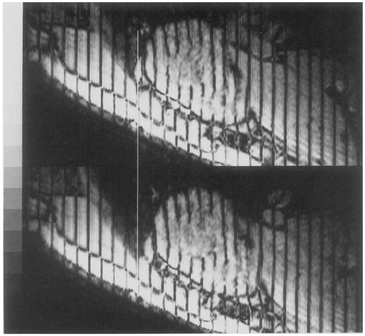
Two end-diastolic coronal images of the heart at (top) inspiration and (bottom) expiration with normal breathing amplitude. These images were obtained in 16 heartbeats with breathing suspended. The heart has translated approximately 1 cm from (top) to (bottom) due only to differences in lung inflation.
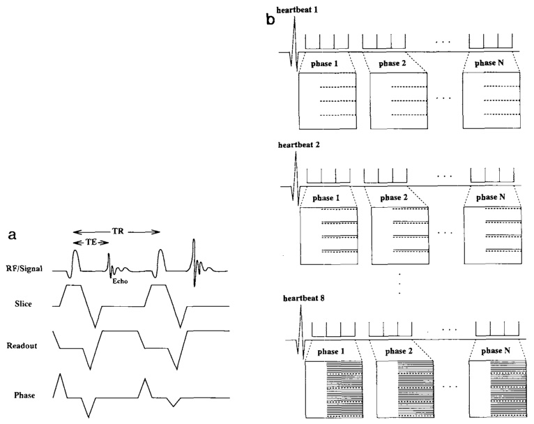
(a) Two echoes from the GRASS pulse sequence used to obtain the cine images. All gradient waveforms use maximum slew rate ramps. The slice selection gradient has maximum amplitude. A “fractional echo” is used to sample in the readout direction. The phase encoding “rewinder” gradient overlaps with the ascending ramp of the slice selection gradient for the next acquisition. (b) The segmented k-space mapping used to obtain the tagged cine images. In the acquisition shown, four lines of k-space are sampled in each movie phase (dashed lines), N total movie phases are acquired, and eight heartbeats are used to acquire the data matrix. For this example a final data matrix of 32 × 160 is obtained for each movie phase. More phase encoding steps can be achieved by using more heartbeats or using more views per movie phase. Note: the k-space map is not to scale.
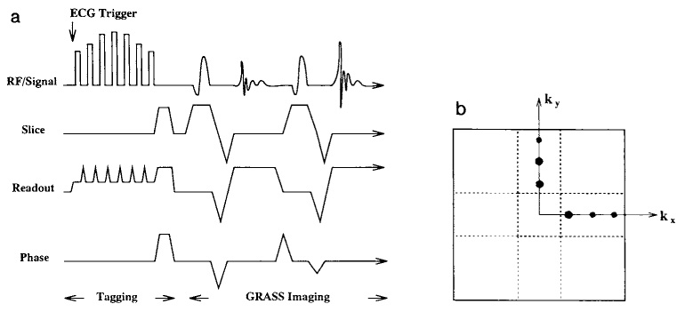
(a) The hybrid DANTE/SPAMM tagging pulse sequence. The parallel line tagging pattern is produced by a sequence of rectangular pulses. The gradient producing the dispersion in the sample is reduced during the rf pulses to reduce the total bandwidth required from the pulse to tag the heart. This permits the use of longer lower amplitude rectangular pulses. The tagging pulses are triggered by the upslope of the QRS complex and the imaging pulses follow immediately after spoiler gradients dephase transverse magnetization from the tagging pulses. (b) The “cross” pattern used to sample k-space for the final grid-tagged images. The Fourier components of the tagging pattern (schematically shown as black dots) are limited to the regions around the axes. The regions sampled with the pulse sequence are enclosed by the bold dashed lines; the regions obtained through symmetry are in the light dashed lines.
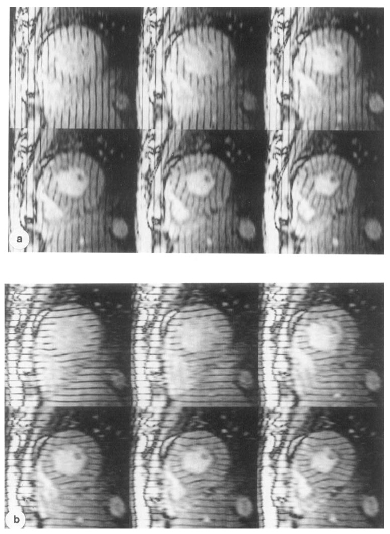
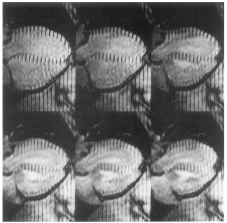
Six phases of the cardiac cycle as imaged in the short axis orientation with the tagged cine protocol. The images were obtained with a time resolution of 48 ms. (a) The readout gradient is oriented in the horizontal direction with the parallel tags in the vertical direction. (b) The readout gradient is oriented in the vertical direction with the parallel tags in the horizontal direction.
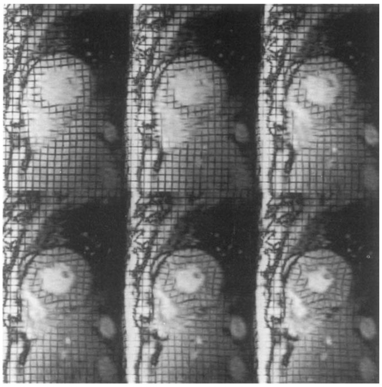
Six phases of the cardiac cycle as imaged in the long axis orientation with the tagged cine protocol. The tags are oriented in parallel planes with the short axis images; the readout gradient is oriented perpendicular to the tags. These images are important for 3D tracking of tissue through the short axis imaging planes.
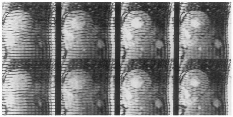
Six phases of the heart cycle with a grid tag pattern derived using Eq. [1] and the parallel tag images shown in Fig. 4.
The effect of reduced phase encoding resolution and k-space segmentation. The acquisition parameters were field-of-view = 28 cm, TR = 6.0 ms, TE = 2.3 ms, α = 20°, 1 NEX, and rectangular field-of-view. (Top) A four-heartbeat acquisition. The number of phase encode views (NPE) is 32, and number of phase encode views per movie phase (NVP) is 8, 48 ms time resolution in the movie phases. (Bottom) An eight-heartbeat acquisition, NPE = 32, NVP = 4, 24 ms time resolution in the movie phases.
References
Publication types
MeSH terms
Grants and funding
LinkOut - more resources
Full Text Sources
Other Literature Sources
Medical
