

Cyclooxygenase enzymes and prostaglandins in pathology of the endometrium
- PMID: 14611628
- PMCID: PMC2695735
- DOI: 10.1530/rep.0.1260559
Cyclooxygenase enzymes and prostaglandins in pathology of the endometrium
Abstract
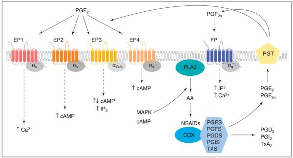
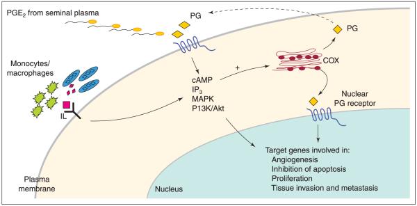
Prostaglandins are bioactive lipids produced from arachidonic acid by cyclooxygenase (COX) enzymes and specific terminal prostanoid synthase enzymes. After biosynthesis, prostaglandins exert an autocrine-paracrine function by coupling to specific prostanoid G protein-coupled receptors to activate intracellular signalling and gene transcription. For many years, prostaglandins have been recognized as key molecules in reproductive biology by regulating ovulation, endometrial physiology and proliferation of endometrial glands and menstruation. More recently, a role for COX enzymes and prostaglandins has been ascertained in reproductive tract pathology, including carcinomas, menorrhagia, dysmenorrhoea and endometriosis. Although the mechanism by which prostaglandins modulate these pathologies is still unclear, a large body of evidence supports a role for COX enzymes, prostaglandins and prostaglandin receptor signalling pathways in angiogenesis, apoptosis and proliferation, tissue invasion and metastases and immunosuppression. Here, an overview is provided of some of the findings from these studies with specific emphasis on the role of COX enzymes, prostaglandin E(2) and F(2alpha) in disorders of endometrial proliferation and menstruation in non-pregnant women.
Figures
References
-
- Adelantado JM, Lopez Bernal A, Turnbull AC. Topographical distribution of prostaglandin E receptors in human myometrium. British Journal of Obstetrics and Gynaecology. 1988a;95:348–353. - PubMed
-
- Adelantado JM, Rees MC, Lopez Bernal A, Turnbull AC. Increased uterine prostaglandin E receptors in menorrhagic women. British Journal of Obstetrics and Gynaecology. 1988b;95:162–165. - PubMed
-
- Aplin AE, Howe A, Alahari SK, Juliano RL. Signal transduction and signal modulation by cell adhesion receptors the role of integrins, cadherins, immunoglobulin-cell adhesion molecules, and selectins. Pharmacology Reviews. 1998;50:197–263. - PubMed
-
- Ashby B. Co-expression of prostaglandin receptors with opposite effects: a model for homeostatic control of autocrine and paracrine signaling. Biochemical Pharmacology. 1998;55:239–246. - PubMed
Publication types
MeSH terms
Substances
Grants and funding
LinkOut - more resources
Full Text Sources
Other Literature Sources
Medical
