. 2003 Dec;23(23):8505-18.
doi: 10.1128/MCB.23.23.8505-8518.2003.

Nonhomologous-end-joining factors regulate DNA repair fidelity during Sleeping Beauty element transposition in mammalian cells

Affiliations

Nonhomologous-end-joining factors regulate DNA repair fidelity during Sleeping Beauty element transposition in mammalian cells

Stephen R Yant et al. Mol Cell Biol. 2003 Dec.

Abstract

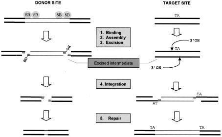
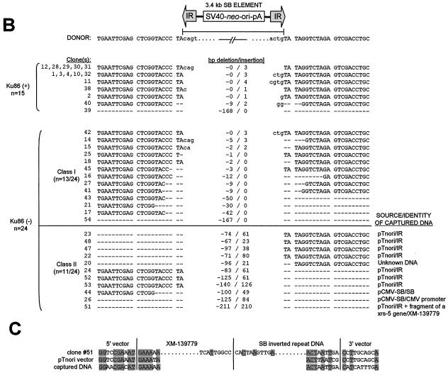
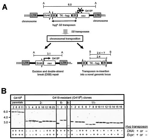
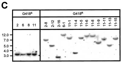
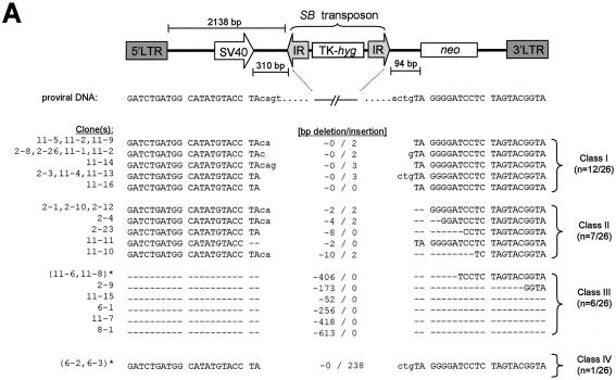
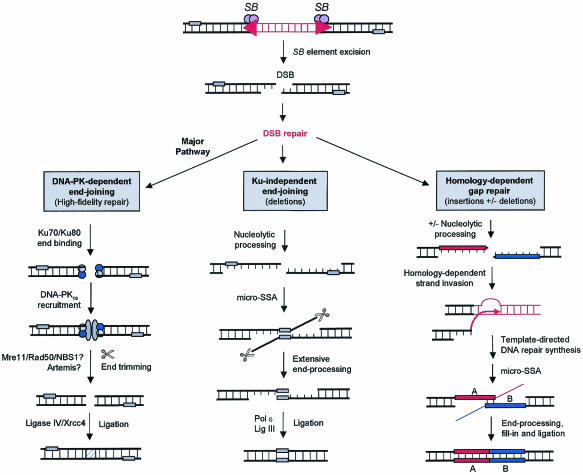
Herein, we report that the DNA-dependent protein kinase (DNA-PK) regulates the DNA damage introduced during Sleeping Beauty (SB) element excision and reinsertion in mammalian cells. Using both plasmid- and chromosome-based mobility assays, we analyzed the repair of transposase-induced double-stranded DNA breaks in cells deficient in either the DNA-binding subunit of DNA-PK (Ku) or its catalytic subunit (DNA-PKcs). We found that the free 3' overhangs left after SB element excision were efficiently and accurately processed by the major Ku-dependent nonhomologous-end-joining pathway. Rejoining of broken DNA molecules in the absence of Ku resulted in extensive end degradation at the donor site and greatly increased the frequency of recombination with ectopic templates. Therefore, the major DNA-PK-dependent DNA damage response predominates over more-error-prone repair pathways and thereby facilitates high-fidelity DNA repair during transposon mobilization in mammalian cells. Although transposable elements were not found to be efficiently circularized after transposase-mediated excision, DNA-PK deficiency supported more-frequent transposase-mediated element insertion than was found in wild-type controls. We conclude that, based on its ability to regulate excision site junctional diversity and transposon insertion frequency, DNA-PK serves an important protective role during transpositional recombination in mammals.


Figures

FIG. 1.
FIG. 1.
Requirement for DNA repair during transposition of SB elements. An SB element is excised by transposase via sequential 3-bp staggered cuts at the ends of the inverted terminal repeats, completely releasing it from its original donor site. This newly excised element then integrates into a TA target site via a series of transesterification reactions such that the TA is duplicated at the ends of the element following repair of the single-stranded DNA gaps. Depending on how the DSB at the site of excision is repaired, a small transposon footprint will sometimes be left behind.
FIG. 2.
FIG. 2.
Stable integration and expression of SB elements in mice in the absence of DNA-PK activity. (A) Schematic of the DpnI-sensitive hAAT transposon injected into mice. The vector pThAAT was purified from bacteria expressing the dam-encoded methylase, making it susceptible to digestion with the restriction endonuclease DpnI. Arrows, locations of DpnI recognition sites, which are substrates for DpnI only when the internal adenine residue is methylated. RSV, Rous sarcoma virus long terminal repeat promoter; hAAT, human α1-antitrypsin cDNA; IR, SB element IR sequence; H, HindIII. (B) Transposon-based human α1-antitrypsin expression in DNA-PKcs-deficient scid mice before and after a surgical partial hepatectomy (PH). Immune-deficient C57BL/6-scid mice were injected with 25 μg of pThAAT together with 1 μg of either pCMV-SB (▴) (contains wild-type SB; n = 6 mice) or pCMV-mSB (▵) (contains inactive SB; n = 3 mice) as a control, and their serum hAAT levels were monitored over time by ELISA. Liver regeneration was induced 10 months after vector administration by surgically removing two-thirds of the mouse liver under anesthesia. (C) Southern blot analysis of DpnI-treated control DNA purified from dam-positive and dam-negative bacteria. We used mouse liver DNA (10 μg) spiked with 0.1 ng of methylated (dam-positive) or unmethylated (dam-negative) vector DNA for Southern blot analysis. Membranes were probed with a radiolabeled hAAT fragment. Asterisks, cleavage fragments derived from DpnI-treated input vector. (D) Methylation-sensitive Southern blot analysis of mouse liver DNA obtained 11.5 months after vector administration. We used 10 μg of total liver DNA digested with DpnI and HindIII for Southern blot analysis and hybridized membranes to a radiolabeled hAAT probe. The left five lanes (5.0 to 0.1 copies/cell) were derived from adding a dam-negative (DpnI-resistant) pThAAT plasmid to mouse liver DNA. Arrow, integrated, DpnI-resistant copies of the transposon in pCMV-SB-treated animals; asterisks, presence of DpnI-sensitive transposon episomes in all samples.
FIG. 3.
FIG. 3.
DNA-PK-independent repair of transposase-induced DSBs in vivo in mouse liver. (A) PCR-based strategy to detect repair of transposase-induced DSBs following transposon excision from donor plasmid constructs. The structure of the pThAAT vector DNA is shown in linear format for simplicity but was delivered to mice as a supercoiled plasmid. Prior to DNA transposition, PCR primers (arrows) are located 2.8 kb apart, which is beyond the PCR capacity. Following SB-mediated transposon excision and DSB repair, the primers are brought close enough together to allow PCR amplification. (B) Transposon excision and DSB repair in mouse liver. Normal C57BL/6 and DNA-PKcs-deficient C57BL/6-scid mice received a donor hAAT vector with (pThAAT) or without (phAAT) transposase binding sites, together with either pCMV-SB or pCMV-mSB as a control. Total liver DNA was isolated at either day 2 or day 30 postinjection and analyzed by PCR for the presence of a 271-bp product of excision and repair. (C) Variety of DSB repair products produced in vivo in the presence (C57BL/6) and absence (C57BL/6-scid) of DNA-PK activity. The total numbers of clones with identical characteristics are shown, as well as the sizes in base pairs of both the resulting footprint and deleted region. Footprints are in lowercase. Dashes, deleted nucleotides; NA, not applicable.
FIG. 4.
FIG. 4.
Repair of transposase-induced DSBs in the presence and absence of mammalian Ku DNA end-binding activity. (A) Transposon excision and DSB repair in the presence and absence of Ku protein. Wild-type (CHO-K1 and V79-4) and Ku-deficient (xrs-5 and XR-V15B) hamster cells were transfected with donor plasmid pTnori (containing a 3.4-kb neo-marked SB element) together with either pCMV-SB or pCMV-mSB. Hirt DNA was isolated 30 h later and analyzed by PCR for the presence of a 271-bp product of excision and repair. (B) Variety of DSB repair products produced in the presence and absence of mammalian Ku. Base pair deletions (dashes) in the region surrounding the excision site are shown. (C) Example of the homologous sequences present at the junctions between the broken DNA molecule and captured sequences. Shaded boxes, positions of microhomologies at each end of the junction and within the region where the two inserts are joined.
FIG. 4.
FIG. 4.
Repair of transposase-induced DSBs in the presence and absence of mammalian Ku DNA end-binding activity. (A) Transposon excision and DSB repair in the presence and absence of Ku protein. Wild-type (CHO-K1 and V79-4) and Ku-deficient (xrs-5 and XR-V15B) hamster cells were transfected with donor plasmid pTnori (containing a 3.4-kb neo-marked SB element) together with either pCMV-SB or pCMV-mSB. Hirt DNA was isolated 30 h later and analyzed by PCR for the presence of a 271-bp product of excision and repair. (B) Variety of DSB repair products produced in the presence and absence of mammalian Ku. Base pair deletions (dashes) in the region surrounding the excision site are shown. (C) Example of the homologous sequences present at the junctions between the broken DNA molecule and captured sequences. Shaded boxes, positions of microhomologies at each end of the junction and within the region where the two inserts are joined.
FIG. 5.
FIG. 5.
Chromosomal excision and reinsertion of a nonautonomous SB element in HeLa cells. (A) Genetic-selection strategy to study DSB repair following rare transposon excision from random chromosomal loci. A neomycin resistance (neo) gene under the control of the simian virus 40 (SV40) promoter is inactivated by the insertion of a nonautonomous SB element containing the HygR gene driven by the thymidine kinase (TK) promoter. Upon packaging into a lentivirus vector, this excision testing construct randomly integrates as a single-copy provirus into the genomes of infected cells. Following transient expression of the SB transposase, the SB element is excised and expression of the neo gene is activated, resulting in resistance to the drug G418. In some instances, the excised hygromycin resistance SB element reintegrates into the genome and acquires novel flanking sequences. Shown are the predicted structures of the testing construct, with approximate distances in kilobases between flanking AvaI (A) and SpeI (S) sites, before (top) and after (bottom) SB-mediated transposon excision. LTR, HIV-1 long terminal repeat. (B) Southern blot analysis confirms excision of the SB element from the testing construct. Twenty micrograms of total DNA from all G418R colonies, as well as the four G418S parental clones, was digested with AvaI, separated by ethidium bromide gel electrophoresis, transferred to nitrocellulose, and then hybridized to a neo probe. The molecular marker (kilobases) is shown to the left. The presence (+) and absence (−) of an expressible transposon in each clone was investigated by both PCR and hygromycin-dependent growth selection. (C) Genomic reintegration of the transposon into novel chromosomal loci following SB-mediated excision. Twenty micrograms of total DNA from each G418S parental clone and 13 of the resulting HygR G418R clones was digested with SpeI (cuts once in the transposon), separated by ethidium bromide gel electrophoresis, transferred to nitrocellulose, and then hybridized to a 32P-radiolabeled hygromycin fragment.
FIG. 5.
FIG. 5.
Chromosomal excision and reinsertion of a nonautonomous SB element in HeLa cells. (A) Genetic-selection strategy to study DSB repair following rare transposon excision from random chromosomal loci. A neomycin resistance (neo) gene under the control of the simian virus 40 (SV40) promoter is inactivated by the insertion of a nonautonomous SB element containing the HygR gene driven by the thymidine kinase (TK) promoter. Upon packaging into a lentivirus vector, this excision testing construct randomly integrates as a single-copy provirus into the genomes of infected cells. Following transient expression of the SB transposase, the SB element is excised and expression of the neo gene is activated, resulting in resistance to the drug G418. In some instances, the excised hygromycin resistance SB element reintegrates into the genome and acquires novel flanking sequences. Shown are the predicted structures of the testing construct, with approximate distances in kilobases between flanking AvaI (A) and SpeI (S) sites, before (top) and after (bottom) SB-mediated transposon excision. LTR, HIV-1 long terminal repeat. (B) Southern blot analysis confirms excision of the SB element from the testing construct. Twenty micrograms of total DNA from all G418R colonies, as well as the four G418S parental clones, was digested with AvaI, separated by ethidium bromide gel electrophoresis, transferred to nitrocellulose, and then hybridized to a neo probe. The molecular marker (kilobases) is shown to the left. The presence (+) and absence (−) of an expressible transposon in each clone was investigated by both PCR and hygromycin-dependent growth selection. (C) Genomic reintegration of the transposon into novel chromosomal loci following SB-mediated excision. Twenty micrograms of total DNA from each G418S parental clone and 13 of the resulting HygR G418R clones was digested with SpeI (cuts once in the transposon), separated by ethidium bromide gel electrophoresis, transferred to nitrocellulose, and then hybridized to a 32P-radiolabeled hygromycin fragment.
FIG. 6.
FIG. 6.
Rejoining of cellular transposase-induced DSBs in wild-type human cells. (A) DNA sequence analysis of the donor sites after excision of the SB element from the testing construct located at four different genomic loci. All examples of recovered excision products are shown together with the original parental sequence. Footprints are in lowercase. Dashes, deleted nucleotides; asterisks, duplicate clones. (B) Microhomology at junctions of repaired genomic DNA breaks in class III G418R clones. Columns A and B show the nucleotides at the edges of the DNA fragment lost during transposon excision and DSB repair in each clone. The shaded regions in columns A and B represent deleted sequences with homology to the right and left junctions of the G418R provirus, respectively. The presence of microhomologies at one end of a clone that acquired a retrotransposon insert is shown below.
FIG. 6.
FIG. 6.
Rejoining of cellular transposase-induced DSBs in wild-type human cells. (A) DNA sequence analysis of the donor sites after excision of the SB element from the testing construct located at four different genomic loci. All examples of recovered excision products are shown together with the original parental sequence. Footprints are in lowercase. Dashes, deleted nucleotides; asterisks, duplicate clones. (B) Microhomology at junctions of repaired genomic DNA breaks in class III G418R clones. Columns A and B show the nucleotides at the edges of the DNA fragment lost during transposon excision and DSB repair in each clone. The shaded regions in columns A and B represent deleted sequences with homology to the right and left junctions of the G418R provirus, respectively. The presence of microhomologies at one end of a clone that acquired a retrotransposon insert is shown below.
FIG. 7.
FIG. 7.
Multiple pathways for DNA repair after SB element excision in mammalian cells. The majority of DSBs introduced during SB element excision are repaired by a DNA-PK-dependent NHEJ pathway which involves the binding of Ku heterodimers to free DNA ends and subsequent recruitment of DNA-PKcs. DNA ends are then unwound and processed, possibly by Artemis or the MRN complex, and then ligated by the DNA ligase IV/XRCC4 complex. In the absence of DNA-PK activity, at least two different Ku-independent repair pathways are activated to fill the DNA gap. The first is a poorly defined error-prone NHEJ pathway that processes DSBs by illegitimate pairing of distant regions of microhomology (small shaded rectangles), followed by ligation and deletion of the intervening DNA sequence. The second route involves strand invasion of DNA sources with homology, which results in the insertion of a short DNA segment at the break site. micro-SSA, microhomology-dependent single-strand annealing.

References

    1. Acel, A., B. E. Udashkin, M. A. Wainberg, and E. A. Faust. 1998. Efficient gap repair catalyzed in vitro by an intrinsic DNA polymerase activity of human immunodeficiency virus type 1 integrase. J. Virol. 72:2062-2071. - PMC - PubMed
    1. Adams, M. D., M. McVey, and J. J. Sekelsky. 2003. Drosophila BLM in double-strand break repair by synthesis-dependent strand annealing. Science 299:265-267. - PubMed
    1. Arca, B., S. Zabalou, T. G. Loukeris, and C. Savakis. 1997. Mobilization of a Minos transposon in Drosophila melanogaster chromosomes and chromatid repair by heteroduplex formation. Genetics 145:267-279. - PMC - PubMed
    1. Baekelandt, V., A. Claeys, P. Cherepanov, E. De Clercq, B. De Strooper, B. Nuttin, and Z. Debyser. 2000. DNA-dependent protein kinase is not required for efficient lentivirus integration. J. Virol. 74:11278-11285. - PMC - PubMed
    1. Beall, E. L., A. Admon, and D. C. Rio. 1994. A Drosophila protein homologous to the human p70 Ku autoimmune antigen interacts with the P transposable element inverted repeats. Proc. Natl. Acad. Sci. USA 91:12681-12685. - PMC - PubMed

Publication types

MeSH terms