. 2003 Dec;23(23):8691-703.
doi: 10.1128/MCB.23.23.8691-8703.2003.

Increased malignancy of Neu-induced mammary tumors overexpressing active transforming growth factor beta1

Affiliations

Increased malignancy of Neu-induced mammary tumors overexpressing active transforming growth factor beta1

Rebecca S Muraoka et al. Mol Cell Biol. 2003 Dec.

Abstract

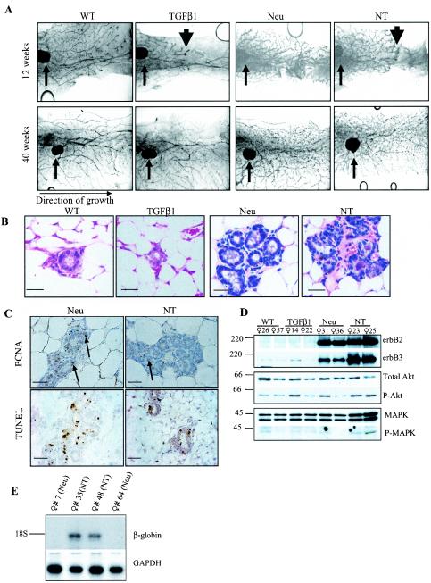
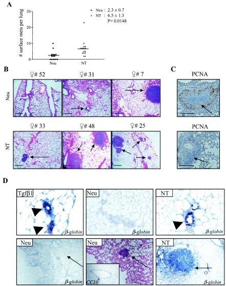
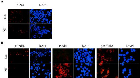
To determine if Neu is dominant over transforming growth factor beta (TGF-beta), we crossed mouse mammary tumor virus (MMTV)-Neu mice with MMTV-TGF-beta1(S223/225) mice expressing active TGF-beta1 in the mammary gland. Bigenic (NT) and Neu-induced mammary tumors developed with a similar latency. The bigenic tumors and their metastases were less proliferative than those occurring in MMTV-Neu mice. However, NT tumors exhibited less apoptosis and were more locally invasive and of higher histological grade. NT mice exhibited more circulating tumor cells and lung metastases than Neu mice, while NT tumors contained higher levels of phosphorylated (active) Smad2, Akt, mitogen-activated protein kinase (MAPK), and p38, as well as vimentin content and Rac1 activity in situ than tumors expressing Neu alone. Ex vivo, NT cells exhibited higher levels of P-Akt and P-MAPK than Neu cells. These were inhibited by the TGF-beta inhibitor-soluble TGF-beta type II receptor (TbetaRII:Fc), suggesting they were activated by autocrine TGF-beta. TGF-beta stimulated migration of Neu cells into surrounding matrix, while the soluble TGF-beta inhibitor abrogated motility and invasiveness of NT cells. These data suggest that (i) the antimitogenic and prometastatic effects of TGF-beta can exist simultaneously and (ii) Neu does not abrogate TGF-beta-mediated antiproliferative action but can synergize with TGF-beta in accelerating metastatic tumor progression.


Figures

FIG.1.
FIG.1.
TGF-β decreases both proliferation and apoptosis in NT mammary glands. (A) Whole-mount hematoxylin-stained inguinal mammary glands from virgin female mice at 12 and 40 weeks of age (n = 3 per time point). Small arrows indicate centrally located lymph node in the no. 4 gland. Large arrowheads demonstrate the delayed progression of ductal epithelium through the mammary fat pads of TGF-β1 and NT mice. (B) Hematoxylin-and-eosin-stained sections of inguinal mammary glands harvested at 40 weeks of age. Scale bars = 50 μm. (C) (Upper panels) PCNA IHC in no. 4 glands harvested from virgin females at 40 weeks of age. PCNA-positive cells are indicated with arrows (n = 5). (Lower panels) TUNEL analysis of no. 4 glands harvested from virgin females at 40 weeks of age. Scale bars = 50 μm. (D) Western analysis of mammary gland lysates at 40 weeks of age. Molecular masses are shown to the left in kilodaltons. The antibodies used are shown to the right. (E) Northern analysis of total RNA extracted from mammary glands at 40 weeks of age. The cDNA probes used are shown to the right. GAPDH, mouse GAPDH.
FIG.2.
FIG.2.
Active TGF-β1 overexpression does not affect Neu-induced tumor latency but increases malignancy. (A) The number of tumor-free days is shown for Neu and NT mice (n = 15; P = 0.47). (B) Total tumor volumes for each Neu and NT mouse (100 days following initial tumor palpation) are shown, along with the average total tumor volume per mouse (n = 15; mean ± standard deviation). Tumor volume was calculated with the formula volume = [(length)/2 × (width)2]. (C to H) Hematoxylin-and-eosin-stained sections of tumors harvested from Neu (C to E) or NT (F to H) mice 100 days following initial tumor palpation. Representative samples from three independent mice per genotype are shown. Panels F, G, and H show poorly differentiated carcinoma. Panels D and E show well-differentiated carcinoma. Panels F and G show highly vascularized invasive NT tumors. Arrows in panel F indicate areas in which the tumor has invaded the basement membrane into surrounding stroma. Scale bars, 100 μm. (I) Western analysis of proteins extracted from tumors 100 days after initial tumor palpation. The mouse identification number and genotype are indicated at the top. Molecular masses are shown to the left in kilodaltons. The antibodies used are shown at right. SMA, smooth muscle actin. (J) E-cadherin and vimentin IHC of representative tumors with immunoblot data in panel I. E-cadherin remains localized at cell junctions and tumor cells are vimentin negative. The vimentin staining in tumor 33 is limited to stromal elements.
FIG.3.
FIG.3.
TGF-β1 increases vascularization and cell survival in tumors but reduces cell proliferation. (A) CD31 IHC in tumor vasculature. Corresponding 4′,6′-diamidino-2-phenylindole (DAPI)-counterstained nuclei are shown at left (n = 5). (B) (Right panels) PCNA IHC in tumor cells. Scale bars, 50 μm. (Left panel) The index of PCNA-positive nuclei was calculated with the formula [(number of PCNA+ nuclei)/(total nuclei)]. n = 30 fields (six random fields of five tumors per genotype). The data point for each field is shown, along with the mean ± standard deviation. (C) TUNEL analysis of tumors. Scale bars, 100 μm. (Left panel) Index of TUNEL-positive nuclei was calculated by using the formula [(no. of TUNEL+ nuclei)/(total nuclei)]. n = 9 fields (3 random fields of 3 tumors per genotype). The data point for each field is shown, along with the mean ± standard deviation.
FIG. 4.
FIG. 4.
TGF-β1 increases mammary tumor lung metastases in MMTV-Neu mice. (A) The number of lung surface metastases per mouse (n = 15) is shown, along with the mean ± standard deviation. Significance was calculated by using the unpaired Student's t test. (B) Low-power magnification of hematoxylin-and-eosin-stained sections of lungs harvested from mice 100 days after initial tumor palpation. Representative pictures of three individual mice per genotype are shown. Lung metastases are indicated with arrows. Scale bars, 200 μm. (C) Representative PCNA IHC in lung metastases. (D) In situ hybridization of a digoxigenin-labeled rabbit β-globin cDNA fragment to RNA in paraffin-embedded mammary gland or lung sections from TGF-β1 and NT mice. Arrowheads indicate mammary epithelium. Arrows indicated mammary tumor-derived lung metastases.
FIG.5.
FIG.5.
TGF-β1 induces motility and invasiveness in Neu tumor cells. (A) PMTCs from Neu or NT mice were cultured in serum-reduced Matrigel supplemented with or without TGF-β1 (2 ng/ml) or Fc:TβRII (20 nM). Representative microphotographs are shown. Scale bars, 200 μm. (B and C) Western analysis of Neu and NT PMTCs incubated in 2.5% serum in the presence of 2-ng/ml TGF-β1, 20 nM Fc:TβRII, 10 μM U-0126, 10 μM SB202190, or 10 μM LY294002 where indicated. The antibodies used are indicated to the right, and molecular masses are indicated to the left. (D) (Left panel) Transwell assays using WT PMECs or 4T1 cells. (Middle and right panels) Transwell assays with WT PMECs, Neu PMTCs, or NT PMTCs. A total of 104 fluorescently labeled cells were plated in the upper chamber of transwells fitted with Matrigel-coated polycarbonate filters (8-μm pores). Medium containing 2.5% FBS with or without 2-ng/ml TGF-β1, 20 nM Fc:TβRII, 10 μM LY294002, or 10 μM SB202190 was placed in the lower chamber. After 24 h, the number of WT PMECs migrating to the lower side of the filter was given the value of 1, such that migration of 4T1 cells, Neu PMTCs, or NT PMTCs is represented as a multiple of WT PMECs. Values shown are the average (± standard deviation) of triplicate transwells in three independent experiments. *, P < 0.01; **, P < 0.001; @, P < 0.001. (E) Rac1 Western analysis of mammary tumor lysates. (F) In situ analysis of Rac1 activity using a PBD-GST fusion protein or with GST, followed by GST IHC as described in Materials and Methods. DAPI-counterstained nuclei are shown below each corresponding panel. (G) NMuMG cells transfected with pCAGA-Lux were treated for 24 h with 2-ng/ml TGF-β1 or with medium conditioned by Neu PMTCs, NT PMTCs, or WT PMECs, each in the presence or absence of 20 nM Fc:TβRII, and luciferase activity was measured as described in Materials and Methods. The ratio of firefly to R. reniformis luciferase activities in untreated NMuMG cells was given a value of 1. Bars represent the mean (± standard deviation) luciferase activities of three experiments each using duplicate wells for each condition compared to untreated NMuMG cells. *, P < 0.001 (Student's t test).
FIG. 6.
FIG. 6.
NT cells exhibit higher active Akt and reduced apoptosis. (A) Immunocytofluorescent detection of PCNA in Neu and NT PMTCs grown in 10% serum. 4′,6′-Diamidino-2-phenylindole (DAPI)-counterstained nuclei are shown to the right of each panel. PCNA+ cells: Neu, 18.6% ± 1.6% versus NT, 4.3% ± 1.6%; n = six ×400 fields. (B) Immunocytofluorescent detection of TUNEL-positive nuclei and P-Akt and p65/RelA expression in Neu and NT PMTCs grown in 0.5% serum for 3 days. DAPI-counterstained nuclei are shown to the right of each panel. Nuclear p65/RelA was found only in NT cells (37.8% ± 3.9%; n = six ×400 fields), whereas p65/RelA was restricted to the cytoplasm in Neu cells.

Similar articles

Cited by

References

    1. Adam, L., R. Vadlamudi, S. B. Kondapaka, J. Chernoff, J. Mendelsohn, and R. Kumar. 1998. Heregulin regulates cytoskeletal reorganization and cell migration through the p21-activated kinase-1 via phosphatidylinositol-3 kinase. J. Biol. Chem. 273:28238-28246. - PubMed
    1. Atfi, A., S. Djelloul, E. Chastre, R. Davis, and C. Gespach. 1997. Evidence for a role of Rho-like GTPases and stress-activated protein kinase/c-Jun N-terminal kinase (SAPK/JNK) in transforming growth factor beta-mediated signaling. J. Biol. Chem. 272:1429-1432. - PubMed
    1. Bakin, A. V., C. Rinehart, A. K. Tomlinson, and C. L. Arteaga. 2002. p38 mitogen-activated protein kinase is required for TGFbeta-mediated fibroblastic transdifferentiation and cell migration. J. Cell Sci. 115:3193-3206. - PubMed
    1. Berking, C., R. Takemoto, H. Schaider, L. Showe, K. Satyamoorthy, P. Robbins, and M. Herlyn. 2001. Transforming growth factor-beta1 increases survival of human melanoma through stroma remodeling. Cancer Res. 61:8306-8316. - PubMed
    1. Bhowmick, N. A., R. Zent, M. Ghiassi, M. McDonnell, and H. L. Moses. 2001. Integrin beta 1 signaling is necessary for transforming growth factor-beta activation of p38MAPK and epithelial plasticity. J. Biol. Chem. 276:46707-46713. - PubMed

Publication types

Substances