. 2003 Dec;23(23):8762-72.
doi: 10.1128/MCB.23.23.8762-8772.2003.

Human immunodeficiency virus type 1 hnRNP A/B-dependent exonic splicing silencer ESSV antagonizes binding of U2AF65 to viral polypyrimidine tracts

Affiliations

Human immunodeficiency virus type 1 hnRNP A/B-dependent exonic splicing silencer ESSV antagonizes binding of U2AF65 to viral polypyrimidine tracts

Jeffrey K Domsic et al. Mol Cell Biol. 2003 Dec.

Abstract

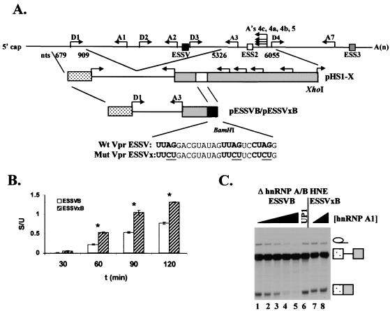
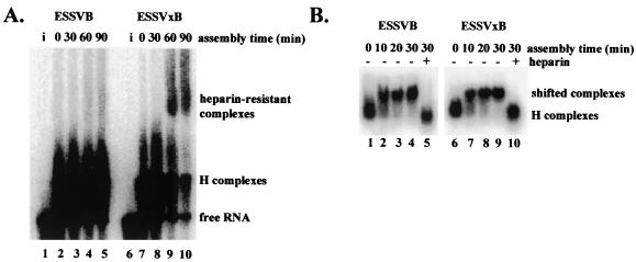
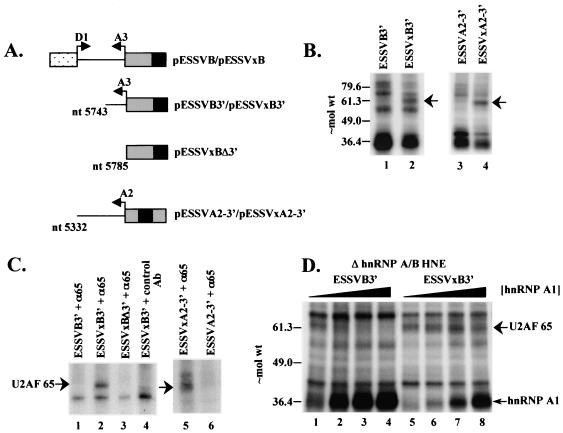
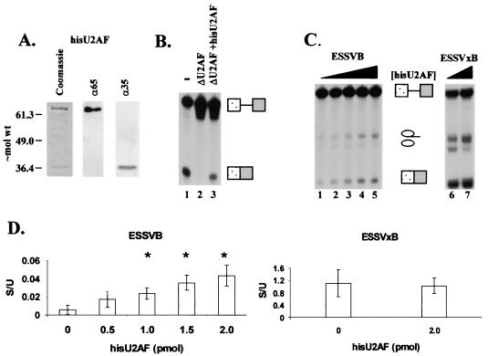
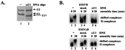
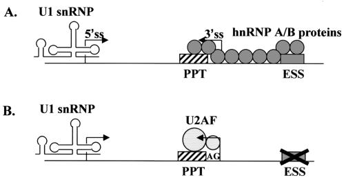
Human immunodeficiency virus type 1 (HIV-1) exonic splicing silencers (ESSs) inhibit production of certain spliced viral RNAs by repressing alternative splicing of the viral precursor RNA. Several HIV-1 ESSs interfere with spliceosome assembly by binding cellular hnRNP A/B proteins. Here, we have further characterized the mechanism of splicing repression using a representative HIV-1 hnRNP A/B-dependent ESS, ESSV, which regulates splicing at the vpr 3' splice site. We show that hnRNP A/B proteins bound to ESSV are necessary to inhibit E complex assembly by competing with the binding of U2AF65 to the polypyrimidine tracts of repressed 3' splice sites. We further show evidence suggesting that U1 snRNP binds the 5' splice site despite an almost complete block of splicing by ESSV. Possible splicing-independent functions of U1 snRNP-5' splice site interactions during virus replication are discussed.


Figures

FIG. 1.
FIG. 1.
Effect of hnRNP A1 on ESSV activity in substrates ESSVB and ESSVxB. (A) Schematic diagram of the in vitro splicing constructs pESSVB and pESSVxB. A minigene containing major 5′ splice site D1 and 3′ splice site A3 was constructed by insertion of a BamHI site immediately downstream of ESSV or ESSVx in pHS1-ESSV or pHS1-ESSVx, respectively. Endogenous locations of ESSV, ESS2, and ESS3 are shown as at the top. Sequences of the wild-type (Wt) ESSV and mutant (Mut) ESSVx are shown below the schematic diagrams. (B) Kinetics of splicing of ESSVB and ESSVxB in hnRNP A/B-depleted HNE. Radiolabeled, capped substrates were transcribed from BamHI-linearized plasmids. Substrates were spliced in hnRNP A/B-depleted HNE. RNAs were electrophoresed on denaturing polyacrylamide gels, band intensity was quantified, and amounts (femtomoles) of unspliced and spliced RNAs were calculated. The ratio of spliced (S) to unspliced (U) RNA is shown. Asterisks, samples with calculated (Student's t test) P values <0.05 compared to results at the 30-min time point. (C) Purified hnRNP A1 or its N-terminal fragment UP1 (0, 0.3, 0.5, 0.7. 0.9, 0.9, and 0, 0.9 μM in lanes 1 to 8, respectively) was added back to hnRNP A/B-depleted extract. Capped, radiolabeled ESSVB or ESSVxB was spliced, and RNAs were electrophoresed on denaturing polyacrylamide gels. The positions of the unspliced RNA, spliced RNA, and intron products are shown on the right.
FIG. 2.
FIG. 2.
ESSV inhibits spliceosome assembly prior to A complex formation. (A) Spliceosomes were assembled with [α-32P]UTP-labeled ESSVB or ESSVxB substrates in HNE in the presence of ATP. Aliquots of reaction mixtures were sampled at the times indicated and electrophoresed on native 4.0% polyacrylamide gels. Lanes i, RNA substrates loaded directly onto gel. The location of the ATP-dependent, heparin-resistant complex is shown on the right. (B) Spliceosomes were assembled as in panel A, except that ATP was depleted from the HNE prior to assembly. Lanes +, addition of heparin to samples at the 30-min time point prior to electrophoresis. Aliquots of reaction mixtures were sampled at the times indicated and electrophoresed on native 1.0% low-melting point agarose gels. The locations of H complexes and heparin-sensitive complexes are noted on the right.
FIG. 3.
FIG. 3.
Inhibition of U2AF65 cross-linking to ESSV-containing RNAs is dependent on hnRNP A/B proteins. (A) Schematic diagram of the 3′ half RNA probes used in the binding studies. Arrows, splice sites; black boxes, ESSV and ESSVx. (B) UV cross-linking of HNE proteins to [α-32P]UTP-labeled ESSVB3′ (lane 1), ESSVxB3′ (lane 2), ESSVA2-3′ (lane 3), and ESSVxA2-3 (lane 4) RNAs. Proteins cross-linked to RNase A-digested samples were electrophoresed on an SDS-10% polyacrylamide gel. Arrow, ∼65-kDa band. Approximate locations of molecular weight (in thousands) markers are shown on the left. (C) Immunoprecipitation of proteins cross-linked to [α-32P]UTP-labeled RNAs. Anti-U2AF65 monoclonal antibody MC3 (α65) and nonspecific murine IgG control antibody (control Ab) used for immunoprecipitations are noted above the lanes. Precipitated proteins were electrophoresed on SDS-10% polyacrylamide gels. The location of U2AF65 is noted on the left. (D) Purified hnRNP A1 (0, 0.3, 0.5, and 0.7 μM in lanes 1 to 4 and 5 to 8, respectively) was added back to hnRNP A/B-depleted HNEs. Cross-linking was performed as in panel B. Locations of U2AF65 and hnRNP A1 are noted on the right.
FIG. 4.
FIG. 4.
Recombinant U2AF stimulates the splicing of a 3′ splice site regulated by ESSV. (A) Characterization of baculovirus-expressed HisU2AF. Left, Coomassie blue stain of purified protein; middle and right, Western blot analysis for purified U2AF65 and U2AF35. Molecular weights are in thousands. (B) Functional activity of purified HisU2AF. U2AF was depleted from HNE based on affinity for poly(U)-Sepharose 4B. −, undepleted HNE; ΔU2AF, depleted HNE. Purified HisU2AF (2 pmol) was added to ΔU2AF HNE (ΔU2AF+hisU2AF). The α-32P-labeled Ad81 substrate was spliced for 2 h in the various HNEs. RNAs were electrophoresed on 4% denaturing polyacrylamide gels. Locations of unspliced and spliced RNA are on the right. (C) In vitro splicing competition assays. hnRNP A/B-depleted HNE was reconstituted with 0.7 μM hnRNP A1. Increasing amounts of HisU2AF were added to the splicing reaction mixtures for ESSVB (left; 0, 0.5, 1, 1.5, and 2 pmol) and ESSVxB (right; 0 and 2 pmol) substrates. Locations of unspliced and spliced RNA species are indicated between gels. (D) Ratios of spliced to unspliced species for ESSVB (left) and ESSVxB (right) substrates. Quantitations are from three independent experiments. Error bars indicate 1 standard deviation. Asterisks, samples with calculated P values <0.05 compared to lane 1 of panel C, as determined by Student's t test.
FIG. 5.
FIG. 5.
Intact U1 snRNA is required for formation of an ATP-independent, heparin-sensitive complex. (A) U1 snRNA in HNE was partially digested with RNase H as described in Materials and Methods. Products of the digestions were electrophoresed on the same 10% polyacrylamide-7 M urea gel and visualized by ethidium bromide staining. Locations of intact U1 and U2 snRNAs (U1 and U2) and partially digested U1 snRNA (U1*) are shown on the right. (B) Analysis of E complex assembly in untreated (−), mock-digested, and U1 snRNA-digested (αU1) HNEs. Locations of H complexes and heparin-sensitive complexes are noted on the right.
FIG. 6.
FIG. 6.
ESSV does not inhibit 5′ splice site splicing efficiency. (A) Schematic diagram of the in vitro splicing constructs pHS1-ESSV and pHS1-ESSVx. A minigene containing 5′ splice site D1 and 3′ splice sites A3, A4c, A4a, A4b, and A5 was generated as previously described. ESS2 was replaced by ESSV or ESSVx sequences as shown. (B) Capped and radiolabeled substrates were transcribed from linearized plasmids and spliced in HNE. RNAs were electrophoresed on denaturing polyacrylamide gels. The identities of the unspliced and spliced RNAs are shown on the right. Note that RNAs spliced at A4a and A4b comigrate on these gels. The positions of introns and exon lariats are in parentheses. (C and D) Splicing kinetics of substrates HS1-ESSV (C) and HS1-ESSVx (D). The amounts of radioactivity in the spliced RNA bands was quantitated on Instant Imager (Packard). The amounts (femtomoles) of the spliced RNA species and the total amount of spliced RNA from each reaction were calculated based on the specific activity of the [α-32P]UTP precursor and the UTP content of each RNA species.
FIG. 7.
FIG. 7.
Working model of the ATP-independent complexes formed in vitro. (A) Assembly of splicing components under conditions of repression. hnRNP A/B proteins (circles) bound to ESSV (gray box) nucleate binding of additional hnRNP A/B proteins to upstream cis sites, resulting in the blocking of the PPT (striped box). Note that, in the absence of splicing, U1 snRNP is bound to the 5′ splice site (5′ss). Only contacts between adjacent hnRNP A/B proteins are shown, although long-range protein-protein interactions may also occur. (B) Assembly of splicing components under nonrepressing conditions. Mutation of ESSV prevents stimulation of binding of additional hnRNP A/B proteins to upstream sites, allowing U2AF65 (large circle) to bind the PPT and U2AF35 (small circle) to bind a region including the 3′ splice site AG. Progression through the spliceosome assembly pathway then results in the splicing of the substrate.

Similar articles

Cited by

References

    1. Abdul-Manan, N., and K. R. Williams. 1996. hnRNP A1 binds promiscuously to oligoribonucleotides: utilization of random and homo-oligonucleotides to discriminate sequence from base-specific binding. Nucleic Acids Res. 24:4063-4070. - PMC - PubMed
    1. Amendt, B. A., D. Hesslein, L.-J. Chang, and C. M. Stoltzfus. 1994. Presence of negative and positive cis-acting RNA splicing elements within and flanking the first tat coding exon of human immunodeficiency virus type 1. Mol. Cell Biol. 14:3960-3970. - PMC - PubMed
    1. Amendt, B. A., Z.-H. Si, and C. M. Stoltzfus. 1995. Presence of exon splicing silencers within human immunodeficiency virus type 1 tat exon 2 and tat-rev exon 3: evidence for inhibition mediated by cellular factors. Mol. Cell Biol. 15:4606-4615. - PMC - PubMed
    1. Arrigo, S. J., S. Weitsman, J. A. Zack, and I. S. Chen. 1990. Characterization and expression of novel singly spliced RNA species of human immunodeficiency virus type 1. J. Virol. 64:4585-4588. - PMC - PubMed
    1. Ashe, M. P., A. Furger, and N. J. Proudfoot. 2000. Stem-loop 1 of the U1 snRNP plays a critical role in the suppression of HIV-1 polyadenylation. RNA 6:170-177. - PMC - PubMed

Publication types