. 2003 Dec;23(23):8795-808.
doi: 10.1128/MCB.23.23.8795-8808.2003.

Retinoic acid receptor alpha fusion to PML affects its transcriptional and chromatin-remodeling properties

Affiliations

Retinoic acid receptor alpha fusion to PML affects its transcriptional and chromatin-remodeling properties

Simona Segalla et al. Mol Cell Biol. 2003 Dec.

Abstract

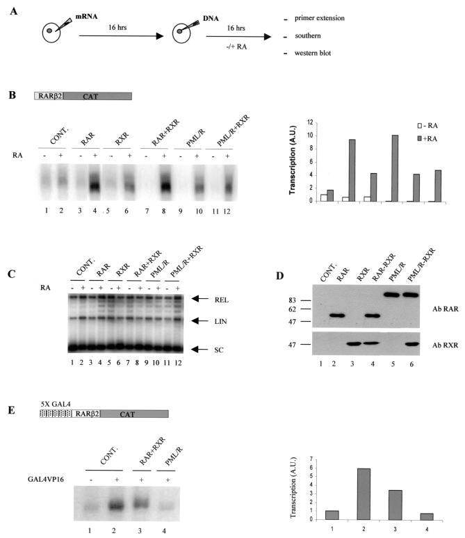
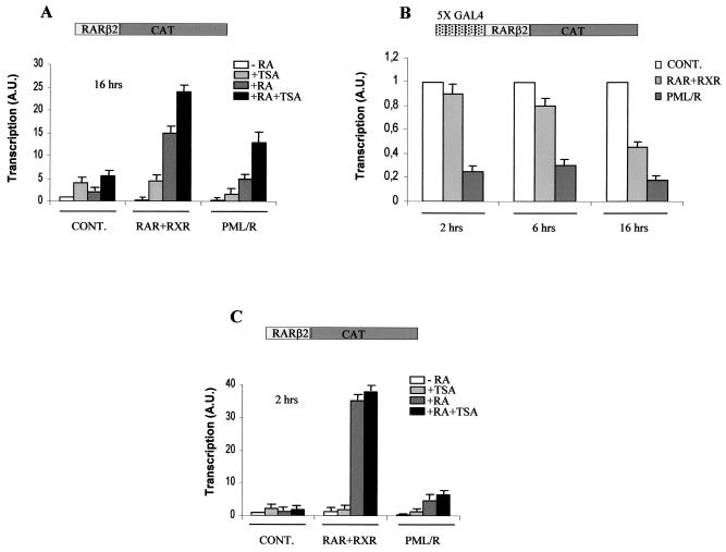
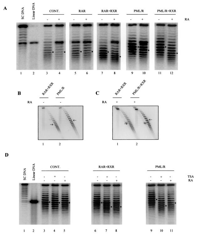
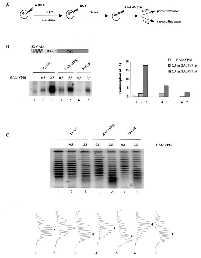
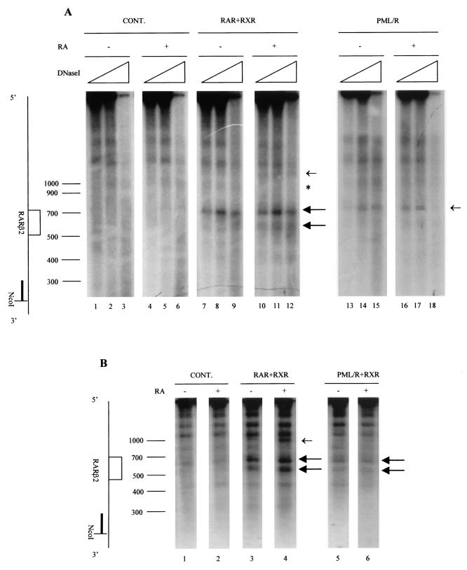
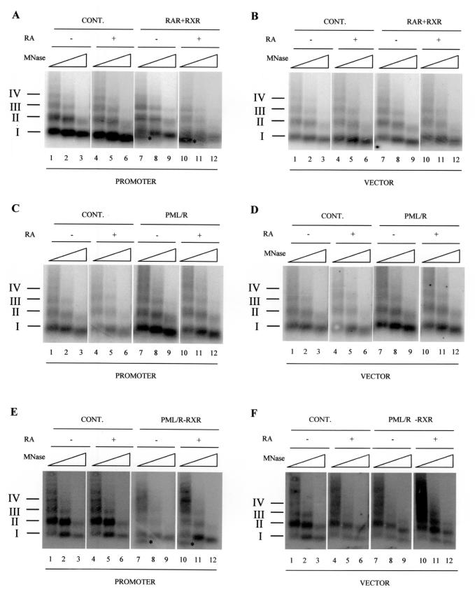
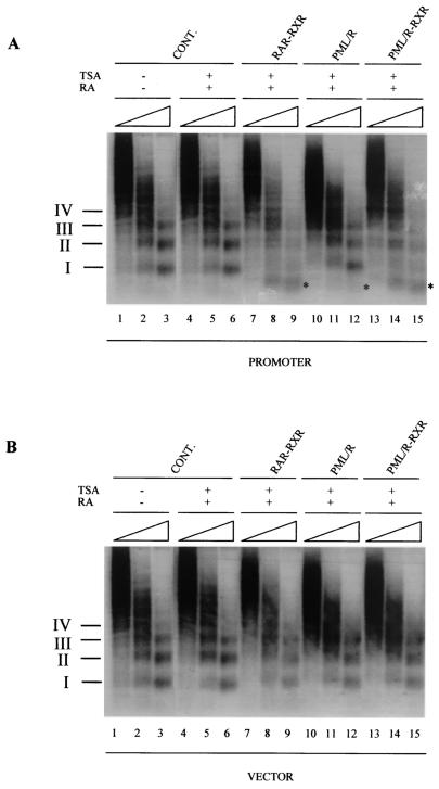
PML-RAR is an oncogenic transcription factor forming in acute promyelocytic leukemias (APL) because of a chromosomal translocation. Without its ligand, retinoic acid (RA), PML-RAR functions as a constitutive transcriptional repressor, abnormally associating with the corepressor-histone deacetylase complex and blocking hematopoietic differentiation. In the presence of pharmacological concentrations of RA, PML-RAR activates transcription and stimulates differentiation. Even though it has been suggested that chromatin alteration is important for APL onset, the PML-RAR effect on chromatin of target promoters has not been investigated. Taking advantage of the Xenopus oocyte system, we compared the wild-type transcription factor RARalpha with PML-RAR as both transcriptional regulators and chromatin structure modifiers. Without RA, we found that PML-RAR is a more potent transcriptional repressor that does not require the cofactor RXR and produces a closed chromatin configuration. Surprisingly, repression by PML-RAR occurs through a further pathway that is independent of nucleosome deposition and histone deacetylation. In the presence of RA, PML-RAR is a less efficient transcriptional activator that is unable to modify the DNA nucleoprotein structure. We propose that PML-RAR, aside from its ability to recruit aberrant quantities of histone deacetylase complexes, has acquired additional repressive mechanisms and lost important activating functions; the comprehension of these mechanisms might reveal novel targets for antileukemic intervention.


Figures

FIG. 1.
FIG. 1.
PML-RAR shows different transcriptional properties. (A) Scheme of the experiment shown in panel B. (B) Oocytes were not injected (CONT., lanes 1 and 2) or were injected with mRNA(s) coding for RAR (lanes 3 and 4), RXR (lanes 5 and 6), RAR and RXR (lanes 7 and 8), PML-RAR (lanes 9 and 10), or PML-RAR and RXR (lanes 11 and 12). Sixteen hours later, RARβ2CAT DNA was injected. The oocytes were then incubated in the presence (lanes 2, 4, 6, 8, 10, and 12) or absence (lanes 1, 3, 5, 7, 9, and 11) of RA. Transcription was assayed by primer extension. A schematic representation of the construct is shown at the top of the panel. Transcriptional signals were quantified with a PhosphorImager, and the transcriptional levels are indicated in the bar graph. RARβ2 expression following the injection of DNA without any subsequent addition is arbitrarily expressed as 1. A.U., arbitrary units. (C) Total DNA was isolated 16 h after injection, purified, and analyzed by Southern blotting. Positions of supercoiled (SC), linear (LIN), and relaxed (REL) DNAs are indicated. (D) Sixteen hours after DNA injection, batches of10 oocytes were collected from the same oocytes of panels B and C, and a whole-cell extract (equivalent to one oocyte) from uninjected (lane 1) or injected (lanes 2 to 6) oocytes were subjected to SDS-10% polyacrylamide gel electrophoresis, followed by Western blotting. Exogenously expressed RAR (lanes 2 and 4) and PML-RAR (lanes 5 and 6) were detected with a specific polyclonal RAR antibody (Ab) (6). RXR expression (lanes 3, 4, and 6) was revealed with a polyclonal serum [RXRα (D-20) Sc:553]. The values on the left are molecular sizes in kilodaltons. (E) Oocytes were not injected (lane 1) or were injected with GAL4VP16 mRNA alone (lane 2) or together with RAR and RXR mRNA (lane 3) or PML/RAR mRNA (lane 4). GAL4RARβ2CAT DNA was injected 16 h later, and the oocytes were incubated, in the absence of RA, for 16 h. Transcription was assayed by primer extension. The quantified signals are indicated in the bar graph. A schematic representation of the GAL4RARβ2 DNA is shown in the upper part of the panel.
FIG. 2.
FIG. 2.
PML-RAR has a chromatin-independent mechanism of transcriptional repression. (A and C) Oocytes were left uninjected (CONT.) or were injected with RAR and RXR mRNAs (RAR+RXR) or PML-RAR mRNA (PML/R). Following DNA injection, oocytes were incubated in the absence or presence of RA, TSA, or RA+TSA for 16 h (A) or 2 h (C). Transcription signals were quantified; the levels of transcription are indicated in the graph. The bars plot the means of triplicate determinations. RARβ2 expression following the injection of double-stranded DNA without any subsequent addition is arbitrarily expressed as 1. A.U., arbitrary units. (B) Oocytes were injected with GAL4VP16 mRNA alone (CONT.) or together with RAR and RXR mRNAs (RAR+RXR) or PML-RAR mRNA (PML/R). After 16 h, GAL4RARβ2CAT DNA was injected. The oocytes were then incubated, in the absence of RA, for 2, 6, or 16 h, and transcriptional levels were tested by primer extension. Error bars represent the standard deviation from the mean of triplicate experiments.
FIG. 3.
FIG. 3.
PML-RAR modifies the overall chromatin architecture. (A) Oocytes injected with the mRNA(s) coding for RAR (lanes 5 and 6), RAR and RXR (lanes 7 and 8), PML-RAR (lanes 9 and 10), or PML-RAR and RXR (lanes 11 and 12) and the RARβ2 promoter or only with DNA (lanes 3 and 4) were treated with or without RA for 16 h as indicated. Purified DNA was analyzed on a chloroquine gel. Supercoiled (SC) DNA (lane 1) and linear DNA (lane 2) were used as markers. Dots indicate centers of topoisomer distribution. (B and C) Purified DNAs from the same oocytes of panel A were resolved on a two-dimensional agarose gel containing chloroquine. In each panel, the arrows indicate centers of topoisomer distribution. (D) Oocytes injected with RAR and RXR mRNAs (lanes 6 to 8), PML-RAR mRNA (lanes 9 to 11), and RARβ2 DNA or injected only with RARβ2 (lanes 3 to 5) were treated with or without RA or TSA for 16 h as indicated. DNA topology was analyzed as described for panel A. The dots indicate centers of topoisomer distribution.
FIG. 4.
FIG. 4.
PML-RAR imposes a more condensed chromatin configuration even in the presence of a strong activator. (A) Scheme of the experiment shown in panels B and C. (B) Oocytes were first injected with RAR and RXR mRNAs (lanes 4 and 5) or PML-RAR mRNA (lanes 6 and 7) or not injected (CONT., lanes 1 to 3). After 16 h, they were injected with GAL4RARβ2 DNA and subsequently reinjected with increasing amounts (0.5 or 2.5 ng) of GAL4VP16 protein or left uninjected (lane 1). After 6 h, transcription was assayed. A schematic representation of the DNA template is shown in the top part of the panel. Transcription signals were quantified, and the results are expressed in arbitrary units (A.U.). One unit corresponds to the transcriptional activity of oocytes injected only with GAL4RARβ2. (C) DNA from the same oocytes used for panel B was purified, and the topology was analyzed. Densitometric scans of the autoradiographs are shown in the lower part. The arrows indicate centers of topoisomer distribution.
FIG. 5.
FIG. 5.
RARα, but not PML-RAR, produces extensive hormone-dependent chromatin disruption. (A) Oocytes were not injected (CONT., lanes 1 to 6) or were injected with mRNAs coding for RAR and RXR (lanes 7 to 12) and PML-RAR (lanes 13 to 18). After 16 h, RARβ2CAT DNA was injected and the oocytes were incubated for a further 16 h with (+) or without (−) RA. DNase I-hypersensitive sites were analyzed as described in Materials and Methods. The position of the promoter (open box) relative to the linearization site (NcoI) is indicated on the left. Hypersensitive sites are indicated by arrows. The asterisk represents a DNase I site that is lost upon liganded RAR/RXR expression. (B) The experiment was the same as that shown in panel A, except that oocytes were injected with PML-RAR and RXR mRNAs; DNase I digestion was carried out with 10 U of enzyme. The values on the left are molecular sizes in base pair.
FIG.6.
FIG.6.
Analysis of the effects of RARα and PML-RAR on nucleosomal organization. A comparison is made between the assembly of the promoter into a nucleosomal array (A, C, and E) and the assembly of vector DNA (B, D, and F). Oocytes were injected only with RARβ2CAT DNA (CONT., all panels, lanes 1 to 6) or preinjected with RAR and RXR mRNAs (A and B, lanes 7 to 12), PML-RAR mRNA (C and D, lanes 7 to 12), or PML-RAR and RXR (E and F, lanes 7 to 12) and incubated for 16 h in the absence (−) or presence (+) of RA. Minichromosomes were digested with MNase, and DNA was purified and resolved as described in Materials and Methods. Hybridization was with an RARβ2 promoter probe (A, C, and E) or a pCAT vector probe (B, D, and F). Positions of mononucleosomal (I), dinucleosomal (II), trinucleosomal (III), and tetranucleosomal (IV) DNAs are indicated on the left. The subnucleosomal band is labeled with a asterisk.
FIG. 7.
FIG. 7.
In the presence of RA and TSA, expression of wild-type RAR/RXR leads to a more dramatic remodeling effect. Oocytes were injected only with RARβ2CAT DNA (CONT., lanes 1 to 6) or preinjected with RAR and RXR mRNAs (lanes 7 to 9), PML-RAR mRNA (lanes 10 to 12), or PML-RAR and RXR mRNAs (lanes 13 to 15) and incubated for 16 h in the absence (−) or presence (+) of RA and TSA. Minichromosomes were digested with MNase, and DNA was purified and resolved as described in Materials and Methods. Hybridization was with an RARβ2 promoter probe (A) or a pCAT vector probe (B). Positions of mononucleosomal (I), dinucleosomal (II), trinucleosomal (III), and tetranucleosomal (IV) DNAs are indicated on the left. The asterisks indicate the migration of DNA smaller than the mononucleosome fragment (subnucleosomal DNA).

Similar articles

Cited by

References

    1. Belikov, S., B. Gelius, G. Almouzni, and O. Wrange. 2000. Hormone activation induces nucleosome positioning in vivo. EMBO J. 19:1023-1033. - PMC - PubMed
    1. Benedetti, L., A. A. Levin, B. M. Scicchitano, F. Grignani, G. Allanby, D. Diverio, F. Lo Coco, G. Avvisati, M. Ruthardt, S. Adamo, P. G. Pelicci, and C. Nervi. 1997. Characterization of the retinoid binding properties of the major fusion products present in acute promyelocytic leukemia cells. Blood 90:1175-1185. - PubMed
    1. Bhattacharyya, N., A. Dey, S. Minucci, A. Zimmer, S. John, G. Hager, and G. Ozato. 1997. Retinoid-induced chromatin structure alterations in the retinoic acid receptor β2 promoter. Mol. Cell. Biol. 17:6481-6490. - PMC - PubMed
    1. Brown, D., S. Kogan, E. Lagasse, I. Weissman, M. Alcalay, P. G. Pelicci, S. Atwater, and J. M. Bishop. 1997. A PMLRARα transgene initiates murine acute promyelocytic leukemia. Proc. Natl. Acad. Sci. USA 94:2551-2556. - PMC - PubMed
    1. Chen, H., R. J. Lin, W. Xie, D. Wilpitz, and R. M. Evans. 1999. Regulation of hormone-induced histone hyperacetylation and gene activation via acetylation of an acetylase. Cell 98:675-686. - PubMed

Publication types

MeSH terms

Substances

LinkOut - more resources