. 2003 Dec;23(23):8809-19.
doi: 10.1128/MCB.23.23.8809-8819.2003.

The DIVa maturase binding site in the yeast group II intron aI2 is essential for intron homing but not for in vivo splicing

Affiliations

The DIVa maturase binding site in the yeast group II intron aI2 is essential for intron homing but not for in vivo splicing

Hon-Ren Huang et al. Mol Cell Biol. 2003 Dec.

Abstract

Splicing of the Saccharomyces cerevisiae mitochondrial DNA group II intron aI2 depends on the intron-encoded 62-kDa reverse transcriptase-maturase protein (p62). In wild-type strains, p62 remains associated with the excised intron lariat RNA in ribonucleoprotein (RNP) particles that are essential for intron homing. Studies of a bacterial group II intron showed that the DIVa substructure of intron domain IV is a high-affinity binding site for its maturase. Here we first present in vitro evidence extending that conclusion to aI2. Then, experiments with aI2 DIVa mutant strains show that the binding of p62 to DIVa is not essential for aI2 splicing in vivo but is essential for homing. Because aI2 splicing in the DIVa mutant strains remains maturase dependent, splicing must rely on other RNA-protein contacts. The p62 that accumulates in the mutant strains has reverse transcriptase activity, but fractionation experiments at high and low salt concentrations show that it associates more weakly than the wild-type protein with endogenous mitochondrial RNAs, and that phenotype probably explains the homing defect. Replacing the DIVa of aI2 with that of the closely related intron aI1 improves in vivo splicing but not homing, indicating that DIVa contributes to the specificity of the maturase-RNA interaction needed for homing.


Figures

FIG. 1.
FIG. 1.
Diagram of in vitro assay for complementation of reverse-splicing activity. Line A illustrates the basic features of the in vitro assay of reverse-splicing activity of RNP particle fractions isolated from yeast mitochondria (46). Each sample has an aliquot of RNP particles in which the IEP, p62, is bound to aI2 lariat RNA. The substrate is a radiolabeled double-stranded DNA (dsDNA) that contains the active aI2 homing site, i.e., the ligated exon 2-exon 3 junction of the COXI gene. In the experiments of Fig. 2 the DNA substrate contains 205 bp of the upstream exons and 35 bp of the downstream exon. The reverse-splicing reaction covalently joins the intron RNA to the sense strand of the DNA target, and the reaction is detected as a shift of fast-migrating labeled substrate to a much slower migrating position on an agarose gel. The product of full reverse splicing is shown, and partial reverse splicing yields slightly shorter products in which only exon 3 is joined to the 3′ end of the intron lariat. Line B shows that RNP particles from strain ΔDV, having the catalytic DV domain deleted, contain p62 bound to unspliced pre-mRNA and that they lack reverse-splicing activity. Line C shows that adding in vitro-made wild-type intron lariat RNA to the ΔDV RNP particles reconstitutes the reverse-splicing activity (15). Line D shows that lariat RNAs having various segments of the intron deleted can be tested for reverse-splicing activity in this complementation assay (Fig. 2). The assay is ideally suited for analysis of deletions of DIV because that large substructure is not essential for self-splicing of two yeast group II introns (18, 22).
FIG. 2.
FIG. 2.
In vitro mapping of the DIV sequences needed for functional p62 binding. (A) Structure of DIV of aI2. DIV extends from the central wheel of the group II intron as the DIV stem, DIVa stem-loop, LIV, and the long DIVb, as indicated. The nucleotides of DIV present in the minimal active substrate DIVbp6 are shown as uppercase letters. The brackets indicate the breakpoints of the ΔDIVa and ΔDIVb constructs. Numbers indicate nucleotide positions counting from the first nucleotide of aI2 from strain ID41-6/161. Also shown are the sequence and predicted secondary structure of the DIVa substructure of the related group II intron aI1 from the same strain. (B) Reverse-splicing assays. Various deletion or mutant RNAs were used to map sequences important for p62 binding, by the in vitro reverse-splicing reconstitution assay (15) (see Materials and Methods for details). Lanes 4 to 12 are overexposed relative to the other lanes to accentuate the low level of activity of aI1-DIVa RNA (lane 12). DIVbp6 (lane 14) is the smallest active substrate in this assay. In most active samples there are three product bands; the slowest-migrating band results from full reverse splicing while the faster-migrating doublet results from partial reverse-splicing reactions (13, 15). (C) Secondary structures illustrating some of the DIVb partial deletions analyzed.
FIG. 3.
FIG. 3.
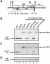
DIVa mutations partially block splicing in vivo. (A) The organization of aI2 and flanking sequences of the COXI gene. The tall rectangles above the line represent COXI exons, and the short rectangles represent intron reading frames. RT, X, D, and En denote domains associated with RT, maturase, DNA binding, and DNA endonuclease functions of the IEP (p62), respectively. The rectangles below the line indicate the positions of intron secondary structure domains; DIVa is the shaded box within DIV. Three consecutive stop codons placed in aI2 in strains 102ΔDIVa-stop and 102aI1DIVa-stop are at nt 840 to 848 of aI2, as indicated. (B to D) RNA blots. Samples of whole-cell RNA from the strains shown were balanced for their level of COB mRNA, separated on a formaldehyde-MOPS-1.5% agarose gel, and transferred to a nylon membrane, which was sequentially probed, stripped, and reprobed with oligonucleotides complementary to COXI exon 6 (B), intron aI2 (C), and COB exon 6 (D). Strains 102+ (lane 1) and 102ΔDV (lane 3) define the positions of COXI mRNA and aI2-containing pre-mRNA, respectively. The faint band migrating slower than COXI mRNA in lanes 1, 2, 4, and 6 of panel B is the pre-mRNA containing unspliced aI4α; the faint band migrating slower than the aI2-containing pre-mRNA in lanes 3 to 5 and 7 contains unspliced aI4α in addition to aI2.
FIG. 4.
FIG. 4.
DIVa mutations block homing. (A) Diagram of donor, recipient, and recombinant COXI alleles. The donor strain contains a short G+C-rich insertion that introduces an HpaII (H) site 387 nt upstream of the 3′ end of exon 1, which provides the upstream marker E1-387. Relevant fragments of HpaII (H)-BamHI (B) digests detected with an exon 1-specific probe are indicated. DdeI (D) sites that distinguish products of RT-dependent homing from RT-independent homing products and donor alleles are shown (see Materials and Methods for further details). (B) Southern blot-RFLP analysis of mtDNA from crosses. The mtDNA is digested with HpaII-BamHI and probed with an exon 1-specific oligonucleotide that distinguishes the three alleles as shown in panel A. Inputs of mtDNA from both parental strains were determined as described in Materials and Methods (data not shown, but see reference for an example) and used to calculate the extent of homing in each cross, reported below the gel.
FIG. 5.
FIG. 5.
Western blots of p62 levels. (A) p62 is present in DIVa mutant strains. Approximately 30 μg of protein from crude mitochondrial fractions of the indicated strains was separated by electrophoresis in an SDS-7.5% polyacrylamide gel, transferred to a nitrocellulose membrane, and subjected to Western blot analysis. p62-3HA is detected with the 6B12 anti-HA monoclonal antibody. An antibody to porin, a nucleus-encoded mitochondrial outer membrane protein, is used as a control for the loading of mitochondrial proteins. (B) Deletion of DIVa shortens p62. Whole-cell extracts of the strains shown were balanced for equal input of p62 and fractionated on the above gel system, but for a longer time of electrophoresis.
FIG. 6.
FIG. 6.
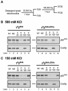
Fractionation experiments. (A) Diagram of the protocol for fractionating lysed mitochondria. Flotation-gradient-purified mitochondria were isolated and lysed as described in Materials and Methods. The lysate was fractionated by differential centrifugation to yield supernatant and pellet fractions as indicated. Pellet fractions were dissolved in SDS buffer, and SDS was added to aliquots of the supernatant fractions prior to gel analysis. (B) Fractionation of p62 from lysates containing 500 mM KCl. Equal aliquots of each fraction from strains with 3HA-tagged wild-type or ΔDIVa alleles of aI2 were analyzed in SDS-7.5% polyacrylamide gels, as described for Fig. 5A. M1 is a sample of mitochondria lysed by SDS and boiled. M2 is a sample of mitochondria lysed with NP-40 and held on ice until the fractionation steps were completed, with SDS added just prior to gel electrophoresis. The lower gel for each strain shows the distribution of the inner membrane protein CoxIIp, demonstrating that the NP-40 lysis completely dissolved the inner membrane under these conditions. Proteins that represent the outer membrane (porin) and matrix (Ilv5p) fractionated the same as CoxIIp for both strains. (C) Fractionation of p62 from lysates containing 150 mM KCl. This experiment has the same format as that in panel B, except that the lysis buffer contained 150 mM KCl.

Similar articles

Cited by

References

    1. Anziano, P. Q., J. V. Moran, D. Gerber, and P. S. Perlman. 1990. Novel hybrid maturases in unstable pseudorevertants of maturaseless mutants of yeast mitochondrial DNA. Nucleic Acids Res. 18:3233-3239. - PMC - PubMed
    1. Arnberg, A., G. V. Ommen, L. Grivell, E. V. Bruggen, and P. Borst. 1980. Some yeast mitochondrial RNAs are circular. Cell 19:313-319. - PubMed
    1. Belfort, M., V. Derbyshire, M. M. Parker, B. Cousineau, and A. M. Lambowitz. 2002. Mobile introns: pathways and proteins, p. 761-783. In N. L. Craig, R. Craigie, M. Gellert, and A. M. Lambowitz (ed.), Mobile DNA II. ASM Press, Washington, D.C.
    1. Bonitz, S. G., G. Coruzzi, B. E. Thalenfield, A. Tzagoloff, and G. Macino. 1980. Assembly of the mitochondrial membrane system. Structure and nucleotide sequence of the gene coding for subunit I of cytochrome oxidase. J. Biol. Chem. 255:11927-11941. - PubMed
    1. Boudvillain, M., A. de Lencastre, and A. M. Pyle. 2000. A tertiary interaction that links active-site domains to the 5′ splice site of a group II intron. Nature 406:315-318. - PubMed

Publication types

MeSH terms