. 2003 Dec;23(23):8846-61.
doi: 10.1128/MCB.23.23.8846-8861.2003.

The proline repeat domain of p53 binds directly to the transcriptional coactivator p300 and allosterically controls DNA-dependent acetylation of p53

Affiliations

The proline repeat domain of p53 binds directly to the transcriptional coactivator p300 and allosterically controls DNA-dependent acetylation of p53

David Dornan et al. Mol Cell Biol. 2003 Dec.

Abstract

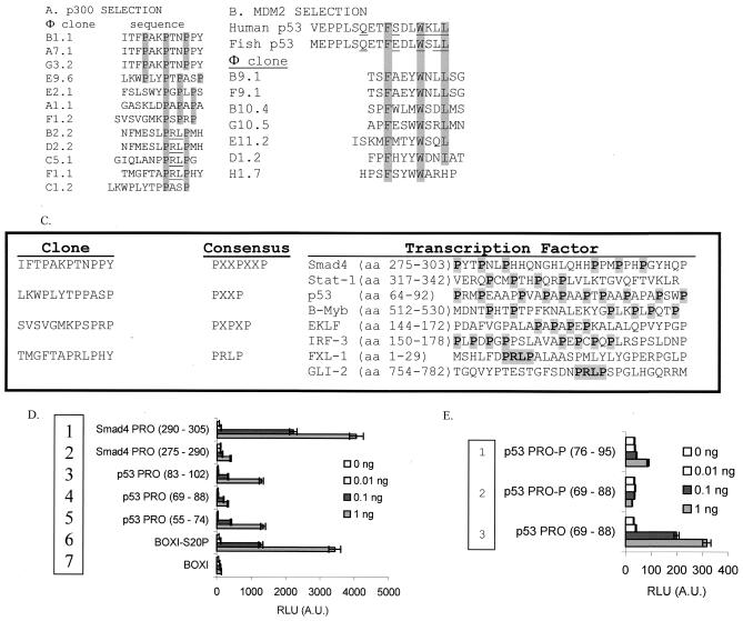
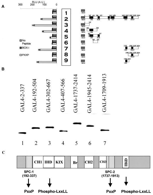
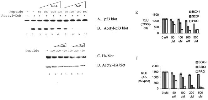
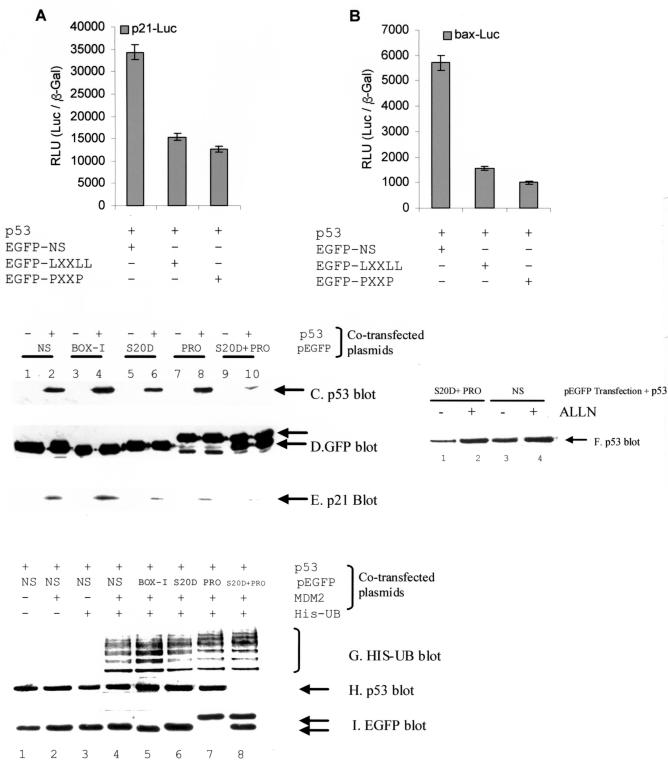
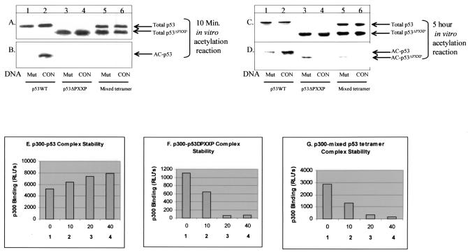
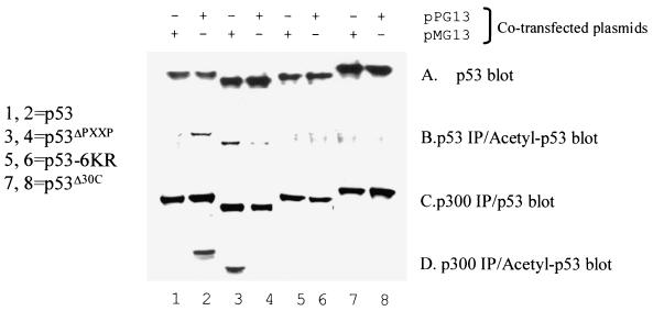
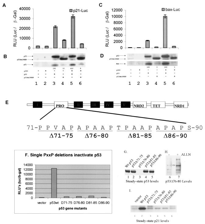
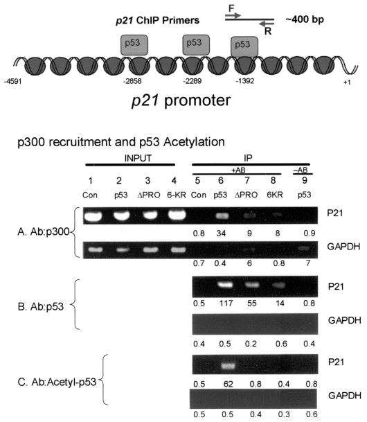
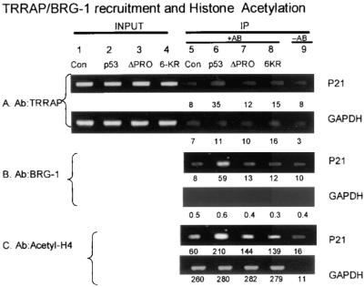
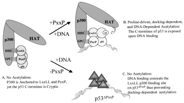
The transcription coactivator p300 cannot acetylate native p53 tetramers, thus revealing intrinsic conformational constraints on p300-catalyzed acetylation. Consensus site DNA is an allosteric effector that promotes acetylation of p53, suggesting that p300 has an undefined conformationally flexible interface within the p53 tetramer. To identify such conformationally responsive p300-binding sites, p300 was subjected to peptide selection from a phage-peptide display library, a technique that can define novel protein-protein interfaces. The enriched p300-binding peptides contained a proline repeat (PXXP/PXPXP) motif, and five proline repeat motifs actually reside within the p53 transactivation domain, suggesting that this region of p53 may harbor the second p300 contact site. p300 binds in vitro to PXXP-containing peptides derived from the proline repeat domain, and PXXP-containing peptides inhibit sequence-specific DNA-dependent acetylation of p53, indicating that p300 docking to both the LXXLL and contiguous PXXP motif in p53 is required for p53 acetylation. Deletion of the proline repeat motif of p53 prevents DNA-dependent acetylation of p53 by occluding p300 from the p53-DNA complex. Sequence-specific DNA places an absolute requirement for the proline repeat domain to drive p53 acetylation in vivo. Chromatin immunoprecipitation was used to show that the proline repeat deletion mutant p53 is bound to the p21 promoter in vivo, but it is not acetylated, indicating that proline-directed acetylation of p53 is a post-DNA binding event. The PXXP repeat expands the basic interface of a p300-targeted transactivation domain, and proline-directed acetylation of p53 at promoters indicates that p300-mediated acetylation can be highly constrained by substrate conformation in vivo.


Figures

FIG. 1.
FIG. 1.
Proline repeat peptides bind p300. (A) Identification of novel p300-binding peptide motifs by phage-peptide display. The clones isolated using full-length recombinant p300 protein as a bait for phage-peptide display are highlighted, with the consensus motifs in grey, and include four representative subsets: PXXPXXP, PXXP, PRLP, and PXPXP. (B) Control reactions using MDM2. Using MDM2 as bait for phage-peptide display, clones containing the canonical MDM2-binding motif, FXXXWXXL, were isolated. (C) Sequence alignments of the four proline repeat motifs found in commonly studied transcription factors. (D and E) p300 binds directly to proline repeat motif peptides. Biotinylated peptides were used as a target for p300 in a microtiter well pull-down assay containing proline repeat peptide sequences from the aligned Smad4 (PXXPXXP, rows 1 and 2) and p53 (PXXP, rows 3 to 5) regions (D). As a control, p300 binds to the Ser20 phosphorylated LXXLL motif from the known BOX-I transactivation domain of p53 (row 6) versus unphosphorylated LXXLL motif (row 7). Phosphovariants of the PXXP domain at Thr81 (JNK site, rows 1 and 2) or the unphosphorylated PXXP domain of p53 (row 3) were used as ligands (E). The amounts of peptide captured are indicated (0, 0.01, 0.1, and 1 ng), and the amount of p300 protein bound to the indicated peptide domain was quantitated as RLU by using a peroxidase-linked secondary-antibody coupled to an anti-p300 antibody.
FIG. 2.
FIG. 2.
Mapping of the proline repeat binding domain of p300 (SPC-1/2). (A) HCT116 p53−/− cells were transfected with 5 μg of GAL4 control (row 1), GAL4-p300 (row 2), or the indicated GAL4-p300 miniproteins derived from the N and C termini of p300 (rows 3 to 9). Lysates were captured onto the solid phase with an anti-GAL4 antibody, and the indicated biotinylated peptide (1 ng) (PXXP peptide [from p53 aa 55 to 74], unphosphorylated LXXLL motif [from p53], or no peptide) was added and the amount of peptide bound to p300 protein was quantitated as RLU by using peroxidase linked to streptavidin. (B) Quantitation of GAL4 fusion protein levels. HCT116 p53−/− cells were transfected with 5 μg of GAL4 control (lane 1), GAL4-p300 (lane 2), or the indicated GAL4-p300 miniproteins derived from the N and C termini of p300 (lanes 3 to 9). Lysates were immunoblotted with an anti-GAL4 antibody. (C) Schematic diagram illustrating the two minimal sites for proline contact (SPC-1 and SPC-2) relative to other p300 subdomains including C/H1, IHD, C/H3, KIX, Bromo, C/H2, and IBiD. The minimal N-terminal SPC-1 domain (aa 192 to 337) is distinct from the phospho-LXXLL p53-binding domain IHD. The minimal C-terminal SPC-2 domain (aa 1737 to 1913) is distinct from the IBiD domain and partially overlaps with the C/H3 domain (aa 1653 to 1817).
FIG. 3.
FIG. 3.
Proline repeat peptides inhibit DNA-dependent acetylation of p53. (A and B) p300-mediated acetylation of p53 is inhibited by PXXP peptides. Acetylation reactions using p300 and p53 were carried out with or without acetyl-CoA and with the addition of various concentrations of peptides (50 to 400 ng): LXXLL peptide (lanes 3 to 6) or the PXXP peptide (lanes 7 to 10; proline repeat derived from p53; aa 55 to 74; see Fig. 1). Shown are results for total p53 (A) and acetylated p53 (B). (C and D) Histone acetylation is not inhibited by PXXP. Acetylation reactions contained 1 μg of histone H4 as the substrate. Acetylation reactions were carried out with various concentrations of peptides (100 to 400 ng): LXXLL peptide or PXXP peptide. Shown are results for total histone (C) and acetylated histone (D). (E) Quantitation of p300-p53 complex stability in the presence of the phospho-LXXLL or PXXP peptide. Acetylation reactions were carried out as described for panels A and B but without acetyl-CoA and consensus site DNA and with various concentrations of peptides as indicated: BOX-I (LXXLL), Ser20 phospho-LXXLL (S20-P), or PXXP peptide (concentrations in micromolar). After capturing p53 with the monoclonal antibody ICA-9, the relative levels of p300 bound to p53 was quantified by using an anti-p300 antibody followed by a peroxidase-conjugated secondary antibody coupled to chemiluminescence. p300 binding is expressed in RLU. (F) Quantitation of p53 acetylation in the presence of the phospho-LXXLL or PXXP peptide. Acetylation reactions were carried out as described for panels A and B with acetyl-CoA, consensus site DNA, and various concentrations of peptides as indicated: BOX-I (LXXLL), Ser20 phospho-LXXLL (S20-P), or PXXP peptide. After capturing total p53 with the monoclonal antibody ICA-9, the relative levels of acetylation of p53 were normalized to total p53 protein levels by using an anti-p53 polyclonal antibody (CM-1) and the anti-p53 acetylation polyclonal antibody (RLU).
FIG. 4.
FIG. 4.
p300-binding peptides attenuate p53 activity and destabilize p53 protein steady-state levels in vivo. (A and B) Competitive inhibition of p53-dependent transcription by p300-binding peptides. pCMV-p53 (1 μg) was cotransfected with the luciferase reporters (1 μg of p21-Luc [A] or bax-Luc [B] and the transfection control [pCMVβ-Gal] and either the GFP-NS [1 μg; control], GFP-LXXLL phosphomimetic [1 μg], or GFP-PXXP [1 μg] peptide). The RLU is expressed as a ratio of Luc to the internal transfection control (pCMVβ-Gal). (C to E) p53 and p21 protein levels are destabilized by cotransfection of GFP fusion p300-binding peptides. pCMV-p53 (1 μg) and the indicated GFP fusion construct (1 μg; NS, nonspecific; BOX-I, the MDM2-binding peptide; S20D, the phosphomimetic LXXLL; PRO, proline repeat) and the levels of the indicated proteins were determined by immunoblotting. (F) Destabilization of p53 by p300 targeting is mediated by the proteosome. Acetyl-Leu-Leu-norleucine (ALLN) was added for 2 h after transfection of the indicated plasmids for 24 h, and the levels of p53 protein are as indicated. (G to I) p53 ubiquitination by MDM2 is not altered after cotransfection of GFP fusion p300-binding peptides. pCMV-p53 (1 μg), the indicated GFP fusion construct (1 μg), pCMV-MDM2, or pCMV-His-ubiquitin was transfected for 24 h, and the levels of the indicated proteins were determined by immunoblotting. MDM2-dependent ubiquitinated products were purified using a His pull-down assay, blotted with p53 antibodies, and are shown in panel G.
FIG. 5.
FIG. 5.
The PXXP transactivation motif of p53 is required for sequence-specific DNA-dependent acetylation by p300. (A and B) The PXXP motif of p53 is required for DNA-dependent acetylation by p300 in vitro. Human recombinant p53 (lanes 1 to 2), p53ΔPXXP (lanes 3 to 4), or p53/p53ΔPXXP mixed tetramers (lanes 5 to 6) were purified from insect cells and incubated in the linear range for the enzyme assay (10 min) with p300 and p53 consensus site oligonucleotide DNA (CON, lanes 2, 4, or 6) or nonspecific DNA (Mut, lanes 1, 3, and 5). Relative acetylation was quantitated by Western blotting using an anti-p53 acetylation antibody (B) and normalized to total p53 protein by using the monoclonal antibody DO-1 (A). (C and D) The PXXP motif of p53 is required for DNA-dependent acetylation by p300 in vitro. Details are as described for panels A and B, except that reactions were carried out for 5 h instead of 10 min. (E to G) The PXXP motif of p53 is required for maximal p300-p53 complex stability in the presence of consensus site DNA. Insect cell-purified human recombinant P53 (E), p53ΔPXXP (F), or p53/p53ΔPXXP mixed tetramers (G) were captured onto the solid phase of a microtiter well with the ICA-9 anti-p53 monoclonal antibody, including a titration of p53 consensus site DNA as indicated in panels 1 to 4 (0, 10, 20, or 40 ng). The captured p53 isoforms were incubated with buffer containing insect cell-expressed recombinant human p300 protein, and the amount of p300 bound to p53 was quantitated using anti-p300 antibodies. The levels of the p53 protein isoforms were determined using an anti-p53 antibody and quantitated as the ratio of p300 bound to total p53 by using enhanced chemiluminescence and expressed as RLU.
FIG. 6.
FIG. 6.
The PXXP motif of p53 is required for sequence-specific DNA-dependent acetylation by p300 in vivo. pCMV-P53 (lanes 1 and 2), pCMV-p53ΔPXXP (lanes 3 and 4), pCMV-p53-6KR (lanes 5 and 6; mutated acetylation sites), or pCMV-p53Δ30 (lanes 7 and 8; lacking acetylation sites) were cotransfected into p53−/− cells in the presence of consensus site plasmid DNA (pG13-CAT, lanes 2, 4, 6, and 8) or plasmid DNA without the consensus site (pMG13-CAT, lanes 1, 3, 5, and 7). (A) The amount of total p53 in each transfection was quantified by direct immunoblotting with the monoclonal antibody DO-1. (B) To quantitate p53 acetylation in vivo, total p53 protein was immunoprecipitated with a mixture of ICA-9 and DO-1 monoclonal antibodies and the levels of acetylated p53 were quantitated by immunoblotting with the acetylation-specific antibody. (C and D) Endogenous P300 protein was immunoprecipitated with an anti-p300 antibody, and levels of total p53 protein isoforms (C) or acetylated p53 isoforms (D) bound to p300 were quantitated by immunoblotting with either the acetylation-specific antibody or a p53 protein antibody to normalize to the total p53 protein.
FIG. 7.
FIG. 7.
Deletion of a single PXXP motif attenuates p53 activity in vivo. (A to D) Deletion of the entire proline repeat domain inhibits p53. The transactivation activity of p53 and p53ΔPXXP on the p21 (A) and bax (C) luciferase reporter promoters (RLU) is expressed as the ratio of p21-Luc or bax-Luc to the internal transfection control (pCMVβ-Gal). Expression levels of transfected p53 forms and endogenous target gene products are shown in panels B and D as follows: (B) transfected p53 protein (lanes 3 and 5) or p53ΔPXXP protein (lanes 4 and 6) and endogenous p21 protein and (D) transfected p53 protein (lanes 3 and 5) or p53ΔPXXP protein (lanes 4 and 6) and endogenous Bax protein were quantitated by Western blotting with an anti-p53, anti-p21, or anti-Bax protein antibody and enhanced chemiluminescence. In each transfection, 1 μg of pCMV-p53 or pCMV-p53ΔPXXP alone or with 5 μg of pCMVβ-p300 was added as indicated. (E) Map of the clustered PXXP repeat motifs in the N-terminal activation domain of p53 and site of the PXXP deletions used in this study. (F) The transactivation activity of p53 and individual p53ΔPXXP deletion mutants on the p21 luciferase reporter promoter (RLU) is expressed as a ratio of p21-Luc to the internal transfection control (pCMVβ-Gal). (G and I) Expression levels of transfected p53ΔPXXP deletion mutant p53 proteins (G) and endogenous p21 protein (I) were quantified by immunoblotting. (H) The unstable PXXP deletion mutant (76-80) was stabilized by ALLN. Twenty-four hours after transfection of p53Δ76-80, ALLN was added for 2 h prior to harvesting of the cells and p53 was blotted with DO-1 monoclonal antibody.
FIG. 8.
FIG. 8.
The PXXP motif is required to acetylate p53 at the p21 promoter in vivo. HCT116 p53−/− cells were transfected with various p53 pCMV DNAs encoding p53 (lanes 2 and 6), p53ΔPXXP (lanes 3 and 7), and the nonacetylatable mutant, p53-6KR (lanes 4 and 8). Following transfection of the indicated construct, cross-linking of protein-DNA complexes with formaldehyde, immunoprecipitation, and processing of the samples as described in Materials and Methods, the released DNA was PCR amplified using primers to the p21 promoter (as indicated in the bottom panel) or GAPDH promoter. The immunoprecipitation was carried out with antibodies specific for p300 (A), p53 (B), and acetylated p53 (C). The data are plotted as input DNA or as immunoprecipitated DNA. Quantitation of the bioluminescence is depicted below each lane. Controls include DNA amplified in reactions processed from cells without antibody in the immunoprecipitation reaction (lane 9) or with vector control only (lanes 1 and 5). The diagram at top depicts the region of the p21 promoter that was focused onto isolate p53-bound transcription complexes.
FIG. 9.
FIG. 9.
The PXXP motif is required to recruit TRRAP and BRG-1 to the p21 promoter in vivo. HCT116 p53−/− cells were transfected with various p53 pCMV DNAs encoding p53 (lanes 2 and 6), p53ΔPXXP (lanes 3 and 7), and the nonacetylatable mutant, p53-6KR (lanes 4 and 8). Following transfection of the indicated construct, cross-linking of protein-DNA complexes with formaldehyde, immunoprecipitation, and processing of the samples as described in Materials and Methods, the released DNA was PCR amplified using primers to the p21 promoter (as indicated in the bottom panel) or GAPDH promoter. The immunoprecipitation was carried out with antibodies specific for TRRAP (A), BRG-1 (B), and acetylated histone H4 (C). The data are plotted as input DNA or as immunoprecipitated DNA. Quantitation of the bioluminescence is depicted below each lane. Controls include DNA amplified in reactions processed from cells without antibody in the immunoprecipitation reaction (lane 9) or with vector control only (lanes 1 and 5).
FIG. 10.
FIG. 10.
DNA induces a conformational change that mediates proline-directed p53 acetylation by p300. (A) In the absence of DNA, p300 can dock to the p53 tetramer via LXXLL and PXXP binding, but conformational constraints in the native p53 tetramer prevent acetylation. (B) Sequence-specific DNA binding changes the conformation of p53 (34), thus permitting acetylation to occur in a PXXP-dependent manner. Thus, DNA binding does not change p300 binding as much as it activates p53 acetylation, suggesting that in the DNA-free state the C terminus of p53 is cryptic with respect to acetylation. (C) When the PXXP domain is deleted, p300 cannot acetylate DNA-bound p53ΔPXXP due to the inability of p300 to form stable contacts with the p53ΔPXXP tetramer. However, the p53ΔPXXP tetramer can be acetylated by p300 in the absence of DNA, with the proline deletion essentially converting the p53ΔPXXP tetramer to a histone-like substrate which can be acetylated in a docking-independent and DNA-independent manner. These latter data also suggest that DNA binding creates a specific interface in p53 for p300 and that the PXXP domain forms an integral part of this interface. Thus, both the PXXP/LXXLL domains and the C-terminal acetylation motif act concertedly after DNA binding to permit p300-catalyzed acetylation. There is a precedent for the C-terminal acetylation domain of p53 being cryptic in the DNA-free state. The p300 acetylation sites in p53 are within the epitope for monoclonal antibody PAb421. When p53 is DNA free, the PAb421 epitope is cryptic and DNA binding exposes the epitope. This led to the postulation that long-range allosteric effects mediate DNA binding by p53 (19). The cryptic nature of the p53 acetylation motif in the DNA-free conformation of p53 builds into the tetramer an intrinsic negative regulatory mechanism to prevent acetylation until the tetramer is promoter bound.

Similar articles

Cited by

References

    1. Ashraf, S. S., E. Anderson, K. Duke, P. T. Hamilton, and Z. Fredericks. 2003. Identification and characterization of peptide probes directed against PKCα conformations. J. Pept. Res. 61:263-273. - PubMed
    1. Avantaggiati, M. L., V. Ogryzko, K. Gardner, A. Giordano, A. S. Levine, and K. Kelly. 1997. Recruitment of p300/CBP in p53-dependent signal pathways. Cell 89:1175-1184. - PubMed
    1. Baptiste, N., P. Friedlander, X. Chen, and C. Prives. 2002. The proline-rich domain of p53 is required for cooperation with anti- neoplastic agents to promote apoptosis of tumor cells. Oncogene 21:9-21. - PubMed
    1. Barlev, N. A., L. Liu, N. H. Chehab, K. Mansfield, K. G. Harris, T. D. Halazonetis, and S. L. Berger. 2001. Acetylation of p53 activates transcription through recruitment of coactivators/histone acetyltransferases. Mol. Cell 8:1243-1254. - PubMed
    1. Bell, S., C. Klein, L. Muller, S. Hansen, and J. Buchner. 2002. p53 contains large unstructured regions in its native state. J. Mol. Biol. 322:917-927. - PubMed

Publication types