Comparative Study
. 2003 Dec;23(23):8878-89.
doi: 10.1128/MCB.23.23.8878-8889.2003.

Cell-type-specific activation of PAK2 by transforming growth factor beta independent of Smad2 and Smad3

Affiliations
Comparative Study

Cell-type-specific activation of PAK2 by transforming growth factor beta independent of Smad2 and Smad3

Mark C Wilkes et al. Mol Cell Biol. 2003 Dec.

Abstract

Transforming growth factor beta (TGF-beta) causes growth arrest in epithelial cells and proliferation and morphological transformation in fibroblasts. Despite the ability of TGF-beta to induce various cellular phenotypes, few discernible differences in TGF-beta signaling between cell types have been reported, with the only well-characterized pathway (the Smad cascade) seemingly under identical control. We determined that TGF-beta receptor signaling activates the STE20 homolog PAK2 in mammalian cells. PAK2 activation occurs in fibroblast but not epithelial cell cultures and is independent of Smad2 and/or Smad3. Furthermore, we show that TGF-beta-stimulated PAK2 activity is regulated by Rac1 and Cdc42 and dominant negative PAK2 or morpholino antisense oligonucleotides to PAK2 prevent the morphological alteration observed following TGF-beta addition. Thus, PAK2 represents a novel Smad-independent pathway that differentiates TGF-beta signaling in fibroblast (growth-stimulated) and epithelial cell (growth-inhibited) cultures.


Figures

FIG. 1.
FIG. 1.
TGF-β activates PAK2 in fibroblast but not epithelial cells. (A, left half) AKR-2B fibroblasts were grown to confluence, serum starved for 24 h, and stimulated with 10 ng of TGF-β per ml for the indicated times. Stimulation with platelet-derived growth factor (25 ng/ml) for 20 min was used as a positive control for PAK1 and PAK2 in fibroblasts (+). Following immunoprecipitation, the in vitro kinase activity of the various PAK proteins was examined with myelin basic protein. (A, right half) Prior to immunoprecipitation and kinase assay, 100 μg of protein was used for Western analysis against the various PAK proteins. (B) The same protocols as described for panel A were used with Mv1Lu epithelial cells. The results shown are representative of three separate experiments. (C) Fibroblast cell lines AKR-2B, Swiss 3T3, and BALB/c 3T3, along with epithelial cell lines Mv1Lu, HeLa, and LLC-pk1, were grown to confluence, serum starved overnight, and either left untreated (−) or stimulated (+) with 10 ng of TGF-β per ml for 45 min at 37°C. Samples were normalized for total protein (within a cell type) and assayed for kinase activity (top) or Western blotted for total PAK2 (bottom). The same protocols were used for human skin fibroblasts (HSF) and human prostate epithelial cells (HPE).
FIG. 2.
FIG. 2.
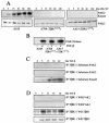
PAK2 activation occurs downstream of the TGF-βR complex. (A and B) Requirement for TGF-βR kinase activity. Stable cell lines expressing either wild-type (A105) or kinase-deficient (A708, TβR1K232R; A615, TβR2K277R) chimeric TGF-βRs were processed for kinase activity and/or PAK2 expression following stimulation with 10 ng of GM-CSF per ml for the indicated times (A) or TGF-β for 45 min (+, B) (1). (C) PAK2 is not a substrate for the TGF-βR complex. AKR-2B cells were grown to confluence and serum starved for 24 h prior to cell lysis and immunoprecipitation (IP) with antibodies to the type I (TβR1) or II (TβR2) TGF-βR after stimulation with TGF-β (10 ng/ml) for the indicated times. Purified PAK2 (top and middle) and Smad2 (bottom) were used as in vitro kinase substrates as previously described (31). (D) PAK2 does not bind the TGF-βR complex. AKR-2B cells were grown to confluence, serum starved for 24 h, and stimulated with 10 ng of TGF-β per ml for 5 to 60 min. Cultures were lysed and immunoprecipitated with the indicated TGF-βR antibody, and associated PAK2, TβR1, or TβR2 was determined by Western blot (WB) analysis.
FIG. 3.
FIG. 3.
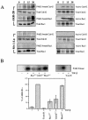
PAK2 activation is independent of Smad proteins. Fibroblast cell lines with a deletion of the gene for Smad2 (Smad2 knockout, vertical columns 1 and 2) or Smad3 (Smad3 knockout, vertical columns 3 and 4) were grown to confluence, serum starved overnight, and either left untreated (−) or stimulated (+) for 45 min with 10 ng of TGF-β per ml. Samples were lysed, normalized for equal protein, split into six aliquots, and tested for PAK2 activation (PAK2 kinase), phospho-Smad2 (P-Smad2), phospho-Smad3 (P-Smad3), or the corresponding total protein. As there is potential cellular redundancy between Smad2 and Smad3, a dominant negative Smad2-GFP construct (Smad2S467A) was transiently transfected into the Smad3 knockout cell line and GFP-positive cells were sorted by flow cytometry (data not shown). Cultures (vertical columns 5 an 6) were left untreated (−) or stimulated (+) with 10 ng of TGF-β per ml for 45 min and processed as described for the other panels.
FIG. 4.
FIG. 4.
Smad phosphorylation is independent of PAK2 kinase activity. (A) AKR-2B cells (Control, vertical columns 1 and 2) or stable AKR-2B clones harboring tetracycline-on regulated wild-type PAK2 (EGFP-PAK2, vertical columns 3 and 4; see Materials and Methods) were grown to confluence, serum starved overnight, and either left untreated (−) or stimulated (+) with 10 ng of TGF-β per ml for 45 min. EGFP-PAK2-expressing clones were grown in the presence of 10 μg of tetracycline per ml. Cells were lysed and assayed for the indicated total or activated proteins as described in the legend to Fig. 3. (B) Studies similar to those in panel A were performed with AKR-2B cells (Control, vertical columns 1 and 2) or stable AKR-2B clones harboring tetracycline-on regulated dominant negative PAK2 (EGFP-PAK2K278R, vertical columns 3 and 4).
FIG. 5.
FIG. 5.
Rac1 and Cdc42, but not RhoA or Rac2, regulate PAK2 activation by TGF-β. (A) AKR-2B fibroblasts or Mv1Lu epithelial cells were grown to confluence, serum starved for 24 h, and stimulated with 10 ng of TGF-β per ml for the indicated times (in minutes). (Left half) PAK2 was immunoprecipitated (IP), and associated Cdc42 or Rac1 was determined by Western blotting. (Right half) GTP-bound Cdc42 or Rac1 was evaluated by binding to GST-PBD. Prior to immunoprecipitation, 100 μg of protein from the same lysate was analyzed for total Cdc42 or Rac1. (B, top left) AKR-2B fibroblasts were transiently transfected with either wild-type EGFP-PAK2 or both EGFP-PAK2 and dominant-negative Rac1 (Rac1N17), Cdc42 (Cdc42N17), or RhoA (RhoAN19). Cells were serum starved for 24 h and either left untreated (−) or stimulated (+) for 45 min with 10 ng of TGF-β per ml. Following immunoprecipitation with an anti-GFP antibody, the in vitro kinase activity of the transfected EGFP-PAK2 protein was determined. (B, top right) AKR-2B cultures were untreated or pretreated for 6 h with Clostridium difficile toxin B (2 ng/ml) prior to stimulation (+) with 10 ng of TGF-β per ml for 45 min. PAK2 kinase activity was assessed as described in Materials and Methods. (B, bottom) TGF-β-stimulated PAK2 kinase activity was quantified following disruption of the indicated Rho pathways and normalized to that of control AKR-2B cells treated with 10 ng of TGF-β per ml as in panel B (top two parts). For the dominant negative Rho mutants, the values represent the remaining transfected EGFP-PAK2 kinase activity while the effect of toxin B reflects endogenous PAK2. Results represent the mean ± the standard deviation of four separate experiments.
FIG. 6.
FIG. 6.
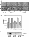
Dominant negative PAK2 prevents TGF-β-stimulated morphological transformation and cell proliferation. (A) Parental AKR-2B cells or AKR-2B clones stably transfected with tetracycline-on wild-type (PAK2-wt) or dominant negative EGFP-PAK2 (PAK2K278R) were grown to confluence in either the absence (−tet) or the presence (+tet) of 10 μg of tetracycline per ml, serum starved for 24 h, and either left untreated (−) or stimulated (+) for 48 h with 10 ng of TGF-β per ml. Representative areas were photographed at a magnification of ×20 by phase microscopy. (B) AKR-2B cells were plated at 2.5 × 10⁵ per six-well dish and grown overnight at 37°C. The serum-containing medium was removed and replaced with serum-free DME alone or containing the indicated multiplicity of infection (MOI) of adenovirus expressing GFP (Control) or dominant negative PAK2 (PAK2K278R). Following 24 h of incubation, TGF-β was directly added to a final concentration of 5 ng/ml (solid bars) and the cell number was determined 48 h later. Dotted bars show results after addition of the indicated virus to serum-free DME. Results represent the mean ± the standard deviation of two separate experiments done in duplicate. (C) AKR-2B cells were plated at 2 × 10⁶ per 100-mm culture dish and cultured as described for panel B. Following 45 min of treatment with (+) or without (−) 10 ng of TGF-β per ml, cell lysates were prepared and PAK2 kinase activity was determined (Materials and Methods).
FIG.7.
FIG.7.
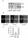
PAK2 antisense oligonucleotides inhibit TGF-β morphological transformation. (A) Cos7 cells were plated at 8.0 × 10⁴ per six-well dish and grown for 2 days prior to transient transfection with either PAK2 antisense morpholino oligonucleotides (PAK2 MO) or invert control (Control MO). Total PAK1 and PAK2 protein was determined as described in Materials and Methods following 48 h of stimulation in the absence (−) or presence (+) of 10 ng of TGF-β per ml. (B) AKR-2B cells were treated as described for panel A. Transfected and total cell populations were visualized by immunofluorescence (IF) and phase-contrast (P) microscopy, respectively, and representative fields were photographed at an original magnification of ×20. (C) Transfected cells were scored as to whether TGF-β (+) stimulated the morphological change where the length was greater than or equal to five times the width shown in panel B. A similar analysis was performed with the untransfected cultures. The data are the mean ± the standard deviation of two separated experiments done in triplicate and represent a total of more than 1,000 cells per treatment.
FIG. 8.
FIG. 8.
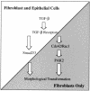
Cell-type-specific PAK2 activation by TGF-β. A model depicting our present understanding of PAK2 activation by TGF-β in fibroblasts is shown. Following ligand binding to the TGF-βR complex, fibroblasts and epithelial cells differentially integrate the signal(s) such that Smad-dependent signaling is activated in both cell types while PAK2 becomes activated only in fibroblasts. Activation of PAK2 is dependent on Rho GTPases Cdc42 and Rac1. While the Smad- and PAK2-dependent pathways are believed to be distinct, both are required for fibroblast morphological transformation and cellular proliferation (Fig. 6 and 7) (13, 17). Arrows do not necessarily indicate a direct interaction and may reflect multiple events.

Similar articles

Cited by

References

    1. Anders, R. A., J. E. J. Doré, S. A. Arline, N. Garamszegi, and E. B. Leof. 1998. Differential requirements for type I and type II TGFβ receptor kinase activity in ligand-mediated receptor endocytosis. J. Biol. Chem. 273:23118-23125. - PubMed
    1. Anders, R. A., and E. B. Leof. 1996. Chimeric granulocyte/macrophage colony-stimulating factor/transforming growth factor-β (TGF-β) receptors define a model system for investigating the role of homomeric and heteromeric receptors in TGF-β signaling. J. Biol. Chem. 271:21758-21766. - PubMed
    1. Bagrodia, S., and R. A. Cerione. 1999. PAK to the future. Trends Cell Biol. 9:350-355. - PubMed
    1. Bhowmick, N. A., M. Ghiassi, A. Bakin, M. Aakre, C. A. Lundquist, M. E. Engel, C. L. Arteaga, and H. L. Moses. 2001. Transforming growth factor-β1 mediates epithelial to mesenchymal transdifferentiation through a RhoA-dependent mechanism. Mol. Biol. Cell 12:27-36. - PMC - PubMed
    1. Bissell, D. M. 2001. Chronic liver injury, TGF-β, and cancer. Exp. Mol. Med. 33:179-190. - PubMed

Publication types

MeSH terms