. 2003 Oct 27;1(1):3.
doi: 10.1186/1479-0556-1-3.

Chitosan IFN-gamma-pDNA Nanoparticle (CIN) Therapy for Allergic Asthma

Affiliations

Chitosan IFN-gamma-pDNA Nanoparticle (CIN) Therapy for Allergic Asthma

Mukesh Kumar et al. Genet Vaccines Ther. .

Abstract

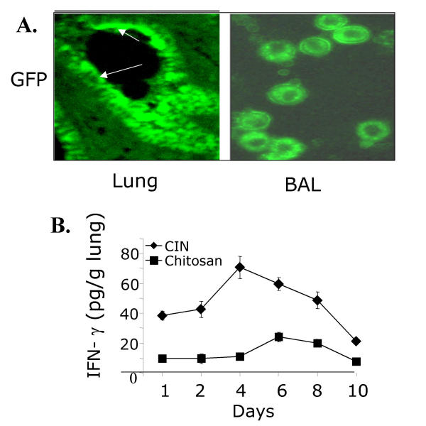
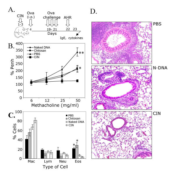
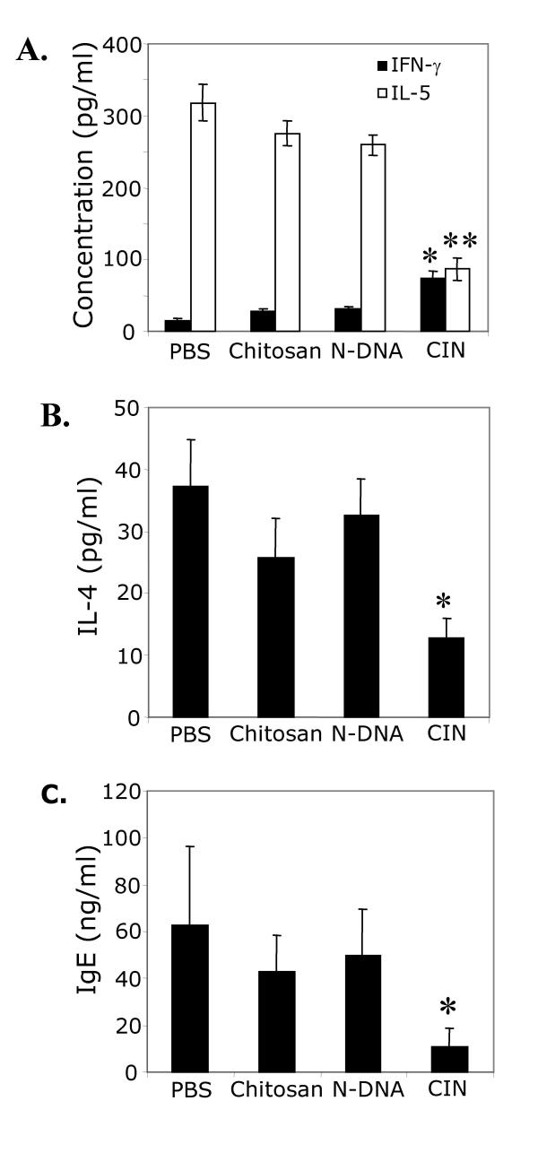
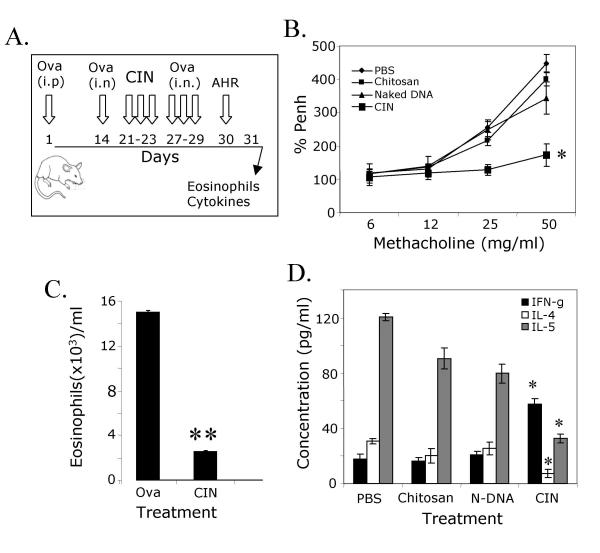
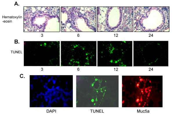
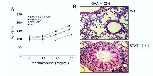
BACKGROUND: Allergic subjects produce relatively low amounts of IFN-gamma, a pleiotropic Th-1 cytokine that downregulates Th2-associated airway inflammation and hyperresponsiveness (AHR), the hallmarks of allergic asthma. Adenovirus-mediated IFN-gamma gene transfer reduces AHR, Th2 cytokine levels and lung inflammation in mice, but its use would be limited by the frequency of gene delivery required; therefore, we tested chitosan/IFN-gamma pDNA nanoparticles (CIN) for in situ production of IFN-gamma and its in vivo effects. METHODS: CIN were administered to OVA-sensitized mice to investigate the possibility of using gene transfer to modulate ovalbumin (OVA)-induced inflammation and AHR. RESULTS: Mice treated with CIN exhibit significantly lower AHR to methacholine challenge and less lung histopathology. Production of IFN-gamma is increased after CIN treatment while the Th2-cytokines, IL-4 and IL-5, and OVA-specific serum IgE are reduced compared to control mice. AHR and eosinophilia are also significantly reduced by CIN therapy administered therapeutically in mice with established asthma. CIN was found to inhibit epithelial inflammation within 6 hours of delivery by inducing apoptosis of goblet cells. Experiments performed on STAT4-defective mice do not show reduction in AHR with CIN treatment, thus implicating STAT4 signaling in the mechanism of CIN action. CONCLUSION: These results demonstrate that mucosal CIN therapy can effectively reduce established allergen-induced airway inflammation and AHR.


Figures

Figure 1
Figure 1
Chitosan nanoparticles target lung epithelial and monocytic cells. (A) BALB/c mice were treated i.n. with chitosan nanoparticles containing pGFP. After 24 h, mice were sacrificed and their lungs were fixed and sectioned by cryotome. Sections (15 micron) were thaw-mounted to slides and viewed for green fluorescent protein ('Lung'). BAL cells were fixed after cytospin on a slide and visualized by fluorescence microscopy to identify GFP-expressing cells ('BAL'). (B) CIN administration induced IFN-γ production in the lung over a period of 10 days. Lung homogenates were prepared from mice after 1, 2, 4, 6, 8, or 10 days of treatment with CIN (25 μg/mouse) or chitosan alone, and IFN-γ levels were determined by ELISA (n = 3).
Figure 2
Figure 2
Prevention of AHR. (A) Prophylaxis protocol. (B) Mice were challenged with methacholine on day 22 to measure airway responsiveness. The values are mean enhanced pause (PENH) expressed as percent of baseline ± SEM (* P < 0.05 and **P < 0.01). (C) On day 24, BAL was performed and differential cell counts were obtained ('mac', macrophages; 'lym', lymphocytes; 'neu', neutrophils; 'eos', eosinophils). (D) On day 24, lungs were removed, sectioned and the sections stained with hematoxylin/eosin ('PBS', phosphate-buffered saline control; 'N-DNA', naked DNA without chitosan; 'CIN', chitosan-DNA complex). Differential cell counts and examination of tissue sections were performed by different persons in a blinded fashion. Representative results are shown.
Figure 3
Figure 3
CIN alters production of cytokines and IgE. On day 23 of the prophylactic procedure (see Fig. 2A) spleens were removed and single-cell suspensions of splenocytes were prepared. Cells were cultured for 48 h with OVA, and the levels of secreted IFN-γ and IL-5 (A) and IL-4 (B) were measured. Total serum IgE was measured on day 23 (C). Values are means ± SEM (*p < 0.05, **p < 0.01).
Figure 4
Figure 4
Reversal of established AHR and eosinophilia. (A) Therapeutic protocol. (B) Mice were sensitized i.p. and challenged i.n. with OVA and treated with CIN as described. AHR was measured 24 h after the last challenge (n = 4). CIN-treated mice exhibited reduced AHR compared to the controls. Data are mean enhanced pause (PENH) expressed as percent of baseline ± SEM (*p < 0.05). (C) On day 31, BAL was performed and eosinophils in BAL fluid were counted (**p < 0.01). (D) On day 23, spleens were removed and single-cell suspensions of splenocytes prepared. Cells were cultured for 48 hours in the presence of OVA and cell supernatants were analyzed for IFN-γ, IL-4 and IL-5. Mice receiving CIN showed more IFN-γ and less IL-4 and IL-5 compared to the chitosan-only control. Data are means ± SEM (*p < 0.05).
Figure 5
Figure 5
CIN treatment induces apoptosis of goblet cells. BALB/c mice (n = 3) were sensitized and challenged with OVA as in Fig. 4 and then treated i.n. with CIN. Mice were sacrificed at 0, 3, 6, 12 and 24 h after CIN treatment and lungs were removed, sectioned and stained with hematoxylin/eosin (Fig. 5A), or unstained sections were analysed for apoptosis by TUNEL (terminal dUTP nick end labeling) assay (Fig. 5B). A final set of lung sections (Fig. 5C, 6 h time point) was stained for the goblet cell-specific protein Muc5a, and for apoptosis by the TUNEL assay. The first panel shows staining of nuclei with diamidinophenylindole (DAPI).
Figure 6
Figure 6
CIN therapy involves the STAT4 pathway. OVA-sensitized BALB/c wild type (WT) and STAT4-/- knockout mice (n = 4) were given CIN therapy intranasally and challenged with OVA. (A) AHR in response to methacholine was measured one day after the last challenge. The values are means ± SEM (*p < 0.05). (B) Mice were sacrificed the day following AHR measurement and their lungs were removed, paraffin-embedded and stained with hematoxylin/eosin.

References

    1. Mosman TR, Coffman RL. TH1 and TH2 cells:ent patterns of lymphokine secretion lead to different functional properties. Ann Rev Immunol. 1989;7:145–173. doi: 10.1146/annurev.iy.07.040189.001045. - DOI - PubMed
    1. Umetsu DT, Dekruyff RH. TH1 and TH2 CD4+ cells in human allergic diseases. J Allergy Clin Immunol. 1997;100:1–6. - PubMed
    1. Ford JG, Rennick D, Donaldson DD, Venkayya R, McArthur C, Hansell E, Kurup VP, Warnock M, Grunig G. IL-13 and IFN-gamma: interactions in lung inflammation. J Immunol. 2001;167:1769–1777. - PubMed
    1. Daines MO, Khurana Hershey GK. A novel mechanism by which interferon-gamma can regulate IL-13 responses: Evidence for intracellular stores of IL-13 receptor alpha 2 and their rapid mobilization by interferon-gamma. J Biol Chem. 2002. [epub ahead of print]. - PubMed
    1. Boraschi D, Censini S, Bartalini M, Ghiara P, Di Simplicio P, Tagliabue A. Interferons inhibit LTC4 production in murine macrophages. J Immunol. 1987;138:4341–4346. - PubMed

LinkOut - more resources