

Bilious pericardial effusion at initial presentation in a patient with lung cancer
- PMID: 14613553
- PMCID: PMC280706
- DOI: 10.1186/1477-7819-1-24
Bilious pericardial effusion at initial presentation in a patient with lung cancer
Abstract
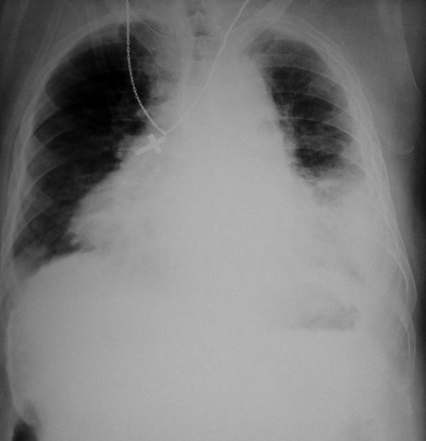
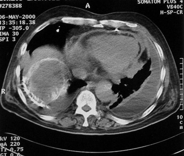
BACKGROUND: Cardiac tamponade as the initial manifestation of metastatic cancer is a rare clinical entity. Furthermore, a thoraco-biliary fistula is another rare complication of echinococcosis due to rupture of hydatid cysts located at the upper surface of the liver to the pleural or pericardial cavity. We report a case of non-small cell lung cancer with a coexisting hepatic hydatid cyst presenting as a bilious pericardial effusion. CASE REPORT: A 66-year-old patient presented with cardiac tamponade of unknown origin. Chest CT-scan demonstrated a left central lung tumor, a smaller peripheral one, bilateral pleural effusions and a hydatid cyst on the dome of the liver in close contact to the diaphragm and pericardium. Pericardiotomy with drainage was performed, followed by bleomycin pleurodesis. The possible mechanism for the bilious pericardial effusion might be the presence of a pericardio-biliary fistula created by the hepatic hydatid cyst. CONCLUSIONS: This is the first case of a bilious pericardial effusion at initial presentation in a patient with lung cancer with coexisting hepatic hydatid cyst.
Figures
References
-
- Frazer RS, Viloria JB, Wang N. Cardiac tamponade as a presentation of extracardiac malignancy. Cancer. 1980;45:1697–1704. - PubMed
-
- Johnson MM, Chin R, Jr, Haponik EF. Thoracobiliary fistula. South Med J. 1996;89:335–339. - PubMed
-
- Haskell RJ, French WJ. Cardiac tamponade as the initial presentation of malignancy. Chest. 1985;88:70–73. - PubMed
-
- DeLoach JF, Haynes JW. Secondary tumors of the heart and pericardium; review of the subject and report of one hundred thirty-seven cases. Arch Intern Med. 1953;91:224–249. - PubMed
-
- Wegener OH. The liver. In: Wegener OH, editor. In Whole Body Computed Tomography. 2. Boston: Blackwell SP; 1992. p. 271.
LinkOut - more resources
Full Text Sources
