Clinical Trial
. 2003 Nov 12;23(32):10224-30.
doi: 10.1523/JNEUROSCI.23-32-10224.2003.

Responses of human motoneurons to corticospinal stimulation during maximal voluntary contractions and ischemia

Affiliations
Clinical Trial

Responses of human motoneurons to corticospinal stimulation during maximal voluntary contractions and ischemia

Jane E Butler et al. J Neurosci. .

Abstract

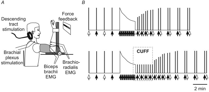
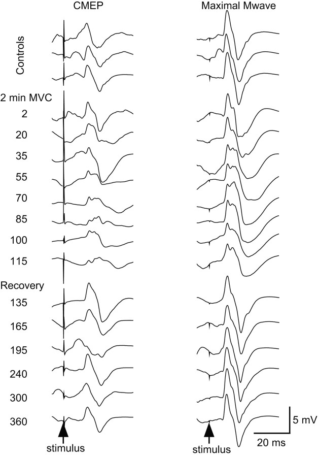
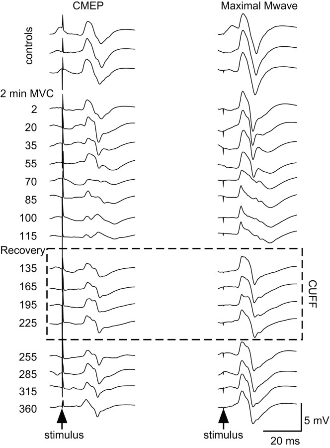
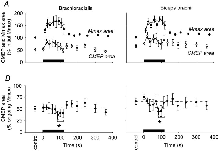
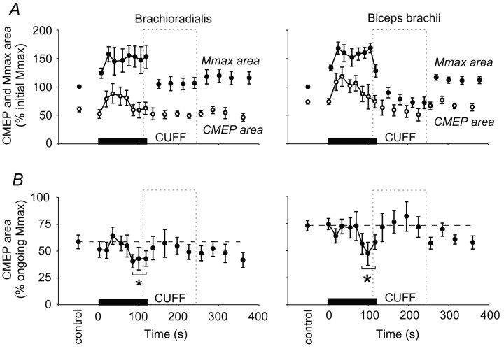
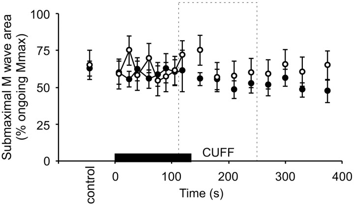
The discharge frequency of human motoneurons declines during a sustained isometric maximal voluntary contraction (MVC) of elbow flexor muscles, but the cause is unresolved. We aimed to determine whether motoneurons were inhibited during a sustained fatiguing contraction of the elbow flexor muscles and whether this inhibition was caused by the discharge of group III and IV muscle afferents. Subjects performed brief MVCs before and after a fatiguing 2 min MVC. During maximal efforts, electromyographic responses recorded from the elbow flexor muscles were evoked by stimulation of the corticospinal tracts at the cervicomedullary level [cervicomedullary motor evoked potentials (CMEPs)] and by supramaximal stimulation over the brachial plexus (Mmax). This revealed a novel decrease in the size of the muscle response to corticospinal tract stimulation during fatigue. During the sustained MVCs, the size of CMEPs decreased to 81 +/- 15 and 78 +/- 15% of the control value for brachioradialis and biceps brachii, respectively (mean +/- SEM; n = 8). This recovered within 15 sec after the fatiguing contraction. In a second set of studies, input from group III and IV muscle afferents was prolonged after the end of the fatiguing contraction by holding the muscle ischemic with a cuff inflated above arterial pressure. Despite the maintained discharge of group III and IV afferents, the CMEPs again recovered within 15 sec of the end of the sustained contraction. These results show a diminished output of spinal motoneurons to stimulation of corticospinal tracts during a fatiguing MVC; however, the mechanisms responsible for this decline are not attributable to activity in group III and IV muscle afferents.


Figures

Figure 1.
Figure 1.
Experimental setup and protocol. A, Experimental setup. B, Two protocols. Brief control MVCs were performed during which stimuli were delivered to the brachial plexus (white arrows) or to the corticospinal tracts (black arrows). The subject then performed a 2 min sustained MVC during which stimuli were delivered alternately to the corticospinal tracts and the brachial plexus. Brief recovery contractions were performed afterward. In the second protocol (bottom panel), the forearm muscles were held ischemic for 2 min after the sustained MVC (region labeled CUFF).
Figure 2.
Figure 2.
Single trials of EMG from a typical subject without maintained muscle ischemia. Single responses from brachioradialis to stimulation of the corticospinal tracts (CMEP, left panels) and the brachial plexus (Mmax, right panels) from a typical subject without a period of postcontraction muscle ischemia. All responses were obtained during maximal efforts: responses during brief control MVCs (top 3 traces), the 2 min sustained MVC (middle 8 traces), and brief recovery MVCs (bottom 6 traces). The time of corticospinal tract stimulation is indicated on the left with brachial plexus stimulation 5 sec later.
Figure 4.
Figure 4.
Single trials from a typical subject with maintained muscle ischemia. Single responses from brachioradialis to stimulation of the corticospinal tracts (CMEP, left panels) and the brachial plexus (Mmax, right panels) from the same subject as in Figure 2 with a period of postcontraction muscle ischemia (dotted box labeled CUFF). Responses are recorded during brief control MVCs (top 3 traces), during the 2 min sustained MVC (next 8 traces), brief recovery MVCs during maintained muscle ischemia (next 4 traces), and during further brief recovery MVCs when the cuff producing ischemia has been released (next 4 traces). The time of corticospinal tract stimulation is indicated on the left, with brachial plexus stimulation 5 sec later.
Figure 3.
Figure 3.
Pooled data for CMEP and Mmax without maintained muscle ischemia. Data from brachioradialis (left panels) and biceps brachii (right panels) before, during, and after a 2 min sustained MVC (0-120 sec; n = 8 subjects). In each panel the solid bar indicates the timing of the sustained maximal effort. A, Panels show the mean area (±SEM) of the responses to stimulation of the corticospinal tracts (CMEP, open circles) and the brachial plexus (Mmax, filled circles) expressed as a proportion of the mean initial maximal M wave (%initial Mmax). Notice the increase in Mmax during the sustained contraction. B, Panels show the mean CMEP area normalized to the Mmax recorded at close to the same time (% ongoing Mmax). During the 2 min sustained MVC the CMEP decreases and then recovers by the first recovery contraction to control size. The broken horizontal lines indicate the mean area of CMEPs during brief control contractions. *CMEPs grouped over the last 40 sec of sustained MVC were reduced compared with those in the control period, in the first 40 sec of sustained MVC and in the first 90 sec of recovery (p < 0.05).
Figure 5.
Figure 5.
Pooled data for CMEP and Mmax with maintained muscle ischemia. Data from brachioradialis (left panels) and biceps brachii (right panels) before, during, and after a 2 min sustained MVC (0-120 sec; n = 8 subjects) with the period of maintained muscle ischemia represented by the dashed box labeled CUFF. In both panels the solid bar indicates the duration of the sustained maximal effort. A, Panels show the mean area (±SEM) of the responses to stimulation of the corticospinal tracts (CMEP, open circles) and the brachial plexus (Mmax, filled circles) expressed as a proportion of the mean initial maximal M wave (% initial Mmax). B, Panels show the mean of the CMEP area normalized to the Mmax recorded at close to the same time (% ongoing Mmax). During the 2 min sustained MVC the CMEP decreases and still recovers by the first recovery contraction to control size despite the maintained period of muscle ischemia. The broken horizontal lines indicate the mean area of CMEPs during brief control contractions. *CMEPs grouped over the last 40 sec of sustained MVC were reduced compared with those in the control period, in the first 40 sec of sustained MVC and in the first 90 sec of recovery (p < 0.05).
Figure 6.
Figure 6.
Responses to submaximal brachial plexus stimulation. Mean (±SEM) area of responses (% ongoing Mmax) from brachioradialis evoked by submaximal stimulation of the brachial plexus (n = 8 subjects). Data are shown from experiments both without (filled circles) and with (open circles) maintained muscle ischemia for 2 min (indicated by dashed box labeled CUFF) after the 2 min MVC (indicated by black bar). Because the ratio remains constant throughout both of the experiments, the data suggest that sarcolemmal changes attributable to fatigue or maintained muscle ischemia affected all muscle fiber types similarly.

References

    1. Allen GM, Gandevia SC, McKenzie DK ( 1995) Reliability of measurements of muscle strength and voluntary activation using twitch interpolation. Muscle Nerve 18: 593-600. - PubMed
    1. Bergmans J ( 1970) The physiology of single human nerve fibres. Vander, Belgium: University of Louvain.
    1. Bigland-Ritchie B, Johansson R, Lippold OCJ, Woods JJ ( 1983a) Contractile speed and EMG changes during fatigue of sustained maximal voluntary contractions. J Neurophysiol 50: 313-324. - PubMed
    1. Bigland-Ritchie B, Johansson R, Lippold OCJ, Smith S, Woods JJ ( 1983b) Changes in motoneurone firing rates during sustained maximal voluntary contractions. J Physiol (Lond) 340: 335-346. - PMC - PubMed
    1. Bigland-Ritchie B, Dawson NJ, Johansson R, Lippold OCJ ( 1986) Reflex origin for the slowing of motoneurone firing rates in fatigue of human voluntary contractions. J Physiol (Lond) 379: 451-459. - PMC - PubMed

Publication types

MeSH terms

LinkOut - more resources