

Lesions of mature barrel field cortex interfere with sensory processing and plasticity in connected areas of the contralateral hemisphere
- PMID: 14614097
- PMCID: PMC6741019
- DOI: 10.1523/JNEUROSCI.23-32-10378.2003
Lesions of mature barrel field cortex interfere with sensory processing and plasticity in connected areas of the contralateral hemisphere
Erratum in
- J Neurosci. 2003 Dec 3;23(35):following 11269
Abstract
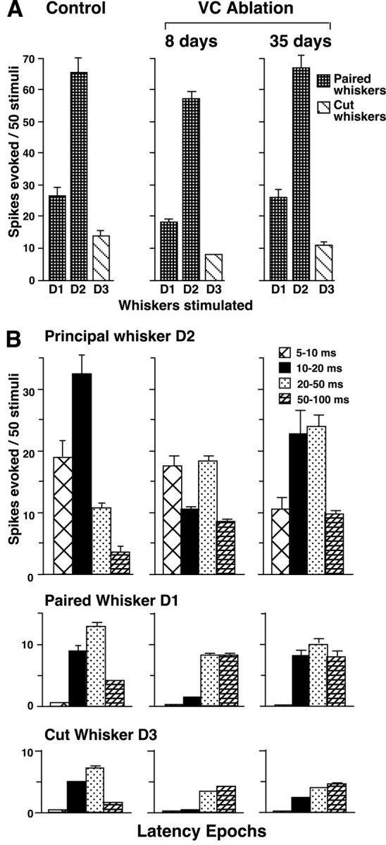
Lesions of primary sensory cortex produce impairments in brain function as an outcome of the direct tissue damage. In addition, indirect lesion effects have been described that consist of functional deficits in areas sharing neural connections with the damaged area. The present study characterizes interhemispheric deficits produced as a result of unilateral lesions of the entire vibrissa representation of S-I barrel field cortex (BFC) in adult rats using single-neuron recording under urethane anesthesia. After unilateral lesions of adult BFC, responses of neurons in the contralateral homotopic BFC are severely depressed. Background (spontaneous) activity is reduced by approximately 80%, responses to test stimuli applied to the whiskers are reduced by approximately 50%, and onset of synaptic plasticity induced by trimming all but two whiskers ("whisker-pairing plasticity") is delayed over sevenfold compared with sham-lesion control animals. These deficits persist with only slight improvement for at least 4 months after lesion. Both fast-spiking and regular-spiking neuron responses are diminished contralateral to the lesion, as are cells above, below, and within the cortical barrels. Enriched environment experience increased the magnitude of responses and accelerated the rate of synaptic plasticity but did not restore response magnitude to control levels. Deficiencies in evoked responses and synaptic plasticity are primarily restricted to areas that share direct axonal connections with the lesioned cortex, because equivalently sized lesions of visual cortex produce minimal deficits in contralateral BFC function. These results indicate that interhemispheric deficits consist of remarkable and persistent decrements in sensory processing at the single-neuron level and support the idea that the deficits are somehow linked to the shared neural connections with the area of brain damage.
Figures


Similar articles
-
Chronic suppression of activity in barrel field cortex downregulates sensory responses in contralateral barrel field cortex.J Neurophysiol. 2005 Nov;94(5):3342-56. doi: 10.1152/jn.00357.2005. Epub 2005 Jul 13. J Neurophysiol. 2005. PMID: 16014795
-
Contribution of supragranular layers to sensory processing and plasticity in adult rat barrel cortex.J Neurophysiol. 1998 Dec;80(6):3261-71. doi: 10.1152/jn.1998.80.6.3261. J Neurophysiol. 1998. PMID: 9862920
-
Balancing bilateral sensory activity: callosal processing modulates sensory transmission through the contralateral thalamus by altering the response threshold.Exp Brain Res. 2006 Jul;172(3):397-415. doi: 10.1007/s00221-005-0337-y. Epub 2006 Jan 21. Exp Brain Res. 2006. PMID: 16429268
-
Intracortical processes regulating the integration of sensory information.Prog Brain Res. 1990;86:129-41. doi: 10.1016/s0079-6123(08)63172-6. Prog Brain Res. 1990. PMID: 1982365 Review.
-
Local cortical interactions determine the form of cortical plasticity.J Neurobiol. 1999 Oct;41(1):58-63. J Neurobiol. 1999. PMID: 10504192 Review.
Cited by
-
Ipsilateral whiskers suppress experience-dependent plasticity in the barrel cortex.J Neurosci. 2007 Apr 4;27(14):3910-20. doi: 10.1523/JNEUROSCI.0181-07.2007. J Neurosci. 2007. PMID: 17409256 Free PMC article.
-
Functional MRI detection of bilateral cortical reorganization in the rodent brain following peripheral nerve deafferentation.Neuroimage. 2007 Aug 1;37(1):262-73. doi: 10.1016/j.neuroimage.2007.03.069. Epub 2007 Apr 25. Neuroimage. 2007. PMID: 17544301 Free PMC article.
-
Reorganization of motor cortex after controlled cortical impact in rats and implications for functional recovery.J Neurotrauma. 2010 Dec;27(12):2221-32. doi: 10.1089/neu.2010.1456. Epub 2010 Nov 22. J Neurotrauma. 2010. PMID: 20873958 Free PMC article.
-
Shaping plasticity to enhance recovery after injury.Prog Brain Res. 2011;192:273-95. doi: 10.1016/B978-0-444-53355-5.00015-4. Prog Brain Res. 2011. PMID: 21763529 Free PMC article. Review.
-
Deficits in Behavioral Functions of Intact Barrel Cortex Following Lesions of Homotopic Contralateral Cortex.Front Syst Neurosci. 2018 Nov 22;12:57. doi: 10.3389/fnsys.2018.00057. eCollection 2018. Front Syst Neurosci. 2018. PMID: 30524251 Free PMC article.
References
-
- Al-Abdulla NA, Martin LJ ( 2002) Projection neurons and interneurons in the lateral geniculate nucleus undergo distinct forms of degeneration ranging from retrograde and transsynaptic apoptosis to transient atrophy after cortical ablation in rat. Neuroscience 115: 7-14. - PubMed
-
- Andrews RJ ( 1991) Transhemispheric diaschisis. Stroke 22: 943-949. - PubMed
-
- Armstrong-James M, Fox K ( 1987) Spatiotemporal convergence and divergence in the rat SI “barrel” cortex. J Comp Neurol 263: 265-281. - PubMed
-
- Armstrong-James M, Millar JM ( 1979) Carbon fibre microelectrodes. J Neurosci Methods 1: 279-287. - PubMed
Publication types
MeSH terms
Grants and funding
LinkOut - more resources
Full Text Sources